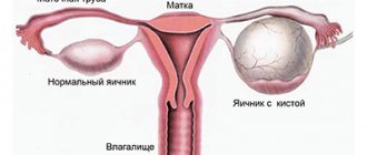
In each organ, completely specific malignant tumors can develop; this is not always cancer, because cancer develops only from epithelial cells, which normally form mucous membranes. In the ovaries, every tenth tumor is not cancer, but germ cell neoplasms. Half of them are malignant, and they affect girls, young women and young women. A similar variant of neoplasms occurs in boys and young men.
Germ cell tumors arise as a result of chromosomal abnormalities; it is known for certain that chromosome 12 loses its long arm, and a second short arm develops instead. Almost a dozen more chromosomes suffer: somewhere the genetic material decreases, somewhere, on the contrary, it arrives, but not chaotically, but strictly according to schedule. Therefore, all girls with disorders of puberty, and boys too, should be sent for genetic testing, which allows for early follow-up and timely detection of the tumor.
Ovarian germ cell tumors
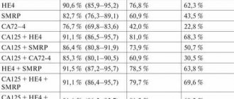
The table below shows the classification of ovarian germ cell tumors
. Benign dermoid cysts are quite common. They are cured surgically. Such cysts are mainly composed of functional thyroid tissue, such as the struma of the ovary.
Less than 5% of struma are represented by malignant tumors
, so-called monomorphic or monodermal teratomas. In rare cases, they develop into secondary malignant tumors, usually squamous carcinoma.
The most common malignant tumor is dysgerminoma
. Histologically, it is a homogeneous tumor resembling a male seminoma. Often the tumor is infiltrated by lymphocytes. Highly malignant is an epidermal sinus tumor (yolk sac tumor), which produces a-fetoprotein (AFP), which accumulates in the tumor itself and in the blood.
tumor often ruptures
. Embryonic carcinoma consists of glandular and papillary areas in which trophoblasts secreting chorionic gonadotropin (hCG) are often found. The tumor can develop in girls and causes premature puberty. The source of a-fetoprotein can also be elements of the yolk sac.
Solid tumors
, are mainly represented by immature teratoma containing areas of various tissues. Basically, these areas consist of primary neuroectoderm, but monomorphic forms represented by thyroid and malignant carcinoid tissue are also found. Due to the large size that the tumor can reach, carcinoid syndrome can occur even in the absence of metastases.
Mixed germ cell tumor
consists of the mentioned cell types. Gonadoblastoma is a small tumor that develops in childhood. It usually contains several elements characteristic of developing gonads. The tumor can calcify and rarely metastasize, but sometimes it causes virilization of the body, since it often includes functionally active Leydig cells.
Clinical picture of ovarian germ cell tumors
Development of dysgerminomas
, like other ovarian tumors, is accompanied by an increase in the volume of the abdominal cavity and pain. However, the average age of patients with these tumors is much lower than for other types of tumors. Dysgerminoma is diagnosed in 75% of women aged 10-30 years (average age 20 years).
If the tumor does not contain teratomatous elements
, then it does not produce marker hormones, and occurs quite often during pregnancy or shortly after its completion.
Other tumors of germ cell origin
(yolk sac tumor, endodermal sinus tumor, teratoma, mixed germ cell tumors) also develop at a young and young age. Many patients experience signs of premature puberty, menstrual irregularities, and a positive pregnancy test.
When carrying out differential diagnosis
it is necessary to take into account the possibility of an increase in the volume of the abdominal cavity due to pregnancy. Typically, these tumors grow quickly, which is accompanied by abdominal pain.
Treatment of ovarian germ cell tumors
Treatment with dysgermin
depends on the stage of tumor development. Up to 90% of patients in stage I of the disease are cured after unilateral oophorectomy, but the likelihood of recurrence is high if the tumor has reached a large size or contains mixed elements of germinoid cells, as well as with positive results of analysis of peritoneal lavages.
For relapses
or if metastases are detected, as in the case of teratoma, chemotherapy or radiation to the abdominal cavity is usually used. For tumors of the yolk sac, endodermal sinus and mixed-type germ cell tumors, even those at stage I, the use of surgical treatment gives a less favorable prognosis than for dysgerminoma.
Analysis for the presence of tumor markers
, such as AFP and b-hCG, allows you to monitor the effectiveness of the use of cytostatics, which are mainly used today to treat most patients (exceptions are discussed below).
Significant progress in the treatment of testicular teratoma
achieved using platinum group drugs. Since the tumor affects one ovary, unilateral salpingectomy and oophorectomy followed by chemotherapy can be performed. At the same time, reproductive function is preserved. Chemotherapy is not prescribed for teratomas with low mitotic activity, which do not contain embryonic or trophoblastic elements, and also if the test for the presence of tumor markers done after surgery was negative.
In such cases, to treat patients
Only the surgical method is used, but after the operation they must be carefully examined. Ovarian choriocarcinoma, like other nondysgerminomatous germ cell tumors, usually responds to chemotherapy. In this case, a combination of cisplatin, etoposide and bleomycin is used according to the same regimen that is used to treat testicular non-seminomatous germ cell tumors. The effectiveness of modern chemotherapy methods is well illustrated by the results obtained on 59 patients with tumor metastases who were treated in a large London clinic.
Treatment of germ cell tumors
We select treatment taking into account the severity of the disease and the patient’s condition. We not only treat the disease, but also maintain the quality of life of our patients after therapy:
- Surgical treatment -
the extent of intervention depends on the stage of cancer; doctors try to completely remove tumor tissue to avoid relapse in the future. At the Oncology Clinic, minimally invasive endoscopic operations are most often used. Gentle intervention shortens the patient’s recovery time. - Chemotherapy with modern drugs complements
surgical treatment and is prescribed before and after surgery.
What is the difference between the treatment of germ cell tumors at the Oncology Clinic?
❏ Multidisciplinary hospital with the ability to treat concomitant diseases.
❏ Polychemotherapy with modern drugs with low toxic effect.
❏ The clinic’s operating rooms have the latest equipment, including for low-traumatic interventions.
❏ A careful approach to therapy while preserving the function of the reproductive system.
Causes of development and risk factors for occurrence
The source of germ cell tumors of the ovaries is germ cells. These neoplasms copy embryogenesis in a distorted form. Neoplasia develops from pluripotent germ cells, but the exact mechanism of their occurrence and transformation has not been studied.
Normally, germ cells should become mature cells of the ovaries or testes in a child, but under the influence of unfavorable factors they can remain in the embryonic state and cause the appearance of gonadal tumors.
Predisposing development factors include:
- existing genetic abnormalities;
- chronic diseases of the mother;
- taking certain medications during pregnancy.
This type of pathology often occurs with Klinefelter syndrome. An existing hereditary predisposition can be combined with chromosomal abnormalities. But this is not a necessary condition for the occurrence of tumors.
Germ cell tumors of the ovaries appear in patients with chromosomal abnormalities. Researchers have found that patients with this disease do not have a long arm on chromosome 12; instead, a second short arm is formed. In addition, the structure of about 10 other chromosomes is disrupted.
Symptoms
Benign germ cell tumors appear predominantly on only one ovary. If cancer has begun, then neoplasms will be detected on 2 ovaries at once.
The clinical picture of germ cell tumors of the ovaries in patients is uniform. Characteristic signs appear due to the rapid growth of tumor formations. Women note the development of abdominal pain and the occurrence of other nonspecific signs of the disease:
- increase in abdominal volume;
- fever;
- general weakness.
Some patients are diagnosed with neoplasms after being admitted to the hospital with symptoms of an acute abdomen. But the pathology is not always accompanied by the appearance of these signs; the disease may be asymptomatic.
In women of reproductive age, pain is often accompanied by menstrual irregularities. Patients who have an enlarged abdomen complain of problems with urination.
In later stages, ascites and dysfunction of other organs are detected.
Classification of germ cell tumors
All germ cell tumors are divided into dysgerminomas and non-dysgerminomas. Each of these species contains several cellular subspecies.
Dysgerminomas are neoplasms that are analogous to seminoma. The sizes of tumor-like formations vary from small (several cm in diameter) to huge (about 50 cm). Dysgerminomas are hormonally inactive. With dysgerminomas, necrosis and hemorrhage are less common than with other types of tumors.
Non-dysgerminomas include:
- yolk sac tumor;
- choriocarcinoma;
- embryonal cancer;
- polyembrinoma;
- mixed types, in which several variants of neoplasia are combined.
Teratomas are also isolated. Mature teratomas are benign tumors that contain cells of cartilage, nervous tissue, non-mucinous and mucinous glands. These types of cells can malignize (become malignant) and transform into squamous cell carcinoma or primitive neuroendocrine tumor.
Among malignant germ cell tumors, the predominant histotype is known as dysgerminoma. Among all germ cell neoplasias, it accounts for 30-40%.
Diagnosis and treatment of germ cell tumors
The diagnosis is established taking into account complaints, the results of a physical examination and additional research data. Depending on the location of the neoplasia, a rectal examination or vaginal examination may be required. Patients are prescribed ultrasound, CT and MRI of the affected area. The content of alpha-fetoprotein in the blood serum is assessed. For malignant germ cell tumors, to exclude lymphogenous and distant metastases, chest radiography, ultrasound and MRI of the abdominal organs, ultrasound of the lymph nodes, scintigraphy of skeletal bones and other diagnostic procedures are performed. The type of neoplasia is determined taking into account histological examination data.
Benign germ cell tumors are excised; for malignant neoplasms, a combination treatment is prescribed, including surgery (for resectable neoplasia), chemotherapy and radiotherapy. In the presence of single metastases in the lungs and liver, their surgical removal is possible. When the effectiveness of therapy for aggressive seminomas is low, in some cases high-dose radiotherapy is performed followed by bone marrow transplantation, however, the effectiveness of this method in germ cell tumors is still difficult to assess due to the insufficient number of observations.
The prognosis for benign neoplasia is usually favorable. Malignant germ cell tumors were previously considered to have a poor prognosis, but the use of combination therapy has increased the five-year survival rate for this pathology to 60-90%. Survival is influenced by the type and extent of germ cell tumor, the radicality of surgery, and the presence or absence of metastases.
Original text and images from the site: https://www.krasotaimedicina.ru
Diagnosis and treatment
To establish a preliminary diagnosis, the doctor must examine the patient and conduct a survey. If characteristic complaints appear and a moderately mobile round node is detected during palpation, which has clear contours, the doctor may suspect the formation of a germ cell tumor.
For diagnostic purposes, ultrasound, MRI, and CT are prescribed. It is important to evaluate the content of α-fetoprotein, β-hCG, LDH, CA-125 in the blood. If gonadal dysgenesis is suspected, karyotyping is prescribed. When detecting malignant neoplasia, a chest x-ray, MRI or ultrasound of the abdominal cavity, scintigraphy of skeletal bones, and ultrasound of the lymph nodes are required. With the help of these studies, metastases in the body can be detected.
Treatment tactics are selected depending on the type of disease. For benign neoplasms, surgery is required. For malignant types, radiotherapy, chemotherapy and surgery are prescribed if the patient has resectable neoplasia. When single metastases are detected in the tissues of the liver or lungs, their surgical removal is practiced.
Diagnostics
Since germ cell tumors can produce specific antigens, the levels of hCG and AFP markers are determined before treatment and during treatment, as well as during dynamic observation throughout life. When the markers are normal, LDH is also determined before, during and after treatment. Normalization of the level of markers or enzyme is considered evidence of the absence of a tumor; accordingly, an increase in concentration is interpreted as progression of the malignant process.
During preoperative examination, and always in case of high levels of hCG or multiple metastases in the lungs, MRI of the brain with contrast is used. In a widespread process, asymptomatic metastases may exist in the brain. A CT scan from the neck to the small pelvis is required, which makes it possible to identify tumor screenings and specifically look for them during surgery, or even abandon surgery in favor of chemotherapy for metastatic lung disease.
Complications that the disease can cause
With the formation of germ cell tumors of the ovaries, a deterioration in health may occur, which is caused by the progression of the disease or torsion of the pedicle of the tumor.
Lack of timely treatment of malignant neoplasia can cause:
- infertility;
- menstruation disorders;
- pain in the lower abdomen.
There is a possibility of malignancy of benign germ cell tumors.
If malignant neoplasia is not treated in a timely manner, it is possible:
- worsening of the oncological process;
- metastasis;
- an increase in the size of the tumor, its germination into neighboring organs and tissues;
- reducing the likelihood of a favorable outcome.
When malignant neoplasms appear, death is possible.
Germ cell tumors of the ovaries are detected mainly in adolescent girls and young women, but they may appear in late reproductive age. These neoplasms are characterized by high rates of growth and spread to neighboring organs. You can understand the types of tumor-like formations on the ovaries by watching the video
Germ cell tumors
Germ cell tumors
– a group of neoplasias developing from the primary germ cells of the gonads. They can occur in the testes or ovaries, or extragonadally. Manifestations depend on location. With superficially located neoplasms, visible deformation is observed; with nodes in the ovary, pain, dysuria and menstrual irregularities are noted. With germ cell tumors of the mediastinum, shortness of breath occurs; with intracranial lesions, focal and cerebral symptoms are detected. The diagnosis is made taking into account symptoms, X-ray data, ultrasound, CT, MRI and other techniques. Treatment – surgery, chemotherapy, radiotherapy.
Signs of germ cell tumors
Tumors of this type grow asymptomatically for a long time; the first symptoms of the disease —
slight pain in the localization area: in the abdomen, scrotum area in boys, lower back. There may also be a general deterioration in health and weight loss.
In these cases, you should consult a doctor as soon as possible to clarify the diagnosis. At the Oncology Clinic, you can undergo the necessary examinations in a short time: from the first visit to the doctor to the diagnosis, usually no more than 14 days pass.
Stages and types of germ cell tumors
Based on the type of cells, there are more than 5 types of germ cell tumors; based on their location, they are divided into gonadal, located in the gonads, and extragonadal, located in other organs.
The tumor is also classified according to the TNM system, where T—
tumor size, N
-
metastases to lymph nodes, M
-
metastases to other organs.
We treat germ cell tumors of any type and at any stage, selecting effective treatment at a consultation with the participation of specialized doctors. Even complex cases are now treatable using modern chemotherapy and surgery techniques
How can we help with germ cell tumors?
We offer compulsory medical insurance treatment in accordance with international standards. In each case, we hold a consultation with the participation of oncologists, surgeons, radiologists and chemotherapists.
We individually select conservative and surgical treatment to help eliminate the tumor, maintain comfort and quality of life, and quickly recover after surgery and chemotherapy.
We use fasttrack surgery - priority to endoscopic operations, active recovery after treatment, maximum manipulations at a time and preservation of organ function. This approach shortens the rehabilitation period and reduces the risk of complications.
General information
Germ cell tumors are a group of benign and malignant neoplasias that arise from primary germ cells, which are the precursors of the testes and ovaries. Due to the migration of such cells during embryogenesis, germ cell tumors can develop outside the gonads: in the mediastinum, sacrococcygeal region, brain, retroperitoneum and other anatomical areas. Primary extragonadal neoplasms account for 5% of the total number of germ cell tumors.
The ratio between the number of extra- and intragonadal neoplasia changes with age. In young children, lesions of the sacrococcygeal zone predominate; as they grow older, the frequency of neoplasms in the testicles and ovaries increases. Germ cell tumors of all localizations account for 3% of the total number of oncological diseases in children, germ cell tumors of the ovaries - 2-3% of all malignant neoplasia of the ovaries in women, germ cell lesions of the testicle - 95% of the total number of testicular tumors in men. Treatment is carried out by specialists in the field of oncology, gynecology, urology and other fields of medicine.
Treatment of germ cell tumors
Germ cell tumors are more common in children and young people; this is a very rare disease -
only 4-6 people per million are affected. They are formed from embryonic germ cells that are found in the ovaries in women and testicles in men. It is extremely rare that such tumors are detected outside the gonads, for example, in the pelvic cavity, kidneys, or in the sacrum or coccyx area.
Most patients can make a full recovery; if a complete cure is not possible, life expectancy can be significantly extended. To achieve such results, doctors at the Oncology Clinic use all modern technologies: high-precision surgical techniques for tumor removal and chemotherapy with the latest drugs. This allows you to destroy cancer cells and preserve the functions of the reproductive system as much as possible.
We understand how important it is to start treatment in a short time: oncology patients are seen by appointment without waiting in line. You can see an oncologist the very next day after the call, and begin therapy within 5-7 days if the necessary tests are available. A team of oncologists, radiologists, chemotherapists, and rehabilitation specialists works with each patient.
Causes of germ cell tumors
Germ cell tumors arise from germ cells, which in the initial stages of embryogenesis are formed in the yolk sac and then migrate throughout the body of the embryo to the urogenital ridge. During the migration process, some of these cells may linger in various anatomical zones, which subsequently causes the formation of germ cell tumors of extragonadal localization. Normally, germ cells turn into mature cells of the testes and ovaries, however, under certain conditions, such cells can remain in their embryonic state and, under the influence of negative external and internal factors, give rise to neoplasms of the gonads.
It has been established that germ cell tumors are often diagnosed in patients with various genetic abnormalities, for example, Klinefelter syndrome. A hereditary predisposition is identified, which may or may not be combined with chromosomal abnormalities. A characteristic feature of germ cell tumors is the isochromosome, which results from the duplication of the short arm and the loss of the long arm on chromosome 12, however, other chromosomal abnormalities may also be detected. There is a frequent combination of germ cell tumors with other oncological lesions, including leukemia, lymphoma and neuroblastoma. The likelihood of testicular germ cell neoplasia increases with cryptorchidism.
The histological type of germ cell tumors depends on age. Benign teratomas are more often diagnosed in newborns, neoplasia of the yolk sac is detected in young children, malignant teratomas and dysgerminomas are detected in adolescents, seminomas in adults, etc. Factors contributing to the activation of growth and malignant transformation of germinal germ cells have not yet been clarified. It is assumed that the impetus for the development of germ cell tumors in children may be chronic diseases of the mother or the mother's taking certain medications.
Morphological types
Experts distinguish the following types of tumor formations of germ cell origin into this group.
Germinoma (dysgerminoma, seminoma)
Germinoma is a rare disease. Tumor formation occurs in the prenatal period. The main provoking factor in the appearance of pathology is the abnormal development of embryonic tissue.
The period of manifestation of the pathological condition occurs in childhood, in particular, by 12 years. It is rare for the disease to be diagnosed at 30 years of age.
According to statistics, germinomas are most often detected in the male half of the population and are localized in the brain.
On this topic
- General
What is a cancer examination
- Natalya Gennadievna Butsyk
- December 6, 2021
Dysgerminoma is also a rare type of tumor formation and accounts for approximately 2 percent of all primary ovarian lesions. This type is one of the most common among all germ cell tumors. It is diagnosed at any age in both children and adults.
Seminoma is a malignant neoplasm. In rare cases, it affects the retroperitoneum and mediastinal area.
Formation occurs from the primary cellular structures of the embryo. When localized in the testicle, it manifests itself as compaction, pain and discomfort in the scrotum.
If the process is accompanied by the spread of metastases, the patient will complain of pain in the abdomen, swelling of the legs, the development of intestinal obstruction and difficulty urinating. Most often, seminoma is detected at the age of 20-40, mainly in males.
Embryonic cancer
This variety belongs to oncological diseases and poses the greatest danger to human health and life, since it is distinguished by an increased degree of malignancy. Tumor development occurs from a highly specialized part of the germ cell type epithelium.
The structure of the cells has a demarcated shape and round nuclei. In most cases, the testes and ovaries are affected. Also, the site of localization can be the silky body, ventricular plexuses, retroperitoneal space, nasal cavity and jaws.
Among all the possible causes of embryonal cancer, experts highlight excessive use of diethylstilbestrol during pregnancy.
Yolk sac neoplasia
The advantage applies to germ cell tumors in children. This is a rare disease that is diagnosed before the age of three.
In some cases, it can also be detected in adults, mainly in development with other types of tumors of this variety.
Spermatocyte seminoma
The structure of the neoplasm consists of 3 types of testicular germ cells. Makes up about 4.5 percent of the total number of seminomas. It is more often found in people 52-58 years old. It can be asymptomatic for a long time.
Chorionic carcinoma
This is a tumor of trophoblastic origin. Formation occurs when the chorionic epithelium becomes malignant.
The location of the neoplasm is the uterine body. Rarely, primary lesions may be located in the fallopian tubes, ovaries, lungs, or vagina. Brain damage cannot be ruled out.
Polyembryoma
In most cases, the tumor is unilateral, which occurs in extremely rare cases. It is detected mainly in young and middle-aged women. They can reach different sizes.
Teratoma
A neoplasm of embryonic cell origin, the formation of which occurs from the meso-, endo- and exodermis. With a benign course of the pathological process, there is no clinical picture. When the tumor reaches a large size, pressure occurs on nearby organs and systems, which is accompanied by corresponding symptoms.
Mixed germ cell tumor
These are compactions, the structure of which contains two or more germinogenic histological types of malignant nature. One of the most common elements is dysgerminoma.
A tumor of the yellow sac and an immature teratoma are also detected. In rare cases, choriocarcinoma and embryonal carcinoma can be found.
Classification of germ cell tumors
There are several classifications of germ cell neoplasia, compiled taking into account the morphological characteristics of the neoplasm, location and characteristics of the course of the disease. According to the WHO classification, the following morphological types of germ cell tumors are distinguished:
- Germinoma (dysgerminoma, seminoma)
- Embryonic cancer
- Yolk sac neoplasia
- Spermatocyte seminoma
- Chorionic carcinoma
- Polyembryoma
- Teratoma, including mature, immature, with a certain direction of tissue differentiation (carcinoid, ovarian struma), malignant.
- Mixed germ cell tumor, which is a combination of several histological variants of neoplasia.
Diagnosis of germ cell tumors
We use modern high-precision diagnostic methods that help make the correct diagnosis in a short time:
- Ultrasound of the pelvis and abdomen helps assess the location and size of the primary tumor.
- MRI, CT are
diagnostic methods for assessing the shape and size of the lesion, tissue changes. prescribed for certain forms of germ cell cancer. - A blood test to look for tumor markers that are elevated in this type of malignancy.
- PET/CT is
a detailed examination of a tumor on a modern scanner and detection of the smallest metastases from 2 mm. - Biopsy with histology -
taking a tissue sample to determine the type of cancer cells and their malignancy. If germ cell tumors are suspected, the procedure is performed only in specialized oncology clinics.
An oncologist may prescribe other tests or examinations for you: each case is unique, and an individual diagnostic regimen is needed to make an accurate diagnosis.
Symptoms of germ cell tumors
Features of the course of the disease are determined by the location, size and degree of malignancy of the neoplasia. Typical symptoms of ovarian germ cell tumors are abdominal pain of varying intensity in combination with menstrual irregularities. In children, the latter sign is absent, which causes a lack of alertness regarding damage to the internal genital organs in the initial stages of the disease. As germ cell tumors progress, the listed symptoms are accompanied by abdominal enlargement and urination problems. Upon palpation in the initial stages, a round, moderately mobile node with clear contours is determined. Subsequently, the node increases in size, and enlargement and deformation of the abdomen occur. At later stages, ascites and dysfunction of various organs due to distant metastasis are detected.
Germ cell tumors of the testicle are manifested by an enlargement of the corresponding half of the scrotum, a feeling of heaviness and distension. Soreness or increased sensitivity of the affected area is noted by about 25% of patients. Palpation will reveal a tumor-like formation or uniform enlargement of the testicle. In 5-10% of patients with germ cell tumors, hydrocele is detected, in 10-14% - gynecomastia. With lymphogenous and distant metastasis, enlargement of the inguinal lymph nodes, neurological disorders, pain in the bones, back and abdomen are possible.
Germ cell tumors of the mediastinum are usually localized behind the sternum. Benign neoplasms (teratomas) are characterized by slow growth, while malignant neoplasms (teratoblastomas and other neoplasias) are characterized by aggressive spread and rapid germination of nearby organs. The most common manifestations of germ cell tumors are shortness of breath, cough and chest pain. When the superior vena cava is compressed, noise in the head, headache, tinnitus, disturbances of consciousness, drowsiness and visual disturbances occur. Convulsions are possible. With malignant germ cell tumors, hyperthermia, fever, weight loss and dysfunction of various organs due to germination or distant metastasis are observed.
Retroperitoneal germ cell tumors are asymptomatic for a long time. May manifest as dyspepsia, abdominal pain, dysuria, shortness of breath, edema and varicose veins of the lower extremities. With malignant lesions in the later stages, symptoms of cancer intoxication are revealed. Germ cell tumors of the sacrococcygeal zone are usually diagnosed in young children and have a benign course. With large neoplasia, pain and weakness in the lower extremities, defecation disorders and dysuria are observed. Bleeding and necrosis are possible. Intracranial germ cell tumors are most often localized in the area of the pineal gland, sometimes in the area of the hypothalamus or pituitary gland. Manifested by headache, nausea, vomiting and disorders of eyeball movements.
Forms of germ cell tumors
Oncologists distinguish two existing classes of germ cell tumors:
- seminomas, accounting for 60% of the incidence;
- nonseminomas, accounting for 40% of diseases.
Seminoma is formed from germ cells that produce sperm. In 70% of patients, the growth does not extend beyond the testicle; in 25%, lymph nodes are affected; in 5%, metastases are observed.
There are 2 types of seminoma formations:
- typical - observed in 95% of patients - men after 40 years of age;
- spermatocyte - rare, slowly growing, practically does not spread metastases, “pure”, occurs in men after 55 years, contains 3-7% of all seminoma lesions. It is asymptomatic and is noted only by an increase in the size of the gonads.
Non-seminoma types of cancer usually occur in adolescents; they can develop in men up to 40 years of age.
The main 4 types of nonseminoma are:
- damage to the yolk sac;
- teratoma, including 2-3 germ layers - ento, ecto, mesoderm;
- dermoid cyst;
- embryonal carcinoma;
- dysgerminoma.
- chorionic carcinoma.
These types of germ cell tumors have the ability to metastasize to lung tissue and lymph nodes. At the time of contacting a doctor, the patient is already diagnosed with stages 3-4 of the metastasis process.
Compared to other types of oncological pathologies among children and adolescents, germ cell tumors are considered rare, accounting for 3-8%. Girls are more susceptible to diseases than boys. Having reached the transitional, adolescent period, the incidence of testicular germinomas increases in boys.
Serious attention should be paid to the fact that an egg that has not descended into the scrotum poses a particular danger to a child - having reached puberty, such a teenager has a huge risk of acquiring oncological formation of the organ.
The group of neoplasms developing from a population of germ cells is extremely diverse in terms of localization of the lesion site and histological structure.