Causes
To date, medicine has not yet been able to establish the exact causes of the appearance of malignant and benign tumors in the pelvis. However, experts identify a number of negative factors that predispose to the development of this pathology.
The most common of them are:
- genetic inheritance;
- exposure to chemicals in the household or in hazardous production conditions;
- the effects of radiation or exposure (including frequent visits to the solarium);
- entry into the body of harmful bacteria or viral infections;
- decreased level of immunity;
- disorders of the endocrine system and hormonal imbalance.
In addition, it is necessary to highlight the presence of tumors of different types, taking into account age groups.
Thus, newborn girls may continue to be exposed to maternal placental hormones for several months. As a result, cystic formations often occur on the ovaries.
During puberty, disturbances in the development of the vagina, uterus and hymen are sometimes observed. For this reason, there are difficulties with the outflow of fluid during menstruation and its subsequent accumulation.
Tumor formations arising in the pelvic region are considered depending on their structural origin.
Many of them appear to come from the pelvis when in fact they come from the abdominal cavity. Ultrasound scanning improves the identification of formations that are not always possible to detect by palpation during vaginal or rectal examination. The origin of tumor-like formations can be described using five components (5 “Fs”):
- fat (fat);
- liquid (fluid);
- faeces;
- flatulence;
- fruit (fetus).
Diagnosis requires a thorough history, clinical examination, and appropriate imaging. Many of the tumor-like formations come from certain organs. Therefore, this part provides a brief overview and links to the relevant sections of the book. Tumor-like formations are examined anatomically from front to back, finally moving to the bones surrounding the pelvis.
What is a uterine cyst? Description, reasons for formation
A benign formation that occurs on the cervix or its appendages is referred to in medicine as a uterine cyst. Its formation is the result of blockage of the uterine glands and filling them with mucous secretion, which leads to their stretching.
Another name for a cyst of the gland of the uterine cervix is Nabothian cyst, and most often it is multiple, several millimeters in size.
A woman with a cyst usually does not have any sensations, and the pathology is detected during a routine gynecological examination.
A uterine cyst looks like a yellowish-white, rounded formation. The main cause of cyst formation is inflammation of the cervix and cervical canal, which occurs due to various injuries and infections during childbirth, abortion, and curettage.
The causes of cysts on the cervix may also lie in hormonal imbalances in the female body. The occurrence of inflammatory diseases such as endometritis, salpingitis and colpitis can occur due to the accumulation of infection in the cervix.
This means that Nabothian cyst requires attention and appropriate treatment.
A cyst on the cervix may have the following reasons for its formation:
- early onset of menstruation;
- malfunction of the thyroid gland;
- colds;
- imbalance of hormonal levels and endocrine system;
- inflammatory diseases of the genital area;
- sexual infections;
- traumatic birth;
- mechanical interventions in the uterine cavity.
Is a cyst on the uterus dangerous?
The pathology of a uterine cyst usually does not show symptoms. However, in some cases it can become filled with pus, requiring surgical removal. There is no point in hoping that the tumor will disappear on its own - in some women they grow so quickly that they deform the cervix.
Experts recommend removing the cyst before it suppurates and grows, preventing the development of inflammatory diseases of the woman’s genital area.
Considering the issue of the danger of cysts for women’s health, it is worth noting that the opinion of experts is not unanimous.
Thus, some doctors believe that the cyst not only does not pose a danger, but is even considered a normal phenomenon that does not cause complications and does not require treatment. Other experts are convinced of the need for surgical intervention in the neoplasm, since purulent infiltrate accumulates in it.
The opinion of third experts is intermediate - removal of the cyst is not a mandatory measure, but if such an opportunity exists, then it is better to remove it and clean the secretory ducts.
Diagnosis of a cyst
Gynecology has several diagnostic methods confirming the presence of a uterine cyst:
- medical examination;
- examination for urogenital infections;
- extended colposcopy;
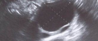
- Ultrasound of the pelvis;
- smear for oncocytology;
- PAP smear (Papanicolaou smear).
Thanks to the latest technique, specialists have the opportunity to timely pay attention to initial precancerous changes. A PAP smear is performed with a specially selected composition of paints and fixatives. For prevention purposes, doctors recommend that women do it annually.
Treatment in different ways
Unfortunately, there are no conservative methods to combat cysts. However, the variety of surgical treatment methods allows you to choose the most optimal option for each specific patient. The techniques that have proven to be the most effective are:
- electrocoagulation, or cauterization with electric current;
- cryodestruction (evaporation with liquid nitrogen);
- laser cyst removal;
- radio wave method.
Radio wave surgery is the best option for most women. The operation looks like removing a tumor using a special Surgitron device. It is carried out in several stages:
- puncture of the cyst shell;
- deleting content;
When diagnosed with a cervical cyst, treatment with the radio wave method is the most rational, since it eliminates the possibility of bacterial infection and bleeding. The undeniable advantage of radio wave surgery is almost instantaneous healing.
If purulent-inflammatory fluid has accumulated in the cyst cavity, it is removed by puncturing.
Subsequently, having determined the sensitivity of the pathogen to antibiotics, appropriate treatment is prescribed.
If the “cryo” technique was chosen as therapy, then the diseased area is simply treated with liquid nitrogen. This painless and gentle treatment of cervical cysts does not leave behind scar changes.
As for laser therapy for cysts, a number of its advantages are also great.
Removal of inflamed tissue, coagulation of blood vessels, absence of bleeding, painlessness, tissue sparing, accessibility for nulliparous women and lack of complications make it a rival to radio wave surgery. Another good thing about laser cyst removal is that the beam penetrates exactly to the depth that is necessary in each specific case.
Features of the treatment of recurrent (recurring)
A combination of homeopathic remedies and herbal medicine helps to cope with relapses.
Painless administration of rectal and vaginal suppositories is complemented by physiotherapeutic procedures such as vacuum sanitation of the cervical canal and vaginal thermal irrigation with solutions of special substances.
The course of such treatment is usually one week. At the same time, the effectiveness of a one-time course is quite high, and its stability is determined by the presence and treatment of indolent endocervicitis, if any.
To achieve success in the fight against cervical cyst pathology, its treatment must take into account many factors, including hidden infections that cannot be detected in a timely manner.
However, most often the success of a particular type of treatment depends on the regenerative abilities of the tissues of a particular patient.
Be that as it may, in any case, there remains a small percentage that falls on repeated procedures associated with follow-up treatment.
Treatment of cysts on the cervix with folk remedies
Traditional types of treatment for cysts, in agreement with the doctor, can be supplemented with traditional medicine - affordable in composition and easy to prepare. The most effective recipes to help get rid of cysts are:
- 300 g of seedless raisins are poured with half a liter of vodka and left in a warm, dark place for 15 days to infuse. At the end of this period, the drug is taken 3 times a day before meals, 1 tablespoon. This must be done for a month, and if there is not enough medicine, it is prepared again.
- A glass of pumpkin seeds, ground to flour, is combined with seven hard-boiled egg yolks. 500 ml of vegetable oil is added to the resulting composition and heated in a water bath for half an hour with continuous stirring. Use the resulting remedy for 5 days, 1 teaspoon, then take a break and resume taking the medicine again. They drink it until the portion is gone.
- Freshly squeezed juices – potato and pumpkin – have a good healing effect in the treatment of cervical cysts. Decoctions of rosehip branches, plantain leaves, and dandelion will also be useful. You can add burdock petioles to salads and snacks. During the day, it is advisable to eat 3 - 4 calendula flowers.
- 250 ml of refined vegetable oil is placed in an enamel container and a piece of beeswax (the size of a matchbox) is added. After waiting until the wax melts in the oil, half the yolk from a hard-boiled and mashed egg is added to the mixture. Add the yolk while the oil is boiling. The resulting mixture is filtered to remove lumps, and it is used to impregnate a tampon, which is inserted into the vagina at night. The course of treatment with tampons lasts from 1 week to a month. At this time, the suspension is stored in the refrigerator, and before use it is heated in the required quantity over a fire.
- The mashed garlic clove is wrapped in sterile gauze, tied to a thread, and a homemade tampon is inserted into the vagina. The procedure is repeated at night for a month.
Source: https://zen.yandex.ru/media/id/59a106263c50f7d16aaaf7ec/5b8eaa75a2313f00ab0ab6ec
Bladder
- Simple stretching and urinary retention.
- Transitional cell carcinoma.
The most common difficulty that arises when diagnosing tumor-like formations of the pelvis is the need to identify a distended bladder, a pregnant uterus, an ovarian cyst or uterine fibroids. This is usually where errors occur. The easiest way is to identify a distended bladder - inserting a catheter eliminates this issue. Until now, neglect of this simple procedure leads to the opening of the abdominal cavity.
Vagina
- Hematocolpos.
- Hydrocolpos.
Distension of the vagina by menstrual blood is difficult to confuse, if we take into account the absolute closure of the atretic membrane that causes this condition. It is often called an “imperforate hymen,” but this is incorrect because vaginal atresia occurs above the level of the hymen, which always has a hole.
Hematocolpos (vagina filled with blood) is actually the only centrally located formation that is defined between the rectum and bladder from the level of the hymen to the upper edge of the pelvic inlet.
The disease is observed in girls 16-17 years old, who often present with acute urinary retention due to the fact that a tumor-like formation fills the pelvis, and the distended bladder, located in front, is forced to move upward. There is primary amenorrhea (absence of menstruation), although for some time the girl may feel the characteristic monthly symptoms without loss of blood. There are two tumor-like formations in the lower abdomen and pelvis - a painful, distended bladder that can reach the level of the navel, and a distended vagina filled with menstrual blood. The uterus is usually defined as a float, movable in the area of the upper edge. The lower pole of hematocolpos appears as a bluish tumor in the vulvar area.
In rare cases, such a tumor-like formation can be found in newborn girls. The vagina is filled with milky fluid (hydrocolpos).
Symptoms of tumors of the female genital organs
Some types of tumors may not produce any symptoms at all, while others, depending on the nature, size, and location of the tumor, may present with local or general symptoms. Local symptoms of tumors are enlarged regional lymph nodes and a palpable tumor.
General symptoms of tumors of the female genital organs are otherwise called “minor signs” of tumors. Tumors of certain organs may have individual symptoms, for example, with uterine cancer, women may complain of uterine bleeding outside the cycle, ovarian dysfunction, etc. With a long-standing large tumor, pain in the lower abdomen may be experienced, radiating to the lower back, perineum, rectum and other organs.
Common symptoms of cancerous tumors are fatigue, rapid progressive loss of body weight, loss of appetite, decreased performance and mood, and low-grade fever.
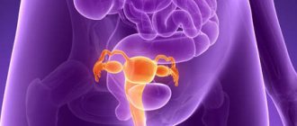
Uterus
- Pregnancy-related enlargement, normal or pathological, with or without associated uterine or ovarian tumors.
- Increase not associated with pregnancy.
- Benign - most often fibroids (leiomyoma). Other causes are hematometra and pyometra (blood or pus in the uterine cavity, respectively). When diagnosing, it is necessary to foresee and exclude a malignant tumor.
- Malignant - most often endometrial cancer. Rare tumors include mixed Müllerian tumors, sarcoma and choriocarcinoma.
A comprehensive medical history is always important, and in women of reproductive age, the possibility of pregnancy and uterine tumors should not be forgotten. Pregnancy and fibroids are the two most common causes of uterine tumors, and a more complete description along with other causes is given in the section Tumors and tumors of the uterus.
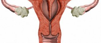
Ovaries
The ovary, as a reproductive organ, consists of three types of cells:
- forming eggs (totipotent cells);
- producing (secretion of sex hormones);
- the remaining cells connect these cells together (epithelial cells).
Ovarian tumors arise from any type of cell. Totipotent cells give rise to dermoid/germ cell tumors; hormone-secreting sex cord cells synthesize excess amounts of hormones. Excess secretion of estrogen leads to uneven endometrial detachment, excess testosterone leads to hirsutism and virilization. Most ovarian tumors arise from epithelial cells.
- Benign - cystodenomas and fibromas.
- Malignant - primary epithelial tumors (85%), sex cord cell tumors (6%), germ cell tumors (2%) and rare - sarcoma and lymphoma. Secondary (6%) originate from the intestines, mammary gland, lungs and thyroid gland.
Ovarian cancer is the most common genital cancer in the UK, although overall it is much less common than breast cancer (6:1 ratio). It is estimated that a GP sees a new case of ovarian cancer every 5 years.
A cyst appears in the ovary every month in the form of a follicle that releases an egg. These follicles reach 25 mm in diameter. It has been empirically found that ovarian cysts up to 5 cm in diameter can disappear without any intervention. Repeated ultrasound examinations are necessary after 2-3 menstrual cycles to ensure its resorption. If the cyst is >5 cm in size, it may need to be removed. The main complications of ovarian cysts are torsion, rupture and hemorrhage. The largest cystodenoma removed in the UK weighed 63 kg, the largest cystodenoma in the world weighing 145 kg was removed in the USA in 1905. If the tumor reaches a very large size, it is most likely benign or possibly borderline. Typically, until the ovary has enlarged to at least 5 cm in diameter, it is not palpable during vaginal examination. In postmenopausal women, the ovaries should not be palpable. Any ovarian tumor is highly suspicious of malignancy until proven otherwise.
Why are pelvic cysts dangerous?
The pelvic space is limited by bones. Its cavity contains the uterus, appendages, ovaries, bladder, and intestines. A cyst can form on any of these organs. Such tumors are most often discovered during an ultrasound examination. At the same time, the neoplasm itself may not show itself in any way and exist asymptomatically for a long time.
A pelvic cyst is called a serosocele. It is an accumulation of fluid, limited by walls of peritoneal tissue or adhesions.
The shape of a pelvic cyst can be oval, round, or irregular. The size of the neoplasm can vary, ranging from a few millimeters to 30 cm in diameter.
Most often, cystic formation of the pelvis in women is diagnosed in the ovarian area. The tumor develops from non-cancerous cells and has a connection with the peritoneum, which lines the internal organs and the walls of the pelvis.
There are several types of pelvic cysts:
- Cysts can be single or multiple. As a rule, the more tumors, the smaller their size.
- Cysts can be superficial or submersible. In the first case, they are located on the outer wall of the pelvic organs or on the wall of the peritoneum, and in the second case, they grow into the stroma of the ovary, uterus or intestines.
Depending on the type of tumor, the prognosis for recovery will vary. It is most favorable for patients with a single small superficial cyst.
There are many risk factors that lead to the development of pelvic tumors. It has been established that most often such neoplasms are diagnosed in elderly patients after entering menopause. Although it cannot be ruled out that a cystic formation of the pelvis in a woman will be detected during her reproductive years and even in adolescence.
Diseases such as endometritis, oophoritis, salpingitis, parametritis, pelvioperitonitis can provoke the appearance of a tumor. Risk factors that can lead to the development of these pathologies
- long-term wearing of an intrauterine device;
- frequent abortions;
- diagnostic curettage;
- previous sexually transmitted infections.
Any prolonged inflammatory process in the pelvis will lead to fibrin plaque appearing on the peritoneum. It glues tissues located in close proximity to each other, which leads to the launch of the adhesive process. It is in such spaces that cysts most often begin to form.
If a woman has undergone surgery on the pelvic organs at least once in her life, then we can consider that she is at risk for cyst formation. In this regard, the danger is posed by cesarean section, removal of the fallopian tubes, uterus, appendicitis, intestinal surgery, etc. The mechanism of tumor formation is based on the initiation of adhesive processes.
With endometriosis, the fluid that the ovaries produce during ovulation is absorbed with certain disturbances. As a result, it gradually accumulates in the peritoneal area, which becomes a prerequisite for the formation of a cystic neoplasm. Most often, such tumors are localized precisely on the ovaries; they are surrounded by a membrane of endometrial cells and filled with menstrual blood.
Suspecting a pelvic cyst based only on its symptoms is quite problematic. Moreover, in approximately 10% of women the disease has a hidden course. Therefore, it is possible to detect a cystic cavity by chance, during a clinical examination or during the diagnosis of other gynecological diseases.
Depending on the severity of the adhesive process or the activity of the ovaries, the growth rate of the cyst will differ. If the connecting cords in the pelvis are rough, or the cyst develops against the background of endometriosis, then it will increase in size quickly.
In this case, the woman complains of abdominal pain of a pulling and aching nature. They can radiate to the lower back, to the sacrum. During menstruation and sexual intercourse, pain tends to intensify.
source
A cyst is a small bubble filled with fluid. It can appear on any organ - lungs, liver, kidneys, pancreas and thyroid glands. And in the reproductive system of women, cysts also form - in the fallopian tubes, uterus, vagina, vulva, and especially often in the ovaries. If moisture accumulates, the cyst grows. If not, it may resolve on its own. But you can’t do this without a doctor’s supervision.
Even during intrauterine development, almost 2 million follicles are formed in the girl’s ovaries, each of which contains an egg in its infancy. By the time they reach reproductive age, no more than 200 thousand remain.
At the beginning of the menstrual cycle, under the influence of estrogen, one follicle enlarges, ruptures and is sent into the fallopian tube to meet the sperm, and in its place a corpus luteum is formed in the ovary.
If fertilization has occurred, it nourishes the child until the placenta takes over the same function (12–14 weeks). And if conception does not happen, it dies, and after 10–14 days menstruation begins.
But there are other options. For example, the follicle has matured, but there has been no ovulation, and then a cyst can form in it, which is called follicular, or functional.
Or ovulation and fertilization have taken place, and the corpus luteum continues to function, but at the same time accumulates excess fluid, and then a luteal cyst is formed.
Each of these types of cysts has the same outcome: they will most likely resolve, luteal cysts - after a couple of months, functional ones - by the beginning of the next menstrual cycle.
Doctors have not yet fully figured out the reasons for the formation of cysts. When you ask your doctor this question, you will probably hear about the individual characteristics of the functioning of the ovaries and pituitary gland, which produce sex hormones.
There is a version that contraception is to blame for the formation of cysts; they say, in the old days, a woman gave birth 5-7 times, so menstruation occurred less frequently, and the ovaries rested more. Now the load on them has increased, and this is the result.
There are other assumptions: it is believed that cervical cysts appear only after inflammation or erosion. Some doctors insist that the lack of regular sex life and orgasm also favors the appearance of cysts, since it provokes stagnant processes in the pelvis.
It is difficult to test these hypotheses. The same cannot be said about endocrine problems (hypothyroidism, obesity), which, of course, affect hormonal processes.
Recently, an expanded EFORT test has appeared in the arsenal of researchers, which determines the tendency to form ovarian cysts, including several at once: on the 3rd–4th day of the menstrual cycle, you need to take a blood test for hormones twice.
The first is done in the morning on an empty stomach, it indicates the level of hormones - follicle-stimulating hormone (FSH), which is responsible for the proper development of eggs in the follicles, and luteinizing hormone, which starts the process of the release of a mature egg from the follicle.
After the test, the woman is given a special drug and her blood is tested again 24 hours later. They get two indicators, compare them and identify risks.
There are many types of ovarian cysts - each with its own structure and growth rate. Some can develop into a malignant tumor. Luteal and follicular cysts are less of a concern than others: they hardly grow and often resolve on their own. The body usually signals the presence of others, but with a delay.
The first are hormonal: cysts always intensively produce hormones, and this can disrupt the menstrual cycle. Menstruation comes irregularly, and in the interval between them there is spotting.
The second category of symptoms indicates physical discomfort. If the ovarian cyst grows (a paraovarian cyst can reach 20 cm), then the ovary increases in size and “invades” other pelvic organs, for example, it compresses the rectum, causing constipation.
The bladder is also attacked and the urge to urinate increases. As well as a feeling of heaviness, nagging or aching pain in the lower abdomen, on the right or left side. In some situations (sexual intimacy), the discomfort intensifies, radiates to the rectum, causing nausea, vomiting and other complications.
The most dangerous of them is twisting of the pedicle (some cysts have this), its rupture and hemorrhage.
During a routine examination, the gynecologist, examining the vagina, discovers a protrusion from the ovary, reminiscent of an inflated bladder, and may assume that it is a cyst. This happens in almost 1/3 of young women under 40 years of age. This symptom is best noticeable in the 2nd phase of the menstrual cycle, a week or two after the end of bleeding.
To confirm or refute the diagnosis, the doctor prescribes blood and urine tests for hormones and sends for an ultrasound. This study is quite sufficient to establish the type of cyst, its essence, location, size, structure, contours, contents.
When a danger is identified, various measures are taken. If the cyst does not exceed 3 cm, medications are prescribed and its “behavior” is observed for 2–3 months. When its parameters are greater, an operation is required.
As is the case if the formation contains seals or its walls are deformed. The situation is clarified by testing the blood for the tumor marker CA-125 - malignant cells.
If medications do not help, surgery will be required to remove the cyst.
Aspiration is considered the most gentle method - this is a type of surgical treatment in which, using a vaginal ultrasound sensor and a puncture nozzle, 10-15 ml of ethyl alcohol, which has a sclerosing (freezing) effect, is injected into the cyst cavity. In second place is laparoscopic surgery - 3 incisions of 1 cm each are made on the abdomen. An optical device is inserted into one, and manipulators are inserted into the other to remove the cyst.
If treatment is carried out in a timely manner, the healthy ovarian tissue will remain intact, which means that the follicular apparatus will be preserved, and the woman will be able to become pregnant.
A few hours after the operation to remove the cyst, the patient is already lifted out of bed, fed liquid food, and on the 5th day she is discharged from the hospital and is recommended not to lift heavy objects, not to exercise, and to exclude sexual activity for a month. If you do not violate the regime, there will be no complications.
Some cysts (follicular) can be treated with oral contraceptives. Vitamin complexes are prescribed along with them. If a woman is overweight, she is recommended to lose weight through exercise and a balanced diet.
Infusion of mature chaga mushroom (the mushroom should sink in water). Rinse the raw materials in cold running water, soak in boiled and cooled water, in a ratio of 1:5. When the chaga softens (after 7 hours), grind 1 part, add 5 parts of the water remaining from the previous soaking, heat to 50°C and leave.
After two days, the water should be drained, the sediment should be squeezed out and an infusion of 20 g should be taken before meals for several courses - 3 months each with a break of a week. During treatment, it is necessary to adhere to a dairy-vegetable diet, including more grains, bran, carrots and beets. Antibiotics and aspirin are contraindicated.
Walnut tincture. Take 14 pieces, wash and break them, place the shells in a liter jar, pour in 500 g of vodka and keep in a warm, dark place for 7 days. Then pour the infusion into a bottle and store in the refrigerator. Take 1 tbsp in the morning on an empty stomach. spoon.
source
A cyst is a pathological cavity filled with some content, resulting from retention or excessive secretion of fluid. Over time it may increase or decrease in size. In most cases, the cause of cyst formation is inflammatory diseases of the pelvic organs.
The woman’s well-being during the formation of cysts remains normal; it is very difficult to suspect any changes. Cysts are usually diagnosed during a gynecological examination, colposcopy, or ultrasound of the pelvic organs.
In the uterine cavity, cyst formation can occur as a result of internal adenomyosis or immersion of the serous epithelium into the myometrium.
Adenomyosis (endometriosis) is a pathological process in which inclusions are detected in the muscular layer of the uterine wall or in other genital organs and outside it, causing a cyst to appear on the uterus (they are similar in structure and function to the uterine mucosa).
As a rule, they do not reach large sizes, are located on the wall of the uterus and are discovered only by chance during colposcopy. Glandular-cystic endometrial hyperplasia is an excessive proliferation of endometrial tissue, as a result of which the volume of the uterus increases and blood circulation becomes difficult.
A cervical cyst is a formation filled with purulent or serous fluid. It is located on the side wall of the cervix and often passes into the broad uterine ligament. There are both single and numerous cysts that have the appearance of beads. Very rarely they reach a large size (up to 10 cm or more). The most common cause of cysts on the cervix is inflammatory processes.
As a rule, the symptoms are extremely scarce. No discharge from the genital tract is observed. Cysts also do not cause a delay in menstruation or any changes in the menstrual cycle.
If they are large in size, they can cause difficulties during sexual intercourse. Also, in the presence of large cervical cysts, a narrowing of the cervical canal is observed, which leads to infertility for mechanical reasons.
A cyst on the cervix does not affect hormonal levels, the course of pregnancy or the development of the fetus, and does not lead to miscarriage.
But in the cervical cyst, viruses and bacteria can persist, multiply and accumulate, which are the source of regularly recurring inflammatory processes in the mucous membrane of the uterus, fallopian tubes, cervix, ovaries and vagina.
It is the inflammatory process that causes infertility and ectopic pregnancy. The list of reasons is extensive, so experts recommend undergoing an examination before planning a pregnancy.
Source: https://03-med.info/kista/kisty-malogo-taza-chem-eto-opasno.html
Fallopian tubes
Fallopian tube enlargements are classified as follows.
- Pregnancy-related tubal pregnancy, or progressive ectopic pregnancy (ectopic).
- Inflammatory - salpingitis, which leads to hydrosalpinx or pyosalpinx.
- Malignant - fallopian tube cancer, very rare.
Diagnosis of small formations limited to the small pelvis or slightly elevated above the edge of the pelvic inlet is often difficult. Despite this, the diagnosis of an ectopic pregnancy with subsequent formation of a blood tumor, which manifests itself exclusively as a tumor, must be carried out immediately for its successful treatment.
Before rupture or abortion, a tubal pregnancy looks like a small tumor in one of the posterolateral corners of the pelvis, adjacent to the uterus, of uncertain consistency, and very painful. Sometimes it is accompanied by short amenorrhea and attacks of acute pain in the pelvis. There may be no obvious signs of pregnancy, but the pregnancy test is positive. Tubal pregnancy can be confused with chronic salpingoophoritis, a small ovarian cyst, a small pedunculated fibroid, or a small ovarian dermoid.
Differential diagnosis is difficult, and it is unlikely that any of the above conditions will cause attacks of pain not associated with menstruation. Typically, pain occurs as a result of overstretching and stretching of the tube due to hemorrhage into the wall or lumen around the fertilized egg, except in cases where the formation is painless (often very painful), this is unlikely to be a tubal pregnancy. When tubal abortion or tubal rupture occurs, symptoms of internal bleeding occur, accompanied by sudden pain and collapse, with bleeding from the uterus or discharge of a cast of decidual tissue, which leaves no doubt about the diagnosis. When a tube ruptures, the bleeding is stronger and more profuse than with a tubal abortion. If the patient regains consciousness after the initial bleeding, the clinical picture is dominated by signs of a retrouterine or peritubar hematocele. The uterus is pushed forward and upward towards the pubic symphysis and behind it a blood clot can be palpated, which causes bulging of the posterior vaginal vault and the anterior wall of the rectum. Vaginal examination is very painful. A tubal miscarriage is often confused with a regular miscarriage. A painful mass on the side of the uterus with a buried cervical canal, the absence of a fertilized egg in the uterine cavity on ultrasound, the absence of uterine contractions or expulsion of any products of conception allow an accurate diagnosis to be made.
Any woman complaining of irregular bleeding and abdominal pain should be considered pregnant and ask whether it is a uterine or ectopic pregnancy. No two cases are alike, and with this disease, as with no other, there are many exceptions to the rule in clinical manifestations. Risk factors for ectopic pregnancy include a history of pelvic inflammatory disease, tubal surgery, including sterilization, progesterone contraception, and a history of infertility. Among rare maternal deaths in the UK, the leading cause remains ectopic pregnancy.
Progressive ectopic pregnancy is rare. It occurs as a result of the continued growth of the embryo after partial separation from the tube as a result of rupture or expulsion through the fimbrial end (abortion). The most typical is a continuous increase in the formation around the uterus with amenorrhea and progressive signs of pregnancy. A typical symptom is abdominal pain in late pregnancy. The uterus in the pelvis is palpated separately from the fetal sac. However, diagnosis is difficult because some blood is always shed, which obscures the contours of the uterus and makes the uterus appear to be part of a pelvic tumor. The fetus is often located high above the pelvis and lies transversely face down. X-rays reveal the fetus in a characteristic unusual position with hyperextension or severe flexion of the spine, and the head and limbs are located at unusual angles to the body.
If the lateral x-ray shows parts of the fetus superimposed on the image of the mother's spine, it is an ectopic pregnancy. Ultrasound establishes the absence of intrauterine pregnancy; The size of the uterus never corresponds to more than 5 months, even with full-term abdominal pregnancy, and there is no sufficient softening of the cervix. In cases where the fetus lies in front of the false sac, during palpation, due to the absence of the anterior wall of the uterus in front of it, it is felt close to the surface. However, it often happens that it is difficult to palpate the fetus due to the location of the placenta anteriorly, while a loud vascular noise occurs immediately medially from the anterior superior iliac spine on the side from which the placenta receives its main blood supply (through the ovarian vessels).
Differential diagnosis of tumor formation in salpingoophoritis is usually not difficult. With this disease, a fixed painful formation is formed in the pelvis, rarely with a definite outline, but sometimes having a typical retort-shaped shape, in which the narrow end is located near the uterus, since the fallopian tube, stretched by fluid, takes this shape. A history of acute illness is identified, usually with bilateral pelvic pain, fever, and peritoneal irritation. Such patients are sexually active. As a rule, this is preceded by discharge from the uterus and severe vaginal bleeding. Due to occlusion of the fimbrial ends of the tubes, this inflammatory disease in women is accompanied by long periods of infertility. In chronic disease, pelvic pain, congestive dysmenorrhea, dyspareunia, vaginal discharge, menorrhagia and infertility are observed. There are no symptoms of suppuration, hyperthermia or leukocytosis, and there is no daily sweating. Pshy in the fallopian tubes is sterile. It is necessary to take smears for chlamydia, including from the endocervix. The patient and her partner should be referred to a dermatovenerology clinic for continued treatment.
A large pelvic abscess accompanies salpingoophoritis or occurs in isolation without affecting the tubes, as is sometimes observed with postpartum septic infections. In this case, peritoneal symptoms occur. The abscess fixes the uterus in a central position, bulges into the posterior fornix and rectum, and tends to rupture into the rectum. Before this, there is a copious secretion of mucus from the anus. Usually the disease begins acutely with symptoms of local peritonitis, rises in body temperature, leukocytosis and sweating. When the abscess empties, the general condition suddenly improves. The abscess is accompanied by parametritis, which displaces the uterus to the side and fixes it in this position. This tumor-like formation causes bulging of one of the lateral arches, extends perpendicular to the lateral wall of the pelvis, tends to descend along the round ligament into the groin and appears there as a psoas abscess. It may have a slow onset, chronic course and not be accompanied by symptoms of local peritonitis. The disease always occurs after childbirth or abortion, while pelvic abscess of peritoneal origin occurs with salpingoophoritis or appendicitis, not to mention pregnancy. Parametritis is not associated with salpingoophoritis. Resorption of parametritis, usually not accompanied by the formation of an abscess, is prolonged.
Malignant neoplasms of the fallopian tube are extremely rare. It has no obvious local symptoms, behaves like ovarian cancer, and the diagnosis is confirmed only histologically after surgery.
Malignant tumors of the pelvis
•Gynecology •Tumors of the female reproductive system •Malignant tumors of the pelvis
Among malignant tumors, colorectal cancer ranks second in frequency of observations in women.
Although the tumor can occur anywhere in the colon, about 70% of cases occur in the rectum.
In women with tumor-like formations located in the pelvic area, the malignant tumor may be localized in one of these parts of the intestine.
Clinical manifestations of pelvic tumors
Cancer of the cecum and its localization in the rectosigmoid region cause the appearance of relatively different symptom complexes.
The latter depends on many factors, such as the size of the tumor and the presence of complications, including intestinal obstruction, bleeding and intestinal perforation.
Melena and anemia-related symptoms resulting from tumor bleeding are most characteristic of cecal cancer. Often such patients experience dizziness, fatigue, pallor, tachycardia and general weakness.
Pain in the right lower quadrant of the abdomen is often described by patients as dull, aching and constant.
Symptoms of intestinal obstruction are rare, although cecal tumors are usually dense and large. This is explained by the fact that in the area of the cecum the feces are liquid, and the intestine itself has a wide lumen.
Characteristic early symptoms of the disease: loss of appetite, indigestion and weight loss. Significant weight loss, cachexia, hepatomegaly and jaundice indicate progression of the disease.
In contrast to cecal cancer, sigmoid colon tumors are characterized by symptoms of intestinal obstruction. Changes in the composition of feces and a decrease in the intestinal lumen determine the appearance of these symptoms. Blood and mucus may be present in the stool, but significant bleeding and anemia are rare.
With rectal tumors, symptoms of intestinal obstruction are not observed so often, because in this section the latter has a large lumen. Early manifestations of the disease are tenesmus and a feeling of incomplete bowel movement.
Quite often the disease is accompanied by scarlet bleeding, but it is usually not massive. Patients may also complain of cramping pain in the left lower quadrant of the abdomen, but this is rarely severe.
Examination for tumors of the pelvic organs
The results of examination of patients with tumors of the pelvic organs, namely the colon and rectum, depend on the stage of the disease. It is necessary to carefully examine the lymph nodes, especially in the supraclavicular region.
Abdominal examination data are not very informative, with the exception of advanced cases of the disease.
Palpable tumor-like formations in the right lower quadrant of the abdomen can be detected only in 10% of patients with cecal cancer.
Abdominal tumor-like formations, metastases in the anterior abdominal wall, hepatomegaly, dilation of the veins of the abdominal wall and ascites appear in the later stages of the disease.
Vaginal and rectal examinations can detect tumors in patients with rectal or sigmoid colon cancer. Palpable lesions of the rectum are found in 30% of patients with colon cancer.
These lesions are characterized by lumpiness, irregular shape, and immobility. Using the Valsalva maneuver, tumors located above the rectum can be identified.
A fecal occult blood test should always be done.
The internal genital organs are usually within normal limits, except in cases where they are involved in the process of primary colon cancer or there is damage to the genital organs unrelated to the underlying disease, for example, uterine leiomyoma.
The detection of large, dense, irregularly shaped tumor-like formations emanating from the uterine appendages suggests the presence of primary or metastatic ovarian cancer (Krukenberg tumor), especially if these formations are observed in women after menopause.
Diagnosis of tumors of the pelvic organs of the small pelvis
Palpation of tumor-like formations located in the right lower quadrant of the abdomen, in the rectosigmoid region or in the rectum in patients with symptoms of colon cancer suggests a malignant tumor. A biopsy usually confirms the diagnosis.
To detect cancer of the rectosigmoid intestine, proctosigmoidoscopy should be performed to a depth of 25 cm.
The tumor appears as an ulcerated, polypoid, nodular, botryomycotic or colloidal mass. Correctly performed biopsy and histological examination of tissue are the decisive factor in diagnosis.
Colonoscopy can provide viewing and facilitate biopsy of tumors located in the cecum.
Proctoscopic examination in combination with irrigoscopy makes it possible to exclude other benign and malignant diseases.
To identify intestinal tumors, especially those originating from the cecum, X-ray examination of the intestine from various angles using gas contrast may be necessary.
Tumor-like formations of the peritoneum, retroperitoneal space and connective tissue
Encapsulated abdominal fluid, hydatid cysts, and retroperitoneal lipomas are usually diagnosed as ovarian cysts and their true origin is only discovered during surgery. There are no specific symptoms to diagnose these diseases. All of them require surgical treatment, and the diagnosis is clarified after surgery. With tuberculous lesions of any location, encapsulation of peritoneal fluid is suspicious for tuberculosis. Tumor-like formations are difficult to distinguish from ovarian cysts, and dullness is often detected on percussion.
Urachal cysts are located in front of the uterus and are closely associated with the bladder, but despite this, they are often confused with ovarian cysts. However, we must remember that only large ovarian tumors are located in front of the uterus, but sometimes small dermoid cysts are also found there. Urachal cysts are embryonic remains and rarely reach a large size.
Colon
Appendicitis is rare during pregnancy and is mistaken for torsion of the ovarian pedicle. A tumor-like formation due to inflammation of the appendix is closely associated with the anterior superior iliac spine and the right iliac fossa. It has unclear contours, and unless there is a large abscess, fluctuation is rarely observed. The acute onset may resemble ovarian peduncle torsion. If a cyst is present, a fluctuating tumor with clear edges is detected; as a rule, some space is palpated between it and the iliac crest. Colon cancer is more common than common gynecological tumors and diverticulitis. Such patients usually present with a history of bowel dysfunction with rectal bleeding.
Bones
Abnormal growth of the pelvic bones is very rare. Any tumors can be cartilaginous or sarcomatous. The detected tumors are integral with the pelvic bones from which they arise. Unlike all gynecological tumors of the pelvis, in which the rectum lies behind, when a tumor grows from the sacrum, the rectum lies in front of the tumor. In most cases, during a bimanual examination, the uterus and appendages are palpated, which are not affected and are free from tumor. The only possible gynecological problem with which this tumor can be confused is a tubo-ovarian abscess. During pregnancy, if the study does not reveal absolute fixation and continuity of the tumor in relation to the pelvic bones, it is difficult to establish its true nature.
The diagnosis is made by X-ray examinations in combination with a biopsy. These diseases are rare, and they are unlikely to be encountered in the practice of a gynecologist.
Other structures
Many of these diseases are not specific to the pelvis but are discussed here because they can be confused with pelvic tumors. Tumors of the kidneys, spleen or pancreas can reach the edge of the pelvic inlet, but the history should show that they grow from top to bottom, and not vice versa. Kidney tumors are accompanied by changes in urine or lack of urine output on the affected side, which is detected by cystoscopy or intravenous pyelography.
Malformation of the genital tract is combined with an abnormal development of the urinary tract. In patients with congenital absence of the vagina and uterus, a single pelvic kidney is often found.
Changes in the hemogram are sometimes associated with an enlarged spleen. A pancreatic cyst is unlikely to be mistaken for a pelvic tumor, but it is difficult to distinguish it from a long-pedunculated ovarian tumor.
Diagnosis depends on the results of imaging tests.
In accordance with the International Classification of Diseases (ICD), pelvic masses are designated by codes from C51 to D29 and are differentiated into malignant (C51-C63) and benign (D25-D30) tumors. Over the past 10 years, the incidence of gynecological oncological pathologies has increased by 8.5%. The most fatal form of neoplasm is ovarian cancer, which ranks 4th among the causes of mortality of all cancers.
Causes of pelvic tumors in men and women
Focal neoplasms in the pelvis can occur in the bones, genitourinary organs, and intestines. The factors that cause tumor formation have not been reliably established. Research has revealed a high incidence of cancer in highly developed countries. This is associated with dietary habits, consumption of fatty, high-calorie foods, alcohol, nicotine, and caffeine, although there is no convincing evidence.
The following circumstances are considered to be the most likely causes of the development of pelvic cancer:
- hormonal factor: infertility, hormonal stimulation and ovulation induction;
- history of malignant formations of the mammary glands;
- Fatall's ovulatory hypothesis;
- hereditary predisposition: familial ovarian, breast and ovarian cancer, as well as Lynch syndrome II.
In newborns, the appearance of ovarian cysts is usually explained by the influence of maternal placental estrogens. During adolescence, girls may develop hematocolpos due to the accumulation of menstrual flow due to the hymen or vaginal atresia. Women of childbearing age more often suffer from cancer due to pathological pregnancy, as well as trauma to the organs of the reproductive system during childbirth or caesarean section. The most common pelvic carcinoma in men is a prostate tumor.
When examined in men and women, congenital formations are sometimes encountered - ovarian hydatides, which arise during the intrauterine development of the genital organs. The pathology is asymptomatic and surgical treatment is extremely rare.
Symptoms of a pelvic tumor
A simple gynecological examination may reveal tumor-like formations in the pelvis.
Pelvic tumors can develop in the organs of the female reproductive system, also in the intestinal tract, bladder, bones, ureter, and skeleton.
In different age groups, tumor-like formations differ in type:
- In girls, during the first months of life, the effect of maternal placental estrogens remains unchanged. In newborns, this can lead to the formation of cysts on the ovaries;
- During puberty, girls may develop hematocolpos. This occurs due to impaired blood flow during menstruation due to fusion of the hymen and defects during the development of the uterus and vagina;
- In women of reproductive age, an increase in the size of the uterus can be observed with fibroids or during pregnancy. At this age, women may experience multifunctional ovarian cysts up to 8 centimeters in volume. Ovulation is not observed. Such cysts often regress within a few months.
Tumors in the area of the uterine appendages can be detected when there are disorders of ectopic pregnancy. Also, if cancer of the ovaries, fallopian tubes, or inflammation of the fallopian tubes is detected.
During menopause, malignant neoplasms of the pelvis are often diagnosed. Endometriosis, fibroids and multifunctional ovarian cysts are less common at this time.
Ovarian oncology
Ovarian tumors refer to neoplasms on the female gonads (ovaries). They are:
- malignant. They grow quite quickly and are capable of growing into neighboring organs and tissues. Together with the blood flow, they metastasize to nearby organs, affecting the lymph nodes;
- benign. They are characterized by slow growth and do not spread to other organs and lymph nodes;
Ovarian tumor, location in the pelvis
- hormone-producing. They are capable of producing sex hormones;
- metastatic. They arise due to the spread of tumor cells that originally originated and were located in another place.
Benign neoplasms without proper treatment or as a result of late detection can develop into malignant tumors.
In childhood, germ cell tumors are more common.
Diagnosis of ovarian tumors
To diagnose ovarian tumors, the following procedures and tests are performed:
No. Useful information
| 1 | The medical history is analyzed and all complaints are taken into account. It is determined when exactly the abdominal pain began, whether it intensifies and spreads |
| 2 | The history of gynecological diseases is analyzed, everything that was suffered by the patient. All operations, pregnancies, abortions are taken into account |
| 3 | the history of menstrual function is analyzed. It turns out exactly when menstruation began for the first time, its regularity, and duration. The pain of menstruation and its abundance are taken into account |
| 4 | An examination by a gynecologist is carried out. It must be carried out in two hands and examined by touch. |
| 5 | Ultrasound of the pelvis and abdominal cavity is performed |
We recommend reading: The first signs of a brain tumor in children
- hormone levels are determined;
- a computed tomography scan of the pelvis and abdominal region is prescribed;
- MRI of the pelvis is prescribed;
- An x-ray is taken on the chest;
- for diagnostic purposes, laparoscopy is prescribed;
- mandatory consultation with an oncologist.
Causes of oncology
Tumor formations can appear precisely in the pelvis, or rather:
- in the bones;
- in the bladder;
- there is damage to the organs of the reproductive system;
- in the intestinal tract;
- on skeletal muscles.
Symptoms indicating the appearance of tumor formations may vary.
The abundance of formations directly depends on the age of the person. Moreover, they are ready to change with age.
Endometrioid heterotopia develops in the form of separate foci in the pelvis, and more in the ovaries.
More dangerous tumors are detected in women during menopause. But diseases such as fibroids, endometriosis and multifunctional cysts are disappearing during this period.
Pelvic examination
Great importance is given to collecting information about the disease. Bleeding in the uterus and pelvic pain may indicate the presence of an ectopic pregnancy. But they don’t always remind us of trophoblastic disease.
Painful periods indicate endometriosis.
Girls who had to experience puberty early and the development of the genital organs, in most cases, will suffer from a hormone-producing ovarian tumor.
If a young woman has virilization, then it is worth assuming a masculinizing tumor. Women who are already postmenopausal may experience feminizing tumors.
To identify a particular disease, a thorough examination will be required. During the examination, the doctor must pay attention to endocrine system disorders, weight and ascites.
Moreover, he is obliged to conduct a gynecological examination. In some cases, it is simply impossible to find a tumor of the appendages or uterus.
When examined by a gynecologist, the tumor is in a state of immobility in the presence of an endometrioid cyst.
On the contrary, tumors that are located in the area of the appendages are mobile upon examination. These tumors can be detected during examination, even with an ectopic pregnancy.
The tender formation of hydrosalpinx may be palpable and move during examination.
In young girls, such tumors can even be palpated through the anterior wall of the peritoneum, since some tumors simply do not have enough space in the pelvis.
We recommend reading: Symptoms of a tracheal tumor
Symptoms of a tumor
The following organs are located in the pelvis:
- intestinal tract;
- bladder;
- reproductive system;
- birth canal;
- ovaries;
- prostate.
All malignant neoplasms in the pelvis can relate to the following diseases:
- bladder disease. Oncological malignant lesions of the mucous membranes. The frequency is within 5% of all cancer cases. In representatives of the stronger sex, bladder cancer develops 4 times more often than in women;
- colorectal education. Damage to rectal cancer cells;
- prostate cancer. Occurs often in men;
- uterine oncology. The most common disease in women affects the reproductive organs;
- cervical oncology. The disease develops on the basis of processes in the appendages, which cannot be cured for a long period. This disease is very rare;
- ovarian oncology in the fairer sex. It occurs quite often among women in big cities.
Signs of a tumor outside the pelvic organs
Despite the existence of a large number of tumors that form outside the pelvic organs, the signs corresponding to formations in the pelvis are largely similar.
This type of cancer is characterized by the absence of symptoms in the initial stages of development.
Increasing in volume outside the organs, tumors can reach very large sizes for some time and at the same time they may not make themselves felt.
Symptoms of pelvic tumors may be insignificant and manifest themselves:
- in the form of difficulty urinating;
- in the form of intestinal obstruction;
- lumbar pain may occur;
- Heaviness in the legs and pain in the abdominal cavity may appear.
The general health and condition of the patient may remain normal for a long time, including if he has already developed a fairly large tumor. The patient may experience the following health problems:
- regular body weakness;
- fast fatiguability;
- weight loss;
- unstable body temperature;
- intoxication.
Pain in the abdomen is one of the primary signs of a tumor of malignant origin. Pain is observed with small tumors. There is compression of the nerve endings connected to the lumbar region or the sacrum.
The tumor is detected by palpating the abdomen. This may be quite enough to determine the presence of the disease. Tumors that have formed in the abdominal region on the right give early symptoms and manifest themselves as venous stagnation.
We recommend reading: Rectal tumor: classification and treatment
In general, various neoplasms in the abdominal cavity can remain silent for a long period of time. This makes it very difficult to make a timely diagnosis.
Classification of neoplasms of pelvic localization
In accordance with the surgical classification of locally advanced tumors located in the pelvis, 4 types of neoplasms are distinguished:
- The first group includes tumors that slightly affect the adjacent anatomical structures. To operate them, atypical or planar surgical removal of organs is sufficient.
- The second group includes formations that grow into the thickness of nearby structures and form difficult-to-separate conglomerates of organs, causing the formation of a pelvic infiltrate.
- The third unites tumors, the spread of which leads to the formation of fistulas: vesico-intestinal, vesico-vaginal, vaginal-intestinal or combined.
- In the fourth option, neoplasms disseminate to bone or muscle-aponeurotic tissues, compressing large main arteries.
Using the Grade classification, neoplasms are typed according to the degree of differentiation of tumor cells in the following order:
- Gх – it is not possible to determine the degree of cell difference;
- G1 – high degree of differentiation;
- G2 – cells with an average degree of functional differences;
- G3 – poorly differentiated;
- G4 – tumor cells have no differences.
Determining functional differences plays an important role in choosing the method of therapy, as well as the scope of treatment measures.
Organ and extraorgan peritoneal tumors localized in the pelvis develop from the following mesodermal tissues:
- lipomas, lipogranulomas - fatty tissue;
- leiomyosarcoma - smooth muscle;
- fibromas – fibrosis of connective tissue);
- rhabdomyosarcomas - striated muscles;
- lymphangiosarcomas, lymphangiomas affect the vessels of the lymphatic system;
- nodular hyperplasia, lymphoma;
- hemangiomas, hemangiosarcomas.
Retroperitoneal neoplasms are characterized by the detection of blood in the stool, the development of anemia due to blood loss, pain in the lower abdomen, and constipation.
Symptoms of a pelvic tumor in women depend on the type of tumor. The morphological classification of ovarian mass formations divides them into 8 main types:
- epithelial;
- stromal cell, affecting the sex cord and stroma;
- lipid cell;
- germinal;
- gonadoblastoma;
- neoplasms nonspecific to the ovaries;
- unclassified or undifferentiated;
- metastatic (secondary);
- tumor-like formations.
Benign cysts in the pelvis are tumor-like neoplasms, inside of which there is a cavity with liquid contents. They lack proliferative growth and their capsule stretches as fluid accumulates.
Depending on its nature, the following types of cysts are distinguished:
- follicular (most common, up to 80%);
- corpus luteum cyst (luteal);
- Theca-luteal;
- hyperthecosis, stromal hyperplasia;
- endometrioid;
- pyovar (inflammatory);
- paraovarian;
- polycystic ovary syndrome.
Benign ovarian formations also include multi-chamber cysts with a cavity divided by septa into several compartments.
Symptoms
A distinctive feature of the development of tumors in the pelvis in women is the absence of negative symptoms in the initial stages of the disease. Most often, pathology is detected during routine gynecological examinations in a medical institution.
With cystic lesions of the cervix and ovaries, the progression of the disease is accompanied by aching or sharp pain in the lower abdomen, a feeling of pressure in the pelvic area, and disruptions in the menstrual cycle.
In the case of bladder cancer, there is blood in the urine, frequent urge and sharp pain during urination.
On this topic
- Oncogynecology
How long do people live with ovarian cancer?
- Olga Vladimirovna Khazova
- December 4, 2021
The presence of pathology in the intestines is confirmed by regular constipation or loose stools. Blood, mucous or purulent masses are found in the stool.
Malignant tumors may have signs such as bleeding (unrelated to menstruation), constant pain in the legs and lower back, and discomfort during sexual relations.
Uterine cancer promotes the growth of polyps, affecting the appendages and abdominal cavity. This process leads to the appearance of an unpleasant putrid odor.
With tumors of the vulva, a woman feels itching and burning. Externally, there is a proliferation of ulcerative nodes, which over time penetrate deep into the organs.
Advanced stages of the disease are characterized by general weakness, patients experience dizziness, nausea, and increased body temperature. Women begin to lose weight quickly and their skin becomes pale. Blood pressure often changes either down or up. Sometimes hair appears on the mammary glands and face.
Characteristic signs of cystic formations of the pelvis
The development of tumor-like, luteal or follicular cysts in women of reproductive age is indicated by an enlargement of the ovaries. Malignant formations are differentiated according to their clinical and echographic characteristics. Follicular cysts are located on the side of the uterus and tend to move well. They are characterized by the following echo pattern:
- single-chamber, regular, oval shape;
- diameter within 3-10 cm, usually 6 or 7 cm;
- thin walls, smooth inside;
- anechoic contents, there is distal enhancement from the posterior wall;
- There are single vessels in the cyst wall.
Follicular cysts usually disappear within 1-3 months.
Serous or mucinous cystadenomas are small in size, have smooth walls, and are often unilateral. The echostructure can be defined as homogeneous, while in mucinous cysts there is conglomerate content with the presence of a hyperechoic suspension.
Endometrial cysts with heterogeneous contents with numerous inclusions are detected on one side, they are single-chamber tumors, the capsule is unevenly thickened due to blood clots.
Generalized forms of ovarian carcinoma are characterized by irregular outlines of a conglomerate of cysts of a solid structure. Their boundaries are blurred, and growths can be seen along the outer contour. Most are diagnosed with ascites. The tumor grows into the thickness of the uterus in the form of hypoechoic metastatic nodes.
A pelvic cyst should be distinguished from an exostosis, a benign growth of osteochondral tissue. The reasons for its development are various diseases of the musculoskeletal system and injuries.
Types of benign and malignant tumor diseases of the female genital organs
Uterine fibroids, uterine fibroids , are one of the most common tumor diseases of the female genital organs in gynecological practice. In most cases, it may not have pronounced clinical symptoms and is determined by chance during a bimanual examination.
The diagnosis is established on the basis of transvaginal ultrasound, hysteroscopy, colposcopy, laparoscopy, biopsy, cytological or histological examination.
GUTA CLINIC provides all types of effective surgical treatment of uterine fibroids using laparoscopic and hysteroscopic myomectomy - non-invasive surgical treatment of fibroids without incisions, as well as laparotomy with myomectomy, hysterectomy according to indications.
Conservative therapy can be used in young patients with small, slowly growing fibroids and no contraindications to prescribed medications. Fibroids require mandatory surgical treatment due to the possibility of transition to sarcoma, a malignant tumor.
An ovarian cyst is a cavity filled with fluid (the exact nature of the contents is determined directly by a detailed examination). Most often, an ovarian cyst is found in young women of reproductive age; in older women it is extremely rare.
There are endometrioid, paraovarian, mucinous, serous, dermoid, follicular cysts, corpus luteum cysts, etc.
An ovarian cyst may not bother a woman and may be detected by chance during an examination by a gynecologist. In some cases, an ovarian cyst may be accompanied by menstrual irregularities, heavy and prolonged menstruation, pain in the lower abdomen, anovulation, infertility, etc.
Currently, the “gold standard” for the treatment of ovarian cysts is laparoscopy, which allows the patient to recover faster and fully maintain her reproductive function. Ovarian cysts are subject to mandatory surgical treatment, because can become malignant and lead to the development of serious complications (the development of peritonitis, cyst suppuration, etc.)
Diagnostic methods
Identifying various factors is necessary to select effective treatment tactics. Diagnosis of pelvic tumors includes the following measures:
- taking anamnesis;
- physical examination - gynecological examination, palpation of lymph nodes;
- laboratory diagnostics - detailed blood test, general urine test, determination of biochemical parameters, test for tumor markers, coagulogram, collection of cervical smears;
- examination using special equipment.
Instrumental diagnosis of pelvic tumors is based on the use of the following techniques:
- endometrial aspiration biopsy;
- puncture of cavities of formations;
- diagnostic curettage, laparoscopy, which allows you to determine the histological characteristics of the tumor;
- Ultrasound of the pelvic organs and abdominal cavity is performed to determine the extent of the pathological formation and study the condition of the lymph nodes;
- chest x-ray;
- contrast magnetic resonance imaging (MRI) of the pelvis to assess the depth of tumor invasion;
- in case of extragenital spread of the tumor, positron emission tomography (PET) is prescribed;
- All patients undergo electrocardiography (ECG).
Ultrasound is a routine method for diagnosing pelvic tumors.
For small tumors, transvaginal echography is the most informative; if the formation exceeds 6–7 cm, it is more rational to carry out transabdominal echography.
Additional research is recommended to select optimal treatment options. According to indications, a cytological analysis of swabs from the peritoneum is performed, as well as cytoscopy, colposcopy, sigmoidoscopy, colonoscopy, and bone scintigraphy. After surgical removal of the tumor, its histological examination is carried out.
Features of therapy
Patients with suspected functional ovarian cysts need to undergo a control echography after several menstrual cycles. In favorable cases, there is a decrease in the size of the formation over 4 months. If there are complications, pain, cycle disruptions or the cyst exceeds 5-6 cm, surgical removal of the tumor is indicated. In the presence of inflammatory processes, a course of antibiotics is prescribed.
The main principle of treatment is maximum preservation of organs and tissues, as well as minimal invasiveness. Tumor-like neoplasms of small size are treated without complications with the help of combined oral contraceptives, vasoactive agents, and metabolic therapy methods. More radical treatment methods are cystectomy, laparoscopy, endocoagulation and ovarian reposition.
In the presence of benign or malignant tumors in patients of reproductive age, cystectomy, oophorectomy, and biopsy of a healthy ovary are performed. During menopause, surgery is performed with total or subtotal hysterectomy with uterine appendages, and, if necessary, radiation therapy is performed.
In the postoperative and post-radiation period, symptomatic pain relief, proper balanced nutrition, control of intestinal function and prevention of constipation, and light physical activity are recommended.
Treatment methods
Once the diagnosis is confirmed, the oncologist selects an individual treatment regimen for the patient. Typically it includes:
- Surgical removal of pathologically changed structures and affected lymph nodes.
- Radiation therapy is the effect of active radio rays on mutated cells. It can be external or internal. Intravenous injections are made of drugs containing radioactive elements that can destroy cancer cells. Thanks to treatment, single formations often disappear completely. The radiation method is used to reduce the patient's excruciating pain.
- Chemotherapy is the use of cytostatic drugs to prevent cancer relapses. It is aimed at destroying atypical cells and stopping their growth. The technique has a number of side effects, negatively affecting healthy organs, but it cannot be avoided. To reduce the harmful effects of chemotherapy on the patient’s body, the selection of medications, their dosage and duration of use are carried out with special care.
For diseases of the genital area, most often the organ affected by cancer is completely removed. Organ-conserving operations with excision of only pathological tissues are performed only on young women in order to preserve the function of childbearing, but there is a high risk of re-growth of metastases. In case of bone and cartilaginous sarcoma, the most common procedure is to remove the joint and replace it with an endoprosthesis to restore the functionality of the articulation of the pelvis and hip.
In the later stages, surgery is not performed because cancer cells from the excised organs can get onto the edges of the healthy incisions.
Possible consequences and complications
The most common complications of pelvic cysts are apoplexy and torsion of the leg. There is always a risk of rupture of the capsule of the cystic formation and the formation of hemorrhages inside the cavity.
Torsion of the cyst stalk and apoplexy are clinically manifested by signs of an acute abdomen. In case of torsion, echography shows thickening of the walls, sometimes a double contour is determined due to edema and hemorrhages. The severity of the pathological process is indicated by the absence or presence of blood supply to the ovarian tissue. Apoplexy occurs due to rupture of the cystic capsule or hemorrhage.
Due to disruption of the functioning of the lymphatic system of the pelvic organs in the postoperative period, there is a possibility of developing complications in the form of lymphorrhea, lymphocyst, lymphostasis, seroma, and edema.
Methods for preventing pelvic tumors
Regular monitoring of women and men, especially those at risk for cancer, helps prevent the development of gynecological tumors, as well as the transformation of benign tumors into malignant ones.
For the purpose of prevention, it is recommended to conduct a physical examination of patients, including a gynecological examination and analysis of the vaginal microflora. An ultrasound examination of the retroperitoneal space, abdominal cavity, and pelvis should be performed annually. For operated patients, ultrasound scans are performed every quarter 4 times a year for 3 years, and once every six months during the 4th and 5th years.
It is necessary to undergo a chest x-ray at least once a year. If there are complaints or symptoms indirectly or directly indicating a tumor, as well as lumps detected during examination, an in-depth diagnosis should be performed using more informative research methods.
Patient prognosis and survival
One of the most difficult problems in oncology is the diagnosis and treatment of tumors. Most pelvic tumors are detected at late stages of development, and therapeutic measures often do not bring the desired results.
The overall survival rate of patients with genital cancer over a five-year period remains at 35–40%. According to statistics, mortality from ovarian cancer is in 5th place, ahead of malignant tumors of the uterus. During the first year from the moment the pathology is detected, about 35% of patients die. The prognosis for the treatment of tumors at the 1st stage is 75-85%, the second up to 75%, the third up to 25%, the survival rate of patients with stage 4 is no more than 10%.
Recently, there has been a slight increase in the five-year survival rate of approximately 3-4%. This is due not only to the improvement in the quality of diagnostic methods, but also to the use of effective platinum chemotherapy for germ cell tumors (dysgerminomas, non-dysgerminomas, dermoid cysts) and disseminated types of ovarian cancer.
Treatment of tumors of the female genital organs
Depending on the identified disease, its form, stage, nature, characteristics of the course, and individual indications of the patient, surgical or conservative treatment is prescribed. As a rule, surgical treatment of tumors of the female genital organs is carried out in case of heavy bleeding, rapid tumor growth or large size of the identified tumor, etc.
The scope of surgical treatment is different - it can be organ-sparing laparoscopy (for ovarian cysts and cystomas) or radical amputation (extirpation) of the uterus - for large fibroids or uterine cancer without metastases. Preference, as a rule, is given to the first - minimally invasive - method of treating tumors of the female genital organs.
In parallel with surgical treatment, antiviral or antibacterial therapy, immunomodulatory and biostimulating drugs are prescribed according to indications.
GUTA CLINIC specialists remind patients that in the absence of treatment, even some benign types of tumors can turn into cancer, leading in some cases to a fairly rapid death due to developing complications and the spread of metastases.
That is why tumors of the female genital organs are subject to mandatory treatment, and in some cases, observation (passive uterine fibroids). Considering that the development of most types of tumors is characterized by unexpressed, erased clinical symptoms, gynecologists at GUTA CLINIC recommend regular preventive examinations by specialists, even if nothing bothers the woman.