Ultrasound of the pelvic organs, performed in women, is a study through which the doctor can diagnose any pathology that has arisen in the female reproductive organs. Echography can also be prescribed to study diseases of the urinary system. Women should pay enough attention to their health. It is very important to detect the disease in a timely manner and take measures to cure it.
When a specialist recommends a pelvic ultrasound scan, women may ask when is the best time to do an ultrasound. Men can undergo this study on any day. With women everything is different. The specialist should know when to do a pelvic ultrasound in women and on what day of the cycle.
The essence of ultrasound
Ultrasound is based on the “transillumination” of tissues using ultrasonic waves. As a result, they reflect differently from structures. It depends on their density. Scanning is carried out using a sensor that picks up response signals and transmits them to the computer. After instant processing, they are displayed on the monitor as an image. Sensors can be of different shapes and are selected depending on the type of examination. For example, for the transabdominal method, wide ones are taken at the end, for the vaginal method, narrow (“finger”) ones are taken, on a long handle.
Types of ultrasound in gynecology
In gynecology, if an ultrasound of the uterus is prescribed, the ovaries and fallopian tubes are simultaneously checked. Organ scanning can be done in four ways:
- Transvaginal. Most often used. The sensor is inserted directly into the vagina at a distance of up to 7 cm. The method is one of the most informative, since the device is very close to the organs being studied. However, the transvaginal method is not used for virgins, after the first trimester of pregnancy, with heavy bleeding.
- Transrectal. The sensor is inserted a short distance into the small intestine through the anus. The device is also located next to the uterus and appendages, which allows them to be examined as much as possible. However, the method is not as accurate as the transvaginal method, since the sensor is separated from the organs by the intestinal wall. Typically, this method of examination is used for virgins who cannot undergo a vaginal examination due to the protective hymen.
- Transabdominal. This is a type of ultrasound that is usually used to examine the abdominal organs, but can also be used to scan the reproductive system. The sensor slides over the stomach. However, the method is considered less informative than those described above. Firstly, the device is located away from the uterus, ovaries and tubes. Secondly, between them there is a wall of the peritoneum, a possible accumulation of gases, which can also distort the results. However, only this method is suitable for examination of pregnant women, starting from the second trimester. It can also be applied to virgins.
The fourth method is most rarely used - intrauterine ultrasound. An oblong sensor is inserted directly into the organ cavity. This scan is usually done to evaluate changes in the endometrium or for uterine fibroids.
How does an ultrasound work and what will it show?
Ultrasound examination can be carried out in two ways:
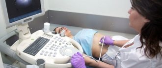
- transabdominal (After lubricating the dermis of the abdomen with a special gel, the specialist places an abdominal sensor above the womb. An image of the uterus is displayed on the screen);
- transvaginally (The woman should take off her underwear and lie down. Her legs should be spread wide enough for the doctor to have access to the genitals. The specialist inserts a thin sensor into the vagina, on which a special condom is placed).
To do an ultrasound, you need to take with you a diaper, a towel, and a referral for diagnostics. If the examination is performed transvaginally, you need to take a condom. No special preparation is required.
For an abdominal examination, you need to fill your bladder. An hour before the diagnosis, the doctor advises drinking about a liter of water (still).
What will a pelvic ultrasound show? If everything is normal for a woman, the uterus will be visible on the screen, which is small in size, has a well-developed muscle layer, and mucous membrane. The mucous membrane should correspond in thickness to the day of the menstrual cycle.
When examined, the fallopian tubes are similar to hollow cords that have oval cellular formations at their ends. These formations are ovaries.
Normally, there should be no formations or inclusions inside the uterine cavity (exception is pregnancy).
Ultrasound diagnostics allows a specialist to detect any disease of the female reproductive system, from inflammation of the ovaries to malignant tumors of the uterus.
The diagnosis takes only about 30 minutes.
The price of such a diagnostic procedure can range from 1,500 to 6,000 rubles. Many factors influence the determination of cost: the qualifications of the specialist performing the diagnosis, the status of the clinic, the purpose of the diagnosis.
Advantages of Ultrasound Diagnostics
Pelvic ultrasound is very popular. This diagnostic method has some advantages over other methods of examining the pelvic organs. Its advantages are:
- absence of ionizing radiation;
- non-invasiveness;
- allowed during pregnancy;
- diagnosing various diseases of the reproductive system and urinary tract;
- image of tissues in “real time” mode;
- providing a clear image of soft tissues that are difficult to see with x-rays.
On what day of the menstrual cycle is it better to do a pelvic ultrasound?
What time is an ultrasound examination performed?
The uterus is scanned on days 5-7 of the cycle. Immediately after menstruation, the endometrial layer is very thin, which helps to most accurately identify any pathological processes - fibroids, hyperplasia, etc. If the examination is carried out later, the mucous membrane becomes denser every day and begins to hide small formations and barely noticeable defects. As a result, the scan results will be of poor quality or some diseases will not be noticed at all.
However, in the 2nd half of the cycle, follicles or a cyst on the corpus luteum can be identified. However, it is difficult to assess its nature (pathological or self-absorbing). Even nascent cysts are easier to identify at the beginning of the cycle. To check whether the ovaries are functioning correctly, an ultrasound scan of the uterus is prescribed on days 8-10, 14-16 or 23-24 of the cycle. The examination is repeated to exclude cysts if the test does not show conception, and the temperature in the small intestine remains high for more than 10 days.
Dependence of the day of ultrasound on the purpose of the study
The exact timing of an ultrasound scan in the area of the pelvic organs depends on what kind of suspected disease needs to be diagnosed and investigated.
For example, with endometriosis and fibroids, diagnosis should be carried out on days 16–20 of the cycle.
Let us consider in more detail in the table the recommended days for performing a pelvic ultrasound for diagnosing specific diseases.
| Procedure name | Suspected pathology | Day of the menstrual cycle |
| Ultrasound diagnostics in the area of the pelvic organs in women | Myoma | 16 – 20 days (second phase of the menstrual cycle) |
| Endometriosis | 16 – 20 days (second phase of the menstrual cycle) | |
| Tumor, cyst | After the end of menstruation | |
| Complication after abortion or surgery on the pelvic organs | any day of the cycle | |
| Inflammation of the uterine appendages | any day of the cycle | |
| Prevention | in the first half of the cycle | |
| Assessment of the condition of the ovaries, follicles in the ovaries | carried out three times per cycle: after menstruation; on days 14–16; for 20 – 24 days. |
Indications and prohibitions for ultrasound
Ultrasound in the field of gynecology is recommended to be performed at least once every 2 years, and after 40 years - annually. However, if alarming symptoms occur, you should consult a doctor earlier:
- very short or too long periods;
- at the first signs of conception;
- pain during sexual intercourse;
- discomfort and disturbances after intrauterine devices;
- bleeding;
- cycle disorders;
- purulent or mucous discharge (including bloody discharge);
- pain in the appendages or uterus, lower abdomen;
- very painful or heavy periods.
Ultrasound examination is also performed in case of suspicion (or presence) of diseases:
- infertility;
- endometriosis;
- myoma;
- salpingitis;
- polycystic disease;
- cancer;
- polyps;
- cysts;
- inflammation of the ovaries;
- endometritis.
Ultrasound of the uterus and appendages is mandatory before planning pregnancy, IVF. The examination is performed at least three times during pregnancy. An ultrasound is done if inflammation of the female genital organs is suspected, to monitor the treatment of uterine fibroids.
Contraindications
A rectal examination is not carried out in case of intestinal obstruction or wounds in the anus. Transabdominal is not done if ulcers, erosions, or skin rashes appear on the abdomen. Transvaginal is not performed in case of bleeding. From the 2nd trimester, scanning is prohibited by any means other than abdominal. In other cases, ultrasound research has no prohibitions.
What can the doctor see?
Transabdominal examination allows you to determine the shape, location, contours, echogenicity (uniformity of structure) and size of the uterus. Indicators of the endometrium, cervix, ovaries, and the structure of muscle tissue are also assessed. If they want to diagnose pregnancy, the specialist additionally evaluates the tone of the uterus and the location of the fertilized egg.
Using ultrasound, you can detect benign and malignant tumors, cysts, malformations, inflammatory diseases, ectopic pregnancy, polyps and other pathological conditions of the reproductive organs.
Interpretation of ultrasound: normal indicators
In a healthy woman, a transabdominal ultrasound of the uterus will reveal the following indicators:
- 4.5–6.7 cm in length, 5–6.4 cm in width, 3–4 cm in thickness in women 15–40 years old;
- the contours are smooth and clear;
- homogeneous echogenicity of the walls;
- cervical canal 2-3 mm;
- the cervical canal is filled with homogeneous fluid;
- normal tone;
- endometrial thickness depending on the day of the cycle (1-15 mm).
Healthy ovaries should be 3-4.1 cm long, 2.0-3.1 cm wide and 1.4-2.2 cm thick with a homogeneous echostructure. In them, the dominant follicle is determined by the middle of the cycle, and an uneven contour is also visible. The fallopian tubes are not recognized.
Normally, it is possible to determine a small amount (several milliliters) of free fluid in the pelvis on the 14-15th day after the next menstruation. At other times it should not be recognized.
Doctors often write in their conclusions “decidualization of the endometrium.” This indicates pregnancy.
Ultrasound signs of uterine pathologies
Ultrasound primarily helps diagnose cancer at an early stage of its development. Affected tissues have a different echogenicity than healthy ones, however, a malignant tumor can be confused with fibroids. To make an accurate diagnosis, the woman is referred for a biopsy.
Endometriosis of the uterus can be suspected by the heterogeneous structure of the organ, its increased size and the discrepancy between the thickness of the endometrium and the days of the menstrual cycle. Characteristic is the proliferation of the mucous membrane in the muscular layer of the uterus and the formation of cysts.
During the examination, the doctor can detect formations of any size, characterized by increased density and spongy structure. It can be single or multiple, and also occupy the entire uterine cavity or some part of it. This is a uterine polyp. It is also characterized by smooth and clear edges.
Doctors often diagnose abnormalities in the development of the uterus. The most common of them are:
- hypoplasia (violation of organ proportions);
- saddle uterus (a depression in the bottom of the organ);
- bicornuate uterus (symbolic image of the heart);
- duplication of the uterus, etc.
If the doctor’s report states that the uterine cavity is dilated, hyperechoic inclusions are visualized, and the structure of the endometrium is heterogeneous, then this means that the woman has chronic endometritis. The acute phase of inflammation is characterized by a decrease in the echogenicity of the mucous membrane.
Ultrasound helps to quickly determine a woman’s pregnancy, both normal and pathological (ectopic). Already at a period of 4-5 weeks, the doctor is able to recognize the embryo in the lumen of the fallopian tube or uterus. An indirect sign of an ectopic pregnancy is the absence of a fertilized egg in the uterus.
Signs of ovarian pathology
Transabdominal ultrasound determines the size of the ovaries, their structure and the presence of pathological formations. Cancer can be suspected by the asymmetry of the organ, the appearance of zones of hypervascularization, the determination of fluid, as well as the visualization of any formation during menopause. Consultation with an oncologist is required if two of the above symptoms are present.
An ultrasound will immediately detect an ovarian cyst. It looks like a round bubble with clearly visible contours. The doctor will determine the contents of the formation and can approximately determine its type (dermoid, follicular). With polycystic ovary syndrome, an ultrasound will show an increase in the size of the appendages and thickening of their capsules. Many follicles ranging from 0.2 to 0.9 cm will be visible in the ovary.
The inflammatory process in the ovaries is characterized by an increase in the size of the appendages, the boundaries become unclear, and the echostructure increases. In conclusion, the doctor will note that the walls of the fallopian tubes are thickened, which indicates their involvement in the pathological process.
Ultrasound examination of the uterus and its appendages is an informative method for determining gynecological diseases. The procedure is performed in private and public medical institutions and outpatient clinics. When choosing an organization, you should pay attention to the quality of the ultrasound machine and the qualifications of the doctor performing the diagnostics.
Preparing to Scan
Preparation for an ultrasound of the uterus depends on its type. Before transabdominal, it is necessary to fill the bladder with fluid. To do this, drink 1 liter of water 60 minutes before the procedure. With a full bladder, the uterus will be more clearly visible.
Also, 2-3 days before the scan, you need to abstain from foods and drinks that cause gas and take remedies to improve digestion. The last meal before the examination should be 12 hours before the scan (it is done on an empty stomach). For constipation, it is advisable to perform an evening bowel cleansing with an enema.
During transvaginal and intrauterine ultrasound, the bladder for collecting urine should be empty so as not to obscure the organs in the pelvic area. You need to take a condom with you that fits over the sensor. If there is an allergic reaction to latex, the doctor should be warned about this in advance.
Before a rectal ultrasound of the uterus and ovaries, the intestines must be clean. It is emptied in the evening and six hours before the scan (for example, using a Microlax microenema). Before it, you can take laxatives or put glycerin suppositories.
Pathologies
Regardless of how an ultrasound of the uterus is performed, the doctor must identify the main pathologies that are encountered in medicine.
Various tumors are quite common diseases that are detected during ultrasound diagnostics, both transvaginal and abdominal.
There is a benign tumor - leiomyoma or fibromyoma, as well as a malignant one - leiomysarcoma. Unfortunately, it is not possible to distinguish cancer from a benign tumor with this study. This should be done using other tests, such as duplex scanning.
Uterine cancer also occurs in the form of endometrial carcinoma, when the growth of the mucous membrane is malignant.
In this case, the endometrium can be very thick, and multiple blood vessels wrap around its structure.
Endometrial hyperplasia is a growth of the mucous membrane, but it is benign. In this case, an ultrasound will show that the thickness of the endometrium is more than 16 mm.
Uterine prolapse and prolapse can usually be diagnosed by palpation, however, ultrasound is also performed to confirm the conditions.
If during an ultrasound examination the doctor discovers that the uterus is enlarged and its structure is heterogeneous, he can diagnose adenomyosis. With this disease, small cysts can also form in its cavity; the walls of the organ in front and behind are asymmetrical.
If there is hyperechogenicity during the examination, the doctor can diagnose polyps on the endometrium.
Also, during the examination, polyps on the cervix can be detected. But they can also be detected during normal palpation.
A very common disease is cervical cancer. And it is transvaginal ultrasound examination that can help identify it.
A precancerous condition is considered to be dysplasia of the organ neck, in which the epithelium inside it develops abnormally.
Very often, during an ultrasound, an ovarian cyst can be detected - a neoplasm on an organ that is filled with fluid. The diameter of the cyst is quite large and can reach up to 20 cm. An ovarian cyst can also cause subsequent development of cancer.
Polycystic ovary syndrome is a common disease. During an ultrasound examination with polycystic ovaries, the ovary is not visible, since it is completely covered with cysts.
Apoplexy of the ovary (its rupture with bleeding) is an acute and extremely dangerous phenomenon. It can occur spontaneously, and the cause can be injury and even sexual intercourse.
Carrying out a uterine ultrasound examination
During a transabdominal ultrasound, the woman lies on a couch, on her back, exposing only her stomach. A special gel is applied to it, and the doctor begins to move the sensor along it. The examination takes no more than 15 minutes.
For the transvaginal method, the woman undresses as if for a gynecological examination and lies on the couch, on her back. A condom is then placed over the sensor and the device is inserted into the vagina. The examination takes no more than 20 minutes.
During transrectal ultrasound of the uterus and appendages, women also undress, but lie on the couch on their side. Then the sensor is inserted into the anus. For this purpose, a thinner device is selected. The examination lasts no more than a quarter of an hour.
During an intrauterine examination, the woman undresses from the waist down, then lies down on a special chair. A speculum and catheter are inserted into the vagina. Then the first device is replaced with a special ultrasound sensor, which moves into the uterus itself. A saline solution is injected into it to expand the walls of the organ from the inside. If it is necessary to check the fallopian tubes at the same time, then the liquid is injected along with air and the image evaluates how the bubbles pass through the channels. This examination lasts approximately half an hour.