A solid tumor does not always mean a terrible diagnosis. It could be a benign lump under the skin or in internal organs, but it could also be solid cancer. Cancer is especially dangerous in children. Timely detection of ominous symptoms during periodic examinations helps to initiate treatment at an early stage and achieve significant improvement and stabilization. Modern treatment tactics consist of a combination of various methods and often improve the prognosis of the patient's condition.
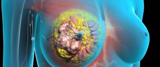
Solid cancer is one of the most aggressive types of epithelial cancer
The name solid cancer comes from the Latin word solidum, which translates to “solid.” Under a microscope, solid cancer looks like clusters of cells arranged in the form of plates (trabeculae), between which there are layers of connective tissue.
One of the most important features of solid cancer is that it consists of undifferentiated cells. They have changed so much that they no longer resemble normal cells at all. Their only occupation is constant reproduction. Therefore, under a microscope, many dividing cells can be seen in a tissue sample. Solid cancer grows quickly and metastasizes early.
What other microscopic types of cancer are there?
Solid cancer is only one of eight possible types of epithelial malignant tumors. Other possible options:
- Carcinoma in situ is a malignant tumor that does not grow into neighboring tissues, in other words, it is non-invasive. In fact, cancer in situ is the initial stage of cancer. Over time, the tumor will begin to grow deeper.
- Squamous cell carcinoma develops where there is squamous epithelium: on the skin, mucous membrane of the mouth, esophagus, vagina, cervix. Sometimes tumor cells can become keratinizing; depending on this, squamous cell carcinoma is divided into keratinizing and non-keratinizing.
- Adenocarcinoma is a tumor that develops from glandular tissue. It can occur both in the mucous membrane and in the internal organs.
- Mucous cancer consists of cells that produce a huge amount of mucus and themselves “choke” in it and die.
- Small cell carcinoma is a fairly aggressive tumor. The cells in it greatly lose their normal features and become similar to each other. The structure of normal tissue also disappears. Small cell cancer grows quickly and metastasizes early.
- Fibrous carcinoma is another aggressive tumor. It differs in that it contains many strands of connective tissue.
- Medullary carcinoma looks like brain tissue under a microscope. It consists of poorly differentiated cells and metastasizes early.
How important is it to know which of these tumor types is?
Some of them, for example, solid cancer, contain undifferentiated cells, that is, they have almost completely lost the features of normal cells. They grow and multiply quickly, so such tumors spread more aggressively in the body and metastasize earlier. Accordingly, the prognosis for the patient worsens, and the doctor is forced to plan more aggressive treatment.
In addition, other parameters of a malignant tumor are also important. For example, in order to prescribe the correct treatment, the oncologist needs to know whether the tumor has grown into neighboring organs, whether it has managed to spread to the lymph nodes and metastasize. These aspects are reflected in the generally accepted TNM classification.
Thanks to modern technologies, it has become possible to create a “molecular portrait” of a tumor. An oncologist has the opportunity to choose treatment based not only on the appearance of the tumor under a microscope, but also on knowledge of which genes of the tumor cells have a mutation, how this affects tumor growth and its resistance to various drugs.
Of course, aggressive solid cancer, especially if it has already spread throughout the body, is much more difficult to fight than early stage “cancer in situ.” But there are effective methods that, even in advanced cases, help to significantly improve the quality of life, relieve the patient of pain and other symptoms, and give an extra year, two, or even more. This is the main mission of the doctors of the European Oncology Clinic. We know how to help, even if the oncologist in another hospital threw up his hands and said that treatment is pointless.
Source: www.euroonco.ru
Cystic-solid formation - what is it?
Many people get scared if they suddenly find a tumor in their body. For some reason, most patients associate these formations only with cancer, which certainly leads to death. However, in reality everything is not so sad.
Among the many types of tumors, there are also completely harmless ones that do not have a significant effect on life expectancy. Such “good” tumors also include cystic-solid formations. What it is is not known to every person not connected with medicine.
Some people associate the word “solid” with the concept of “large, voluminous,” which causes even greater anxiety and fear for their lives.
In this article, we will clearly and clearly explain what the above-mentioned pathology means, how and why it appears, what the symptoms are and much other useful information.
Cystic-solid formation - what is it?
Both “good” and “bad” tumors are classified according to their morphological characteristics. Among the neoplasms there are:
- Cystic. The safest ones are usually easily curable. They vary in shape and tend to grow, shrink, and disappear completely for various reasons. They are a cavity filled with a viscous substance.
- Solid. The most dangerous, in a neglected state, incurable. They are characterized by a hard shell, which entails the immutability of shapes and sizes, that is, they do not increase, decrease or disappear. Solid tumors are filled with tissue fragments inside.
- Cystic-solid. They are a cross between the first and second types of tumors. They can appear in any organ, which determines the nature of the substrate in their cavity. In most cases they contain both parts of tissue and liquid.
Symptoms
This pathology can manifest itself in different ways, depending on its location. Thus, for a cystic-solid formation of the medulla oblongata (remember, this section is located in the occipital part of the head and is a continuation of the spinal cord) the following manifestations are characteristic:
- Dizziness.
- Deafness (usually develops in one ear).
- Difficulty swallowing, breathing.
- Sensory impairment in the trigeminal nerve.
- Impaired motor activity.
Tumors in the medulla oblongata are the most dangerous, as they are practically untreatable. When the medulla oblongata is injured, death occurs.
In general, cystic-solid formations in various parts of the brain are characterized by the following symptoms:
- Headaches, even vomiting.
- Dizziness.
- Insomnia or drowsiness.
- Deterioration of memory, spatial orientation.
- Impaired vision, speech, hearing.
- Loss of coordination.
- Frequent mood changes for no apparent reason.
- Muscle tension.
- Sound hallucinations.
- Feeling like there is some inexplicable pressure in the head.
If a cystic-solid formation of the spinal cord occurs, this is manifested by pain, aggravated in the supine position and at night, descending lumbago, impaired motor function, and paresis.
If at least some of the signs from the above list appear, you should immediately go to the doctor.
Cystic-solid formation in the thyroid gland
As a rule, a cystic-solid formation in the thyroid gland is a cavity limited by a dense membrane, filled with cells of the thyroid gland itself. Such cavities are observed single and multiple. The reasons for this may be the following:
- Hereditary factor.
- Frequent stress.
- Hormonal disorders.
- Iodine deficiency.
- Infectious diseases.
Symptoms
A cystic-solid formation of the thyroid gland may not manifest itself at all and may be discovered by chance during a routine examination of the patient. In such cases, the doctor palpates small lumps on the thyroid gland. Many people with this pathology have complaints:
- Difficulty and even pain when swallowing.
- Shortness of breath (which was not there before) when walking.
- Hoarseness of voice.
- Pain (uncharacteristic sign).
The occurrence of a cystic-solid formation in the left or right lobes of the thyroid gland is felt approximately the same. More often they are very small in size (up to 1 cm). However, cases of very voluminous cystic-solid formation (more than 10 cm) have been recorded.
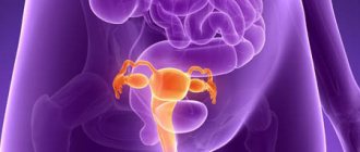
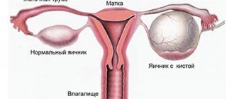
Cystic-solid formation in the kidneys and pelvis
Kidney tumors occur with approximately equal frequency in men and women. But in women much more often than in men, cystic-solid formations appear in the pelvis.
What can this bring to patients? Since this pathology is mainly observed in women of childbearing age, without timely treatment it can lead to infertility.
The main cause of the disease is hormonal disorders caused by:
- Pregnancy.
- Climax.
- Abortion.
- Taking birth control pills.
Tumors manifest themselves as pain in the lumbar region and/or lower abdomen, headaches, and menstrual irregularities.
Cystic-solid formations appear on the kidneys for the following reasons:
- Organ injuries.
- Tuberculosis (developing in the kidneys).
- Infections.
- Operations.
- Stones, sand in the kidneys.
- Hypertension.
- Congenital anomalies of the organ.
Patients complain of pain in the lumbar region, difficulty urinating, and unstable blood pressure.
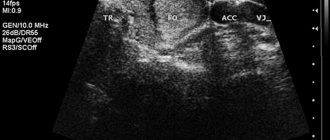
Cystic-solid formations of any location are diagnosed using the following methods:
- Examination by a doctor, palpation.
- Blood analysis.
- CT.
- Ultrasound.
- MPT.
- Biopsy.
If cystic-solid formations occur in the spinal cord, additional radiography of the spine, electroneuromyography, and spinal angiography are performed.
Treatment
The discovery of a cystic solid tumor is not a reason to prepare for death. In the vast majority of cases, this pathology is successfully treated. According to indications, the doctor may prescribe drug therapy or surgery. This mainly depends on the location of the tumor.
Thus, with a cystic-solid formation on the medulla oblongata, operations are not performed; only treatment with tablets and radiotherapy is practiced. If the tumor is localized in other parts of the brain, surgical intervention using laser and ultrasound is usually prescribed.
Chemotherapy and radiation therapy are prescribed only if the tumor is inoperable. For this pathology in the thyroid gland, treatment methods depend on the size of the formation. Small nodules (up to 1 cm) are treated with tablets.
If larger formations appear, a puncture may be prescribed followed by removal of the affected part of the thyroid gland.
Forecasts
Of course, the appearance of a tumor in any organ should be taken seriously. If the patient consults a doctor on time and follows all his instructions, then a solid cystic formation in the kidney, thyroid gland, genitourinary system and some other organs can be cured completely and without complications.
The outcome of treatment for such a pathology in the brain is less favorable, since surgical intervention almost always affects neighboring tissues, which can lead to a number of complications. A tumor in the spinal cord or medulla oblongata is the option with the least favorable outcome.
But even in these cases, timely treatment can save the patient’s life.
Source: https://FB.ru/article/359628/kistozno-solidnoe-obrazovanie—chto-eto-takoe
How does cancer spread?
Some types of cancer are characterized by a high growth rate, the nature of which is infiltrative (as the size increases, the cancer grows into the surrounding tissues). If such a tumor grows into a vessel (blood or lymphatic), then the fluid flow of this vessel contributes to the spread of cancer cells throughout the body. This is called metastasis, and in this case distant tumor foci are formed - metastases. The disease cancer differs from a benign tumor histologically. Often the changes are so pronounced that it is very difficult for pathologists to know from which tissue the tumor was formed. Cancer cells are atypical, have an irregular shape, a large number of nuclei and mitoses. Malignant tumors are divided into cancer (which arises from epithelial tissue) and sarcoma (which arises from mesenchyme). Cancer forms about 90% of all neoplasms. This led to the spread of the terms “cancer neoplasm” and “anti-cancer measures.”
Pathogenesis
The occurrence of R. is always separated from the moment of action of one or another factor, which can be considered as etiological, the so-called. latent period, during which there are completely no clinical or other objective signs of tumor growth. It is assumed that it is during this period that those pathogenetic processes unfold, on the basis of which the formation of tumor growth occurs, culminating in the emergence of a certain clinical and morphological form of cancer (see Oncogenesis).
Disease cancer and sarcoma
The tumor can have a different appearance: nodes, cauliflower, mushroom cap. The surface can be smooth, bumpy, rough, papillary, etc. Cancer is located in an organ differently: from the surface of the organ to its center. A superficial tumor may be located on the feeding pedicles. Malignant neoplasms lead to bleeding and ulceration. The disease cancer can also form from a benign tumor, this process is called malignancy. All processes occurring in the tumor, as well as in the body due to its activity (metastasis, growth, damage to internal organs, bleeding, intoxication with tumor metabolic products) lead to exhaustion of the body - cachexia. It is believed that cancer appears quite late. However, this statement is only half true. The bottom line is that, for example, with stomach cancer in the early stages there is no excruciating pain, high temperatures and similar symptoms. Severe pain, weight loss and severe weakness appear in later stages of the disease. There is cancer that is more common in children and young adults: teratomas (embryonic tumor), tumors of nerve cells, angiomas (vascular lesions), nephroblastoma (renal cancer).
Cancer - tumor growth rate
Tumors are also divided depending on their growth rate. Fast-growing cancers expand over a period of weeks or months, while slow-growing cancers can maintain their size for months or years. The disease cancer can be with or without metastases, the so-called classification according to the presence of dissemination (from the Latin dissemino - to spread). Depending on the direction of cancer growth in relation to surrounding tissues, expansive and infiltrating are distinguished. With expansive growth, cancer does not grow into the surrounding tissues, as if pushing them aside (typical of benign tumors and some malignant ones). In this case, the cells develop and reach a mature state. With infiltrating growth, cancer grows into the surrounding tissues between their fibers. This growth is typical for most malignant neoplasms. With it, cells do not have time to achieve differentiation and contribute to the spread of cancer in the body (formation of metastases). A tumor (cancer disease) has a duration of development, as well as a clinical manifestation, which is determined by the patterns of its location, and also depends on the general condition of the body. For example, breast cancer and skin cancer are characterized by a rather long course. However, regardless of the location of the pathological process, the clinical picture of cancer in the early stages is often very poor. The patient's lack of anxiety, as well as his apparently good condition, often does not allow cancer to be diagnosed in the early stages.
Bibliography
Blokhin NH, Orlov-sky L. V. and Serebrov A.I. Anticancer propaganda, M., i980; Gerasimenko V. N. Rehabilitation of cancer patients, M., 1977; Gershanovich M. L. and Pike and N M. D. Symptomatic treatment of patients with malignant neoplasms in advanced stages, M., 1980; Gnatyshak A.I. Textbook on general clinical oncology, M., 1975; Zilber L. A. Virus-genetic theory of the occurrence of tumors, M., 1968; Zilber JÎ. A. et al. Evolution of the virus-genetic theory of the occurrence of tumors, M., 1975; Kazantseva I. A. Study of the mitotic regime in the morphological diagnosis of tumors, Arkh. pathol., t. 42, no. 2, p. 77, 1980; Clinical Oncology, ed. N. N. Blokhin and B. E. Peterson, vol. 1-2, M., 1979; K r e v s k i y N. A. To the doctrine of the pre-tumor (precancerous) period, Arch. pathol., t. 36, no. 9, p. 3, 1974; Novinsky M. A. On the issue of grafting malignant neoplasms, St. Petersburg, 1877; Tumor growth as a problem in developmental biology, ed. V. I. Gelypteina, M., 1979; Pavlov K. A., Paikin M. D. and Dymarsky L. Yu. Oncology of a polyclinic doctor, M., 1979; Pathological diagnosis of human tumors, ed. N. A. Kraevsky et al., M., 1982; Peterson B. E, Surgical treatment of malignant tumors, M., 1976; S e y c I. F. Biochemical aspects of malignant growth, Vopr. oncol.,t. 24, no. 10, p. 3, 1978; Shab a d L. M. Experimental and morphological aspect of the problem of precancer, Arkh. pathol., t. 35, no. 8, p. 61, 1973; aka, Carcinogenic substances in the environment and the prevention of malignant tumors, Vopr. Oncol., vol. 23, no. 10, p. 11, 1977; Epidemiology of cancer in the USSR and the USA, ed. N. N. Blokhin and M. A. Shneiderman, M., 1979; Yagubov A. S. and Kats V. A. Some features of the ultrastructure of tumor cells, Vopr. onkol., vol. 22, no. 3, p. 64, 1976; Cancer medicine, ed. by JF Holland a. E. Frei III, Philadelphia, 1973; D u 1 be with about R. Transformation of cells in vitro by DNA-containing viruses, J. Amer. med. Ass., v. 190, p. 721, 1964; HuebnerR. Ja To-d a g about GJ Oncogenesis of RNA tumor viruses as determinants of cancer, Proc. nat. Acad. Sei., v. 64, p. 1087, 1969; International classification of diseases for oncology, Geneva, WHO, 1976; Urbach F. Ultraviolet radiation and skin cancer in man, Prev. Med., v. 9, p. 227, 1980. See also bibliogr, to art. Tumors.
Yu. N. Solovyov, L. E. Denisov.
Theory of origin of cancer
Professor K. Nishi, an oncology specialist, is the author of the theory of the development of cancer in humans associated with an excess of carbon monoxide in the body and a lack of oxygen. One of the reasons for this imbalance, according to Nishi’s theory, is wearing outer clothing (after all, cancer is extremely rare in animals), which provokes the formation of not only carbon dioxide, but also carbon monoxide in the human body. Also, an excess of this compound can form as a result of frequent constipation, since when the intestines are disrupted, the acid-base balance in the entire digestive tract is disrupted, and the formation of carbon monoxide increases. Cancer, according to Nishi, can develop due to a lack of vitamin C, which causes an increase in capillary permeability and the occurrence of subcutaneous hemorrhages. Stimulation of cell growth under these conditions can lead to the formation of cancer. Also, consuming large amounts of carbohydrates and alcohol leads to further destruction of the walls of blood vessels, and, as a result, to excessive synthesis of carbon monoxide.
Forecasts
Of course, the appearance of a tumor in any organ should be taken seriously. If the patient consults a doctor on time and follows all his instructions, then a solid cystic formation in the kidney, thyroid gland, genitourinary system and some other organs can be cured completely and without complications. The outcome of treatment for such a pathology in the brain is less favorable, since surgical intervention almost always affects neighboring tissues, which can lead to a number of complications. A tumor in the spinal cord or medulla oblongata is the option with the least favorable outcome. But even in these cases, timely treatment can save the patient’s life.
author: Dr. med. Gesche Tallen, erstellt 2003/12/11, editor: Natalie Kharina-Welke, Print permission: Prof. Dr. med. Dr. hc Günter Henze, Last modified: 2012/08/15
The word tumor means nothing more than compaction or thickening. This does not say anything about its properties. The word itself simply describes a solid (solid, as it is commonly called in medicine) clearly limited growth from its own mature (differentiated) tissue or immature (rudimentary, undifferentiated) tissue. Sometimes this tissue can be as immature (fetal) as the tissue of a child before birth (intrauterine fetus).
In medical terminology, the word part/suffix “om” indicates in the name of the disease that it is a tumor. And the first part of the name of the disease, as a rule, is a term from the Latin language. This part of the word names the specific tissue from which the tumor began to grow. For example, the term “lipoma” means a tumor of adipose tissue, “osteoma” means a tumor of bone tissue.
Squamous cell carcinoma
Squamous cell carcinoma develops on mucous membranes and in the skin where there is squamous epithelium, for example: cervix, oral cavity, vagina, esophagus, etc.). On those mucous membranes that are covered with prismatic epithelium, this type of cancer develops only if there has been a transformation of the epithelial tissue. Most often, squamous cell carcinoma is a highly differentiated tumor and consists of numerous strands of atypical epithelial tissue that grow deep and destroy structures located below. Thus, cancer has the appearance of nested clusters, and along the periphery of such a cluster there are basal cells, and in the center there are more differentiated mature cells, which sometimes retain the ability to become keratinized and form cancer called “cancer pearls.” Depending on the ability of atypical cells to keratinize, keratinizing and non-keratinizing types of squamous cell carcinoma are distinguished.