What is it and is it dangerous?
Fibroma is a benign, highly differentiated tumor that is formed from connective tissue fibers. It is not prone to rapid growth and increase in size. The neoplasm is not dangerous if it is not injured.
Fibroids can develop both on the surface of the body and in internal organs. Its most typical formation is in the uterus and mammary glands.
The tumor can also grow and mix with other tissues, for example, with:
- Muscular (fibromyoma).
- Glandular (fibroadenoma).
- Vascular (angiofibroma).
- Fatty (fibrolipoma).
What does fibroblastoma look like?
It should be understood that fibroblastoma, unlike fibroma, is a malignant tumor. Externally, it looks like a single node on or under the skin, with unclear boundaries.
Fibroblastoma is typical for children and adolescents, since the tumor is formed during the period of intrauterine development of the fetus.
Fibroma looks different, depending on its type.
There are hard and soft tumors.
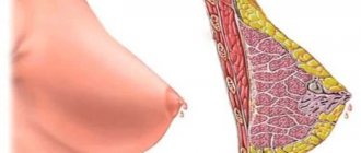
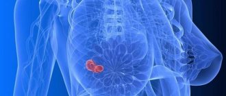
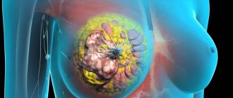
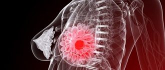
Types of breast fibroids
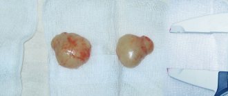
Fibroid formation in the mammary gland is divided into two types: diffuse fibroadenomatosis and fibroadenoma. The first type is characterized by the growth of a tumor throughout the entire volume of the breast with the presence of compaction and the presence of pain in the mammary gland. The second has a local form of manifestation of fibroma. This is a round formation with a dense structure; when palpated, you can feel a moving node not connected to the skin. The size of this fibroma reaches 70 mm in diameter.
Fibroadenoma is further classified into several types based on morphological characteristics. The most common types include: pericanalicular, intracanalicular, mixed fibroadenoma, which combines the features of the first two types. The above tumors are characterized by tissue growth around the ducts and the absence of a tendency to degenerate into a malignant formation. The rarest type of fibroadenoma is phylloides, which is characterized by rapid growth and the ability to degenerate into sarcoma.
Soft
Soft fibroma is a small cutaneous nodule, oval or round in shape. The skin over it is of normal color or slightly pigmented, wrinkled.
Soft fibroids usually form in older women who are prone to excess weight, in the armpits, folds under the mammary glands and on the breast itself, on the eyelids and in the groin area.
The number of neoplasms can vary from a few to several dozen.
Diagnostics
There are several stages to diagnose the disease. First of all, a clinical examination of the woman is carried out. Then, with the help of palpation, the neoplasm is detected. An ultrasound scan of the breast provides a detailed understanding of the location of the tumor, its exact shape and size, as well as its structure. Next, a tumor biopsy is prescribed, which is controlled by ultrasound and performed using a thin needle using the aspiration method. The resulting material undergoes cytological examination to determine the presence of malignant cells.
The diagnosis may require clarification in order to completely exclude diseases such as cysts, cancer and cystadenopapilloma. In such cases, a core needle biopsy, X-ray mammography is prescribed, or a diagnostic resection of the mammary gland is performed, followed by histological examination. The presence of a cancerous tumor can be definitively excluded after its removal and laboratory examination.
Fibromatosis of the skin
Fibromatosis of the skin is the multiple appearance of fibromas on it. This is typical mainly for soft tumors.
The most common locations: eyelids, armpits, groin, mammary glands, less common on the mucous membranes.
Fibropapilloma of soft tissues
Benign tumors in soft tissues form quite often.
Let's look at their most common locations.
Brain
Fibroma in the brain remains asymptomatic for a long time.
The first signs of deterioration may appear if the tumor has reached a very large size and affects any vital center. Brain fibroids are diagnosed using skull x-rays and computed tomography.
On the head
Mostly hard fibroids form on the head due to the fact that the skin in this area is quite thin and fits tightly to the skull.
The neoplasm usually causes discomfort only in aesthetic terms. Pain will occur when the fibroid is touched with a comb or when the tumor is significant.
On the nose
In the nasal area, both soft and hard fibroids form with equal frequency.
The soft tumor is usually small in size and resembles a papilloma.
The hard one can periodically hurt, especially if it is located in places that are constantly injured (for example, on the lower part of the wing of the nose, which lends itself to frequent friction during a runny nose).
On the neck
Fibroids on the neck are mostly soft, but very often cause pain.
New growths are constantly touched by the collar of clothing, which can lead to injury.
Cutaneous angiofibroma
Cutaneous angiofibroma is a neoplasm consisting of connective tissue and blood vessels. It is hard to the touch and round. The color is often pink or closer to red due to rich vascularization.
The tumor is translucent, the vessels are clearly visible. It develops mainly after 40 years.
Angiobioma with frequent trauma can bleed profusely.
Belly
A predominantly soft fibroma forms on the abdomen; it is localized in the skin folds, especially in obese people.
It rarely hurts or causes discomfort.
Shoulder
Both soft and hard fibroids can appear on the shoulder, it all depends on the person’s constitution. The formations interfere only in cosmetic terms, but almost never hurt.
Hips
In the thigh area, soft fibroids are more often formed than hard ones, due to the predominance of adipose tissue in this area.
Tumors are painless unless they are constantly touched or injured.
Backs
Soft fibroids appear on the back, often in groups of several at once. New growths are very often injured due to constant wearing of clothes and as a result can hurt.
Eyes
In the eye area, or more precisely on the upper eyelid, soft fibroids form in groups. Even if they are small, they often get in the way.
They are removed almost immediately.
Benign adenoma in the groin
In the area of the inguinal folds, soft fibromas most often appear, both single and grouped.
They get in the way due to constant friction in this area and are often damaged, especially in obese people.
Uterine fibroid - symptoms and treatment
As a rule, diagnosing uterine fibroids in most cases is not difficult. First of all, it is necessary to correctly identify the medical history and take into account all risk factors for the occurrence of uterine fibroids, and conduct a gynecological examination. The easiest way to diagnose uterine fibroids is a gynecological examination on a chair . During this examination, subserous fibromatous nodes may be palpated separately from the uterus. Most often in the form of separate formations of a round shape, dense, with varying degrees of mobility. The uterus itself can be of various sizes, but more often it is enlarged, and can be of enormous size. The surface of the uterus is palpable with a tuberous, fibromatous nodes of a denser structure. If blood circulation in fibromatous nodes is impaired, palpation becomes painful. In women with interstitial fibroma, an enlarged uterus can be felt, the consistency of which will be dense, the surface may be smooth or lumpy. Malnutrition of the interstitial nodes usually does not occur, so palpation of such a uterus is most often painless.
Ultrasound examination (ultrasound) of the pelvic organs is the gold standard not only for the diagnosis of primary uterine fibroids, but also for their dynamic monitoring.[1] The advantage of the method is its information content, accessibility, and safety. However, it is worth considering the fact that ultrasound is a rather subjective diagnostic method, because the reliability of the results largely depends not only on the qualifications of the specialist, but also on the patient’s preparation for the study.
Ultrasound diagnostics makes it possible to assess the size of the uterus, establish the number of pathological foci and their location, the nature of the shape and contours, size, structure and density; during dynamic observation, compare the data with the results of the previous study, assess the dynamics of the pathological process. To clarify the location of fibromatous nodes, you can use ultrasound tomographs that provide a three-dimensional ultrasound image. Significant diagnostic data can be revealed by color Doppler mapping (CDC) . Using it, you can evaluate not only the echographic picture of the structure of the fibroma, but also evaluate its blood flow.
Along with ultrasound diagnostics, methods such as computed and magnetic resonance imaging . However, methods of radiological diagnosis in women of reproductive age are resorted to under strict clinical indications.
In case of menstrual irregularities, patients are advised to undergo diagnostic hysteroscopy - a highly informative method that allows assessing not only the condition of the uterine cavity, pathological processes of the endometrium, the type of node and its location, but also deciding on the possibility of performing transcervical fibromectomy with endoscopic control.[1] Carrying out such a procedure is possible only in patients with fibroids with an enlarged uterus no more than 12-13 weeks of pregnancy.
In addition, to assess the condition of the endometrium for diagnostic purposes, endometrial curettage with histological examination is used. The results of histological examination can significantly affect the management of the patient. Carrying out diagnostic curettage allows you to decide on the continuation of conservative therapy or the extent of surgical intervention.
In special cases, if it is necessary to differentiate between fibroma and giant ovarian and retroperitoneal tumors, diagnostic laparoscopy is used.
Causes
The exact causes of fibroids are unknown, but there are predisposing factors, which include:
- Hormonal imbalance, especially during menopause and pregnancy.
- Abortion.
- Endocrinological diseases.
- Immunodeficiencies.
- Hereditary predisposition.
- Frequent skin trauma.
- Increased sweating.
- Prolonged exposure to ultraviolet radiation.
- Liver pathologies.
- Chronic somatic diseases.
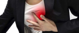
Symptoms
Typically, patients notice a tumor on their body. It rarely hurts and may simply cause minor discomfort depending on the location. The tumor is more of a concern in cosmetic terms, and pain can only appear when damaged or when the fibroid is inconveniently located, for example, on the foot.
If a tumor develops in hollow organs, it does not manifest itself for a long time, until it becomes significant in size and leads to severe dysfunction.
The child has
Fibroids in children do not have any characteristic features. Parents may notice some kind of neoplasm. If it is too small, a child may accidentally damage it or even tear it off. Tumors of internal organs are rare in children.
In children, the following types of fibroids are sometimes encountered:
- Fibrous hamartoma. Multiple tumors that are located on the extremities, groin, armpits and buttocks. They are yellow-gray and have a three-layer structure.
- Gingival fibromatosis. It represents the appearance of multiple hard fibroids on the child’s gums.
- Multiple juvenile hyaline fibromatosis. It appears mainly in boys on the back, head, nose, ears and knees. It looks like dense grayish subcutaneous nodes.
- Infantile digital fibromatosis. Multiple hard fibromas appearing on the fingers and toes.
- Fibromatosis of the neck. A very rare pathology in which fibromatous tissue is located at the site of the sternocleidomastoid muscle. The tumor nodes are quite large, several centimeters in diameter.
Symptoms
The clinical picture of fibroma depends on where the tumor is located. The main sign of pathology is a growth that rises above the skin and has a wide base. Its color may not differ from the color of the skin, only after time it darkens slightly.
Fibroids will not cause pain, discomfort or anxiety to the patient. He regards it as a cosmetic defect. But there are situations when the growth begins to hurt and becomes sensitive. This may be due to the unfortunate location of the fibroid. For example, if it is on the sole, then the pain is felt while walking on the floor, for the arrangement of which you can use linoleum in Yekaterinburg. Empire Flooring offers a wide selection of flooring based on thickness, color and price.
Fibroids affecting internal organs are not felt by patients in any way. And they can be detected during the next examination or during the development of complications caused by tumor growth.
Complications
Typically, fibroma is complicated by trauma, bleeding (especially if it is an angiofibroma), twisting, followed by necrosis. After damage, a secondary bacterial infection may occur.
Fibroids of internal organs, especially the uterus, can cause severe pain and bleeding, and if twisted, they can cause an “acute abdomen” and even peritonitis.
How to cure?
Treatment of fibroma is required when it is subject to constant mechanical damage.
If the tumor does not cause any discomfort, it is better not to touch it. The fibroid is removed surgically.
On the body
There are 4 ways to surgically remove fibroids:
- Traditional (using a scalpel).
- Laser.
- Radio wave.
- Electrocoagulation.
Any of these methods can be used on the body, since the cosmetic aspect is not so important.
On the face
Laser and radio wave therapy are mainly used to remove fibroids on the face. After the operation, no traces are left, which contributes to the rapid healing of the postoperative wound.
The procedure takes about 15–20 minutes.
Traditional surgery is used only to remove large, hard fibroids.
How to treat a child?
Treatment methods for children are the same as for adults. The least possible disruption of tissue integrity is encouraged.
For children under 10 years of age, general anesthesia is required; for older children, only local anesthesia can be performed.
If the tumor does not cause discomfort, it is better to leave it and protect it from exposure to sunlight.
Removal on the face
On the face, the tumor is removed using radio wave or laser methods, or electrocoagulation.