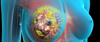
Oleogranuloma of the mammary gland is a tumor-like neoplasm, which, although considered a benign growth, still has nothing to do with tumors either in terms of cellular composition or other characteristics.
A granuloma is formed as a result of the reaction of organ tissue to the action of an external factor or a foreign object introduced into the gland.
Features of development
It is immediately worth noting that this is a pathological condition that requires careful monitoring of changes. Often, during the first examination of the mammary gland and palpation, oleogranuloma is confused with a tumor, and only with the help of instrumental methods can this nature of the neoplasm be refuted.
There are some differences that characterize oleogranuloma and breast tumor.
So, with cancer pathology the following symptoms are present:
- discharge of unnatural exudate from the nipple (purulent, bloody);
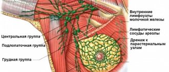
- retraction of halos;
- the skin of the gland thickens and wrinkles.
The clinical manifestations mentioned above do not occur with oleogranulomas. The only symptom that occurs (rarely) is retraction of the areola.
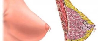
In the internal cavity of the growth, the formation of a fatty cyst often occurs - a benign neoplasm, which is formed from adipose tissue. The latter is surrounded by a dense wall, which can become calcified.
As a rule, the cyst disappears on its own. In case of severe pain, puncture and removal of exudate are required.
Other factors
Breast oleogranuloma forms after surgery, irradiation, or trauma to the gland. Such factors disrupt blood circulation in the organ, as well as oxygen exchange. As a result, the lobes of the gland die, and the body tries to destroy the dead cells by releasing special enzymes.
Due to the functioning of enzymes, an inflammatory process occurs, and scar tissue forms in the area of dead cells. The dead cells release fatty deposits that fill the cystic cavity. Two such processes in combination contribute to the development of compaction - oleogranuloma.
The risk of formation of such a growth increases due to the following interventions:
- biopsy of gland tissue;
- lumpectomies;
- mastectomy;
- paraurethral cyst;
- plastic surgery;
- breast reduction and implant removal surgeries;
- endometrioid cyst;
- rare, but there are still cases of granuloma formation in the following pathological conditions: mastitis, dilated ducts of the gland, panniculitis, polyarteritis nodosa, Weber-Christian disease.
In most cases, the pathology is diagnosed in old age in women with large breasts.
Classification
Mammologists classify lipogranuloma according to the reasons that led to its appearance:
- artificially created or injected - arise at the site of plastic surgery on the mammary gland, performed using materials unsuitable for implantation: paraffin, petroleum jelly, various vegetable oils. Spreading between the fatty layers and lobules of the glandular body of the breast, foreign substances lead to the formation of multiple foci of inflammation and granulation, which subsequently cause the appearance of serious purulent processes;
- after injuries to the mammary gland (post-traumatic) - appear as a result of falls, blows, compression in everyday life or transport, or unprofessionally performed massage;
- previous surgical interventions on this breast (near inflammatory) - a granuloma is formed near the place where there is an inflammatory focus and, as it were, surrounds it. The causes of inflammatory processes can be: lactation mastitis, mastectomy, non-healing postoperative sutures, remnants of unremoved ligature stitching threads or their untimely removal;
- spontaneous (idiopathic), which appeared without obvious reasons, with little noticed, small, local skin infections or minor injuries to the mammary gland;
- as complications after radiation therapy, hormonal imbalance, rapid loss of normal weight;
- with long-term therapy with anticoagulants;
- with Weber's panniculitis.
Symptoms
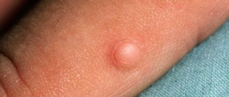
In the ultrasound photo, the first signs of oleogranuloma are the presence of small colorless nodules and seals in the gland, which can disappear on their own after a certain time. They have different sizes, shapes (oval or round), and surfaces (rough, smooth).
As the disease develops, the nodules transform into dense formations that can be seen without magnifying devices and instrumental diagnostic methods.
In advanced cases, severe pain occurs, the oleogranuloma increases in size and acquires a copper color. In some cases, a trophic change forms on the skin in the form of a purulent necrotic ulcer.
In the affected area, there is a disruption in blood circulation, and fistula openings are formed. A general intoxication syndrome occurs: the general temperature rises, general malaise occurs, and causeless rapid fatigue occurs. Such symptoms indicate activation of the inflammatory process.
Diagnostics
Often, oleogranuloma is discovered by the patient herself. In this case, you should consult a doctor who will prescribe a mammogram or ultrasound of the gland. To exclude a cancerous tumor, a puncture biopsy is performed.
On mammography, the formation has a round or irregular shape measuring less than 2 cm. Sometimes it is surrounded by a thin, dense capsule, which makes it possible to preliminarily distinguish it from a cancerous tumor. If such a membrane is thickened or uneven, the doctor must rule out a malignant process. Calcifications are often found - accumulations of lime in the granuloma.
By ultrasound, fat necrosis is defined as a subcutaneous lesion with increased echogenicity (density), uncharacteristic of cancer. Cavities - cysts - can be visualized inside it, or it can be homogeneous. Hyperechogenicity is characteristic of only 0.8% of all malignant breast tumors, among them invasive ductal and lobular cancer, lymphoma, angiosarcoma, liposarcoma.
One of the main methods for diagnosing oleogranuloma is mammography.
An MRI test is not prescribed because it does not provide convincing evidence of the absence of cancer and is more expensive. This study reveals a focus of fat necrosis with a thin rim around it.
Computed tomography is also not included in the standard examination for this pathology. If it is performed for another breast disease, signs of oleogranuloma may be the presence of liquid fat, fibrous tissue around it and inflammation. Calcifications begin to be detected only when they reach a large size.
The main diagnostic methods are mammography and biopsy with histological examination.
Diagnostic measures
If you find an unknown neoplasm on your breast, you should contact a mammologist or gynecologist. The doctor examines the mammary gland in detail and palpates it. If a tumor-like formation is found, the woman is given a referral for mammography and ultrasound examination.
At the initial stage, a granuloma appears as a cancerous tumor on a mammographic image. During this period, a biopsy is prescribed, which consists of collecting biological material from the growth, which is subjected to histological analysis in the laboratory. It is a biopsy that helps to detect an atypical cyst.
After breast cancer surgery: nutrition, prognosis, risk of recurrence
To answer these questions, we analyzed information about 503 patients with stage I–III breast cancer who received treatment at the Federal State Budgetary Institution Russian Cancer Research Center named after. N. N. Blokhin RAMS in 1992-2002. The main group included 124 patients, average age 41.5 years (24–67). Women were operated on with radical mastectomy with preservation of the pectoral muscles in combination with primary breast reconstruction: an expander (n=14) or a skin-fat flap on the latissimus dorsi muscle using an endoprosthesis (n=18), or a transverse rectoabdominal flap on a muscular pedicle (n=92).
The control group consisted of 379 patients, mean age 40.1 years (26–79). 145 patients underwent breast-sparing surgery, 234 underwent radical mastectomy with preservation of the pectoral muscles. The groups were comparable in terms of the main factors influencing prognosis (stage, age, treatment). Drug and radiation treatment was carried out according to general principles. Median follow-up duration was 63.7 (20.4–140.5) months.
The local recurrence rate was:
- in the mammary gland after organ-preserving operations - 4.1%;
- after modified radical mastectomy - 1.7%;
- after modified radical mastectomy with primary reconstruction - 1.6% (p
How to treat oleogranuloma
Not all oleogranulomas are treated. In many cases, such tumors disappear on their own. If pain symptoms are present, a painkiller is prescribed, a course of massage is prescribed, and warm compresses are used. The latter are applied to the sore area for 30 minutes four times a day.
In rare cases, surgery is used to remove the tumor. This is required if the size of the oleogranuloma is large and the woman experiences increased anxiety due to pain. During surgery, the doctor excises the tissue that contains the granulomatous node.
If a cystic formation is present, a puncture biopsy is prescribed to empty the cavity of liquid exudate. This reduces the size of the oleogranuloma.
Before surgery, tests are required:
- blood – general and biochemical;
- blood - for HIV, hepatitis, syphilis;
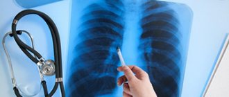
- fluorography, x-ray examination;
- electrocardiography.
Do not take blood thinning medications before surgery. On the day of intervention, they refuse food and water.
Contraindications
As with any other treatment, chemotherapy has contraindications, as it will not always be effective. Age is also important - for older women, chemotherapy for breast cancer may not bring the expected results.
This method is not used for hormonally dependent forms of oncology due to reduced levels of estrogen and progesterone. Radical measures are used - removal of the ovaries. In combination with the use of drugs that block the production of sex hormones, it is possible to achieve stable remission.
Also, a reason that prohibits chemotherapy treatment may be a high level of bilirubin in the blood. Excessive amounts of this substance, which is formed during the breakdown of hemoglobin, is one of the symptoms of a certain disease of the gallbladder or pancreas. This must be treated before anticancer drugs are prescribed.
Like any treatment, chemotherapy has a number of contraindications. They are mainly associated with the ineffectiveness of the procedure or the possibility of using other, safer methods.
For example, if a patient has a hormonal form of breast cancer, it is necessary to try hormone treatment. Side effects include:
- Digestive problems: nausea, vomiting, loss of appetite.
- The appearance of bruises, hemorrhages, anemia.
- Decreased immunity, increased risk of contracting viral infections, increased temperature.
- Hair loss up to complete baldness.
- Changes in the functioning of the endocrine system.
- Disruption in the menstrual cycle.
- Increased fatigue, lethargy, feeling tired.
Doctor's advice: you cannot interrupt the course of chemotherapy due to the appearance of such side effects: after some time the body will be able to adapt, and cancellation will lead to accelerated cell growth.
To alleviate the patient’s condition, the doctor may prescribe vitamins and antibiotics to boost immunity. Treatment of breast cancer has come a long way: today there are quite effective ways to cope with the disease, which have received a huge number of positive reviews.
Prognosis and prevention
Preventive recommendations for oleogranuloma of the mammary gland boil down to avoiding injury to the organ.
If it was not possible to avoid injury, you should not self-medicate, but rather contact a mammologist who will prescribe appropriate treatment to avoid the formation of tumors.
Kinds
Depending on the cause of the development of the pathology, the following types of oleogranulomas are distinguished:
- post-traumatic - occurs after injuries, bruises, massage;
- post-injection - cosmetic procedures (injections of various drugs, implantation of threads and other procedures);
- spontaneous - occur after minor injuries or damage to breast tissue, which women either do not remember or did not notice how they received them.
Spontaneous oleogranulomas are more common than others, so women are advised to take care of their breasts and protect them from any damage.
Treatment
Treatment of mammary granuloma begins after examination for the presence of an oncological process. Therapy can be conservative or surgical. Conservative treatment involves prescribing the patient a course of antibacterial agents, corticosteroids, immunostimulants and vitamins. Its goal is to eliminate areas of inflammation and activate the body's defenses. Conservative therapy gives positive dynamics extremely rarely. Therefore, the surgical method is more often prescribed.
Surgical intervention
The operation to remove granuloma is performed under local or general anesthesia. The area of tissue removed depends on the size of the tumor. After excision of a small nodule, the shape of the breast remains virtually unchanged. When the formation is large, sectoral resection is performed. In this case, a significant proportion of the tissue to which necrosis has spread is removed.
Recovery period
The complex of postoperative therapeutic measures includes a course of drug therapy, consisting of: immunomodulatory, anti-inflammatory, antibacterial medications and vitamin preparations, as well as a rehabilitation course that accelerates rehabilitation, and physical procedures.
You can prevent the occurrence of unpleasant complications in the postoperative period if you follow these rules:
- timely removal and treatment of sutures until they are completely healed;
- wear post-operative compression garments (support top) for as long as the doctor requires;
- you need to inform your doctor about any allergies you have to antiseptic drugs and synthetic materials;
- timely treatment of infectious chronic foci with a full course of antibiotics;
- For at least 30 days it is prohibited: visiting saunas and baths, lifting and carrying heavy objects, not playing sports, not actively sunbathing, not visiting a solarium, not taking baths with hot water.