The lymphatic system is one of the body's main defenders against infection. This system contains lymphatic vessels and lymph nodes, which are located in key areas of the body. Lymph nodes are responsible for filtering lymphatic fluid (lymph) and detecting chemical changes that signal the presence of infection.
Cancer cells can travel to the lymph and then to the lymph nodes. When cancer spreads to the underarm lymph nodes, they become enlarged. The prognosis for breast cancer is good if the cancer cells do not involve the lymph nodes. But most people who have enlarged axillary lymph nodes do not have cancer at all.
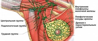
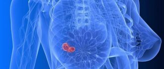
Lymphatic system of the mammary gland
The female breast is a rather complex organ from an anatomical point of view. The breast consists primarily of connective, glandular and adipose tissue. The mammary glands are located above the pectoralis major muscle and are quite mobile. Such anatomical features also determine the structure of the lymphatic system - in the chest and next to it there are many lymph nodes communicating with each other.
There are several separate groups of lymph nodes:
- Paramammary system. Lymphatic ducts are located in the pectoralis major muscle.
- Axillary lymph nodes are located throughout the breast and are connected to each other using the paramammary system.
- Intramarmals are the largest group.
- Regional.
Intramammary, in turn, are divided into several groups:
- Central.
- External.
- Subscapular.
The main load falls on the central nodes, which belong to the intramammary system. It is these nodes that are responsible for the outflow of lymph from the mammary glands into the common (larger) lymphatic ducts.
The easiest way to independently detect inflammation of the regional lymph nodes (axillary). Due to their close proximity to the mammary glands, they quickly respond to any changes in the breast. In case of oncology, it is customary to remove them, since it is to the regional nodes that metastases occur.
When do metastases appear in breast cancer and the main degrees of damage?
In this type of cancer, the growth of abnormal cells into the lymph nodes begins already at stage II of the disease.
It is expressed in 3 degrees, each of which is characterized by specific changes in the peripheral organs of the lymphatic system:
- minimal pathological transformations;
- the appearance of a small number of mutated cells;
- significant damage to the lymph nodes by abnormal cellular structures.
Enlarged lymph nodes
Sometimes the functioning of certain parts of the lymphatic system is disrupted - and individual lymph nodes or groups become inflamed. This condition is called lymphadenopathy. If the lymph nodes under the arm are inflamed, then lymphadenopathy is called axillary - after the name of the group of lymph nodes. Inflammation is manifested by discomfort in the axillary region, enlargement of individual nodes, as well as pain on palpation.
Lymphadenopathy is not a separate disease, it is a pathological condition that indicates some disease in the affected area. Thus, various diseases from mastitis and mastopathy to breast cancer cause inflammation and enlargement of regional lymph nodes.
If this happens, then a series of diagnostic measures are prescribed to establish the root cause of the disease. In some situations, a biopsy of the lymph nodes is required to make sure that there are no malignant cells in them.
Axillary (axillary) lymph node dissection
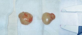
If cancer is found during your sentinel node biopsy, your surgeon may remove some or all of the axillary lymph nodes—those in the armpit area—for additional testing.
Axillary lymph node dissection may be performed as part of a lumpectomy or mastectomy. As many as 10 or 40 lymph nodes may be removed. They will be sent to a pathology laboratory, where they will be carefully examined for the presence of cancer cells.
Your pathology report will indicate how many nodes were removed and how many, if any, contained cancer cells. This important information affects the stage of your cancer and affects your treatment options.
Related article: Axillary lymph nodes and breast cancer
Main causes of inflammation
The intramammary lymph node in the chest is usually the first to take the hit - the lymphoid fluid flows in this direction. Afterwards, the subclavian lymph nodes and regional lymph nodes of the mammary gland are connected to the fight against the pathological process. Therefore, in the presence of inflammation, it is in the area of these lymph nodes that the first symptoms appear.
There are three main causes of a serious inflammatory process in lymphoid tissue:
- Mastitis.
- Mastopathy.
- Oncology
Mastitis is an infectious disease that most often occurs during lactation. But mastitis can also appear in nulliparous girls if the skin of the mammary glands is damaged or if hygiene rules are not followed. For treatment, it is necessary to identify the pathogen and select a suitable antibacterial drug. With mastitis, inflammation of the regional lymph nodes occurs - this is how they react to infection.
Mastopathy is a whole group of diseases characterized by the appearance of benign cysts or tumors. Lymph nodes enlarge towards the end of the menstrual cycle, at which time other symptoms appear: swelling, soreness. The main sign of mastopathy is lumps in the breast tissue, which can usually be felt independently.
Malignant tumors are extremely dangerous to health. Treatment largely depends on the stage, which is why early diagnosis of oncology is so important. In oncology, there is significant damage to all lymph nodes located in the area of the mammary glands. The main danger is that metastases are often localized in intramammary or axillary lymph nodes, so at stages 2-3 of cancer it is often suggested to remove dangerous nodes.
Axillary lymph nodes
Of the existing lymph nodes in the mammary gland in women, the axillary lymph nodes are most susceptible to changes and functional disorders. When they increase, axillary lymphadenopathy occurs.
What is axillary lymphadenopathy of the mammary gland and why is it dangerous?
The cluster of this group of lymph nodes is located deep in the breast tissue, in the so-called axillary region. Axillary lymph nodes in the mammary gland are normally difficult to diagnose. They can only be determined using mammography. Violation of lymph outflow provokes congestion in the glands.
A puncture biopsy will help determine axillary lymphadenopathy of the mammary gland and what causes it. To carry it out, a puncture is made with a thin needle to collect material from the affected area and sent to the laboratory for further cytological examination.
Any inflammation in the nodes is a rather dangerous symptom. If intramammary lymph nodes in the mammary gland or axillary ones are affected, then this usually indicates the presence of cancer (but not always). Affected intramammary lymph nodes often indicate an advanced pathological process, when it is not possible to solve the problem with a conservative method.
Therefore, a modified intramammary breast nodule is always dangerous. Lymphadenopathy of the axillary lymph nodes is included in the list of main symptoms of metastasis in the human body. The localization of the focus of inflammation indicates one or bilateral axillary lymphadenopathy. The doctor uses a mammogram to diagnose whether the lymph nodes are enlarged or not.
Atrophy in the regional lymph nodes of the mammary gland is caused by the following factors:
- any inflammation occurring in the tissues of that area of the body;
- infection with Mycobacterium tuberculosis;
- breast cancer.
Depending on whether it is benign lymphadenopathy or oncology, a treatment regimen is prescribed. Detection of cancer requires a lightning-fast decision on the further form of treatment.
Diagnostic methods
The following research methods help diagnose pathology.
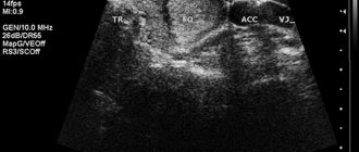
- Ultrasound of the mammary glands and lymph nodes (often used to detect benign axillary lymphadenopathy).
- Mammography.
- Axillography (used to determine the patency of the ducts located in the node).
- Magnetic resonance imaging (to diagnose the type of malignancy).
- Thermography, which allows you to diagnose bioenergetic processes in the body using infrared radiation. The result of the analysis is presented in the form of a thermogram.
- Radionuclide testing (makes it possible to identify malignant tumors and monitor the effectiveness of therapy).
- Radiation diagnostics using x-ray or ultrasound and other methods allows you to get the most accurate picture of what is happening. Its rationality requires compelling reasons.
- Axillography is the most acceptable method of determination for damage to the axillary region.
- Pneumocystography (study of cyst puncture material).
- Ductography (used by administering a water-soluble emulsion containing iodine for nipple discharge).
Symptoms of lymphadenopathy
Regardless of the cause of lymphadenopathy, this condition has a number of typical general symptoms:
- Increased body temperature, increased sweating, especially at night.
- Digestion is disrupted, body weight decreases, and appetite disappears.
- Reduced blood pressure is observed.
- Tachycardia, rapid heartbeat.
- Enlarged liver and spleen.
If one lymph node of the mammary gland is affected, the general manifestations are mild; they appear if the lymphadenopathy has spread to a whole group of lymph nodes. Only a doctor can determine whether there is cause for concern and an accurate diagnosis. Therefore, if the described symptoms appear in connection with inflammation of the lymph nodes, you should contact a mammologist or therapist.
The following symptoms appear from the lymph nodes themselves:
- Obstructed lymph flow can cause pain.
- Pain on palpation of nodes and between nodes.
- Softness and increased mobility of lymph nodes.
- Change in size and shape.
- Asymmetry.
- Painful sensations in the chest.
To establish an accurate diagnosis, a number of diagnostic measures are prescribed.
Diagnosis of the inflammatory process
For diagnostics, use the entire available arsenal of tools:
- Biopsy – required to confirm a malignant process in the lymph nodes. Histological examination of tissues taken during biopsy makes it possible to confirm oncology or refute the diagnosis of stage 2 with metastases.
- Mammography and ultrasound examination. Allows you to find the primary foci of the tumor and determine the extent of the pathological process.
- Axillography is a special study of axillary lymph nodes and ducts. Allows you to find out the state of the lymphatic system and find specific pathologies in the nodes.
- CT and MRI - high-tech studies are prescribed if simpler studies fail to establish an accurate diagnosis.
There are also more rare studies - thermography (a more high-tech analogue of mammography, used when cancer is suspected) and radionuclide testing. These diagnostic measures refer to auxiliary examinations for previously diagnosed tumors.
Treatment
Treatment begins immediately after the final diagnosis is established.
All possible treatment methods are divided into three main groups:
- Conservative.
- Operational.
- Complex oncological treatment.
The treatment method is chosen depending on the disease and its danger to life. Thus, fibroadenoma and malignant tumors are usually removed regardless of size. If a benign tumor is diagnosed, the decision is made on the basis of images and laboratory tests - the location, growth rate, and size of the tumor are taken into account.
If conservative therapy is chosen, hormonal agents are usually used. Together with them, vitamin and mineral complexes, recommendations on diet and lifestyle are used.
Oncology
In breast cancer, the lymph nodes are the first to be affected - it is in them that metastases appear when the cancer reaches stages 2-3. This happens because cancer cells easily enter the lymphatic ducts. They circulate along with the lymph flow and sooner or later settle in the lymph nodes. Initially, lymph is an obstacle to the path of cancer cells to the most important organs. Therefore, at stages 2-3, the patient can be saved by removing the primary tumor and metastatic lymph nodes.
It is customary to remove suspicious lymph nodes and perform a biopsy to confirm malignancy - this is done by histological examination. Removal is not practiced if the metastases have spread beyond the group of nodes and affected the tissues of any organ outside the primary focus. In this case, systemic therapy is used.
How and with what to treat breast cancer with metastases in the lymph nodes?
The effectiveness of treatment for this type of metastatic lesion is directly related to the use of complex therapy. Only a combination of several methods allows a woman to achieve long-term remission or complete recovery.
For patients whose lymph nodes have started to grow due to a cancerous tumor in the breast, mammologists offer the following therapeutic methods:
- Surgical intervention. Most often, complete or partial resection of regional nodes located on the side of the breast affected by cancer is performed. Removal of lymph nodes for breast cancer is preceded by lymphoscintigraphy, with the help of which a specialist determines the extent of the upcoming operation. This intervention is carried out for both therapeutic and diagnostic purposes.
- Chemotherapy. For breast cancer, this is the main therapeutic technique. In antitumor courses, such groups of drugs are used as the antimetabolites Capecitabine, Thiopurine, which stop the development of the tumor, alkylating drugs, Lomustine, Mitomycin, which destroy the DNA molecules of mutated cells, taxanes Yutaxan, Paxen, which block the process of uncontrolled division of cellular elements of a cancerous tumor.
- Radiation therapy. This treatment strategy is used in cases of metastatic lesions of regional lymph nodes. It is quite popular at stage II III of the oncological process, since irradiation of lymph nodes located in close proximity to the tumor structure allows to minimize the number of relapses.
Worth knowing! If a woman has been diagnosed with actively progressing breast cancer and a large number of lymph nodes have been removed, her lymph flow is disrupted, which leads to a certain number of consequences - significant swelling of the arm from the side of the resection, limitation of its movement and numbness of the skin. In order to quickly cope with this unpleasant consequence, in the postoperative period, patients should get proper rest and follow a daily routine, as well as eat a balanced diet. In addition, a complete rejection of bad habits is necessary.