Characteristics of the proliferation process
Seeing the word “proliferation” in the test results, many women worry. They want to understand the meaning of this term. Proliferation means that cells are actively multiplying. Usually this process is clearly expressed in the mucous membranes of the respiratory system, digestive tract, and uterus. In the tissues of these organs it is normal.
In case of injuries and after surgical interventions, the process of proliferation is natural; it is necessary for rapid healing and tissue restoration. Often, intensive cell proliferation indicates the onset of an inflammatory process or the appearance of tumors. In the latter case, the proliferation process becomes non-stop: cells divide continuously, leading to the growth of tumors.
Women who have been diagnosed with this condition should follow all doctor's recommendations. After all, proliferation is observed during tissue growth in neoplasms and during hyperplastic changes. For example, increased proliferation is accompanied by precancerous changes on the cervix, including dysplasia.
With the proliferation of columnar epithelium of the cervix, atypical cells are often detected. This indicates the threat of precancerous changes in tissue cells. With intense proliferation, this is not surprising, because with rapid proliferation, cells do not have time to form correctly, and the likelihood of gene mutations increases.
Breast
Breast changes are quite common, including among young girls and women. The organ constantly experiences the action of sex hormones, undergoes characteristic changes throughout the menstrual cycle, during pregnancy and lactation, and is therefore susceptible to various kinds of pathologies. According to statistics, up to 60% of women of reproductive age have signs of mastopathy.
Mastopathy is considered a benign process, but if it is present, the risk of malignancy increases several times. Proliferative variants are even more dangerous, they increase the likelihood of cancer by 25-30 times.
The presence or absence of proliferation is the most important sign when assessing the type of mastopathy. Non-proliferative forms are represented by foci of fibrous tissue, cystically altered ducts, the epithelium of which does not proliferate and is even atrophic. The risk of malignant transformation is relatively small.
According to the severity of proliferation, several degrees of mastopathy are distinguished. In the first degree, proliferation is not detected, in the second, it is there, but the cells do not show signs of atypia, the third degree of mastopathy is manifested by active proliferation of epithelial cells with atypia.
Thus, proliferation of mammary gland cells is not only a criterion for the form of mastopathy, but also an indicator of the likelihood of cancer, therefore excessive division of the epithelium always attracts the attention of specialists. Diagnosis of this change is carried out by cytological examination of tissue obtained during puncture.
Diagnostic methods
Women should monitor the condition of their reproductive organs. They are required to undergo a gynecological examination annually. If problems are identified, the doctor prescribes extensive diagnostics: colposcopy and cytological examination are performed.
It is a cytological examination that allows us to establish that a woman has developed the following symptoms:
- epithelial damage;
- cell proliferation;
- metaplasia;
- transformation.
The analysis makes it possible to understand why the cervical epithelium was changed. In the process of cytological examination of the material, the following is determined:
- inflammatory processes that have developed due to the negative influence of pathogenic microflora;
- pathological changes that appear as a result of medicinal, hormonal, radiation, mechanical effects on the body;
- conditions in which the likelihood of dysplasia and cervical cancer is high.
Cytological examination is recommended to be carried out once a year, even if no changes in the mucous membrane are visible during a gynecological examination. When it is carried out, it is possible to establish changes in the epithelium and the presence of atypical cells.
Interpretation of endometrial biopsy results
In his conclusion, the pathologist can give a different interpretation of the endometrial biopsy, with or without a detailed description of the morphology. Often the results are accompanied by a pathological diagnosis, which is a brief summary of the descriptive characteristics.
Below is an explanation of the various terms that can be found in the results of an endometrial biopsy.
- Adenocarcinoma is a synonym for uterine cancer.
- Adenomyosis is the growth of the uterine glands in the myometrium (the muscular lining of the uterus). A rare find, mainly in RDV.
- Atrophic mucosa - thinned endometrium with single glands. Quite normal during menopause; at a younger age indicates serious hormonal imbalances.
- Chorionic villi are auxiliary components of the fertilized egg, thanks to which it attaches to the uterine mucosa. Subsequently, they transform into the placenta. The presence of chorionic villi in endometrial scraping is an unambiguous sign of pregnancy.
- Hyalinosis is a thickening and narrowing of the lumen of small uterine arteries.
- Atypical hyperplasia is the proliferation of endometrial glands with signs of malignant transformation. Potentially precancerous condition.
- Simple hyperplasia is the same thing, but without signs of a tumor. Occurs with hormonal disorders due to ovarian cysts and other conditions.
- Hypoplastic mucosa is a thinned endometrium, occupying an intermediate state between mature and atrophic.
- Gravidar mucosa is the endometrium of the pregnant uterus with fields of decidual tissue.
- Detritus is a designation for amorphous masses represented by the remains of dead cells.
- Decidual tissue is a collection of large cells containing an abundance of nutrients. Sign of pregnancy.
- Dysplasia is a change in the structure and cellular composition of the uterine glands, reminiscent of tumor transformation.
- A cyst is an enlarged gland filled with fluid.
- Leiomyoma is a benign tumor of smooth muscle located in the middle lining of the uterus.
- Lymphoid follicles are accumulations of lymphocytes that reflect a long-term inflammatory process (for example, chronic endometritis).
- Menstrual mucosa - endometrium at the height of menstruation.
- Metaplasia is a condition in which the normal epithelium of the uterine mucosa is replaced by epithelium characteristic of another organ.
- Placental polyp - remnants of the placenta after a miscarriage, abortion or natural birth (less often after a cesarean section).
- A polyp is any formation of the mucous membrane protruding into the lumen of the uterus.
- Hydatidiform mole is a pathology of pregnancy, a variant of trophoblastic disease. Characterized by the growth of chorionic villi alone; the fetus is not formed.
- The Arias-Stella reaction is a variant of changes in the endometrium, reminiscent of the transformation of the glands of the cervical canal during pregnancy. Indicates a hyperplastic process.
- Regenerating mucosa - endometrium in the first 1-3 days after menstruation.
- Spiral arteries are small blood vessels that penetrate the lining of the uterus. The main source of its blood supply.
- Stroma is the tissue in which the endometrial glands are embedded.
- Trophoblast is the cellular material from which chorionic villi and other auxiliary structures of the embryo are formed.
- The proliferation phase corresponds to days 5-14 of the menstrual cycle (after menstruation).
- The secretion phase corresponds to days 15-26 of the menstrual cycle (before menstruation).
- Fibrosis is the proliferation of dense connective tissue in the stroma. A sign of chronic inflammation, and also a normal phenomenon in postmenopause.
- Choriocarcinoma is a malignant tumor of atypical chorionic villi, a variant of trophoblastic disease. To put it roughly, choriocarcinoma is a “cancerous hydatidiform mole.”
- Endometritis is an inflammation of the uterine mucosa.
Cervical epithelium
To conduct a cytological examination, a gynecologist or trained nurse takes material from the cervix. If altered tissues are visible, a smear is made from the affected areas.
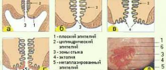
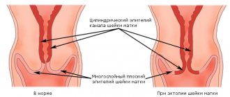
Normally, the exocervix (the part of the cervix that protrudes into the vagina) is covered with squamous stratified epithelium. And the endocervix (the cervical canal that connects the uterine cavity and vagina) is covered with columnar epithelium. In the absence of gynecological problems, the border between the flat and cylindrical epithelium is located in the area of the external os of the endocervix.
In newborn girls, the cervical epithelium can occupy the entire area of the vaginal part of the cervix, up to the vaginal vault. But by the time of puberty, due to the process of squamous metaplasia, the columnar epithelium is completely replaced by flat multilayered epithelium. This is possible due to the presence of special reserve cells in the tissues of the cervix.
When diagnosed, three layers can be detected on the cervix:
- multilayer flat;
- cylindrical (also called glandular);
- metaplastic.
But in some women, abnormalities are detected: the border shifts from the external os, and part of the exocervix is covered with columnar epithelium. With this pathology, a diagnosis of “ectopia of columnar epithelium” is made. For women, this pathology is better known as cervical pseudo-erosion. Normally, the cylindrical layer should not come into contact with the acidic environment of the vagina. This can trigger the appearance of a chronic inflammatory process.
You can understand what it is, the columnar epithelium of the cervix, if you look at what it looks like. Your doctor may notice the problem during a routine gynecological examination or colposcopy. Areas of the exocervix covered with columnar epithelium stand out: against the background of pale pink stratified epithelium, they look like red or bright pink spots.
Symptoms
Hyperplasia of the epithelium of the cervical canal may not be accompanied by any clinical manifestations for a long period of time. That is why it is often detected at a late stage or completely by chance during a routine gynecological examination.
The following symptoms are possible:
- A nagging pain that occurs in the lower abdomen.
- Heavy menstruation.
- Uterine bleeding.
- Pain during intercourse.
- Menstrual irregularities.
Hyperplasia may be accompanied by inflammation, which further worsens the patient’s condition. There is also a risk that the woman will have problems conceiving a child.
Gynecological pathologies
Pseudo-erosion itself, in which the cervix is partially covered with columnar epithelial cells, is not dangerous. But to avoid problems, doctors often prescribe chemical destruction or surgical treatment:
- laser coagulation;
- cryodestruction;
- radio wave coagulation;
- electrocautery.
With this therapy, the lesions are removed, and the cervix, after healing, is again covered with squamous epithelium.
Normally, it is acceptable for there to be moderate proliferation of squamous epithelium. This process is necessary to renew the cellular layer that covers the cervix. Do not panic if pregnant women exhibit proliferation of the columnar epithelial layer. This is possible due to serious hormonal changes taking place in the body of expectant mothers.
Proliferation
Gynecologists diagnose pseudoerosion if part of the exocervix is covered with cylindrical cells. Normally, they should be located only in the cervical canal. Often with this pathology various infectious and inflammatory diseases are detected. They contribute to the development of pseudo-erosion and prevent its normal healing even during treatment.
Based on the results of a cytological study of cells taken from the affected areas, proliferation of not only the cylindrical, but also the flat epithelial layer can be detected. Multilayered squamous epithelium begins to actively multiply in order to neutralize the pathological growth of the cylindrical layer. This condition is considered quite dangerous, because there is a process of active division and formation of atypical cells. At the same time, their correct orientation on the surface of the cervix may be disrupted.
Proliferation of the columnar epithelium of the cervix provokes the appearance of new glands, branches and papillae. If at the same time the flat epithelial layer begins to actively multiply, then dysplasia may appear. This change in cells is considered a precancerous condition.
Proliferation of columnar epithelium located inside the cervical canal can also be diagnosed. A moderate process indicates natural cell renewal. If it intensifies, then polyps appear inside the cervical canal, carcinomas and adenomas. Also, intensive proliferation is possible against the background of activation of the inflammatory process.
In the absence of atypical cells, conservative treatment or monitoring of the condition of the cervix is possible. Proliferation of columnar epithelium is diagnosed not only with erosion, but also with leukoplakia, polyps and inflammatory diseases of the female reproductive organs. If the cytological examination and colposcopy are normal, then a biopsy is not performed.
The structure of the mucosal layer
The endometrium is the inner mucous membrane of the uterine body, lining its cavity. It is abundantly supplied with blood vessels. The mucous layer consists of the glandular and outer layers of the epithelium, ground substance, stroma and vascular bed. It promotes normal homeostasis in the uterine cavity, ensuring optimal functioning of the fetus.
Proliferation is not an independent disease. It is an isolated cytological sign that indicates the presence of a serious pathology. With the proliferation of glandular epithelium, body tissue grows by cell multiplication by division. Their form changes, and so do their functions. It can be localized only in the cervical canal and sometimes located outside the cervix.
A woman's body performs different functions during the three phases of the menstrual cycle, each of which plays a unique role in the reproductive process. Classification of stages:
- proliferative;
- secretory;
- menstrual
To assess cyclic changes, the duration of the menstrual cycle, proliferative and secretory phases, as well as the volume of discharge during menstruation should be taken into account.
Treatment tactics
If it has been revealed that the columnar epithelium has moved from the cervical canal to the vaginal part of the cervix, then it is necessary to fully examine the woman and, depending on the test results, choose the most appropriate treatment tactics.
The tactics for treating proliferation of the glandular epithelium of the cervix depends on the cause of this pathology and its form.
When endocervical polyps appear, the mucous part of the cervical canal is scraped. These formations look like folds of the cervical canal, hanging on a stalk, elongated or rounded in shape: they can be bright red or pale pink. Usually their diameter does not exceed 2 cm.
For leukoplakia, it is necessary to use destructive treatment methods - pathological foci must be removed. The doctor can send the woman for laser, chemical or radio wave coagulation, or electrical cauterization. Leukoplakia is characterized by the fact that the squamous epithelium changes so that the cervix is covered with whitish spots. If left untreated, there is a high probability of malignant neoplasms.
Dysplasia, in which intense proliferation of atypically changed cells is detected, is treated surgically. Depending on the extent of the damage, local destruction or radical intervention can be performed. In severe cases, conization or amputation of the cervix is performed.
Before surgical treatment, it is necessary to carry out antiviral, anti-inflammatory or antibacterial therapy, depending on the identified concomitant problems. Particular attention is paid to the treatment of diseases that are sexually transmitted. But patients should know that proliferation of columnar epithelium does not always require surgery.
Columnar epithelium is the cells that line the uterine cervix in women. Under the influence of negative factors, they are able to grow. What symptoms occur with columnar epithelial hyperplasia, what kind of disease is it?
Painstaking research
For a detailed study of modified structures, colposcopy and cytological analysis are necessary. It is taken into account that proliferation is an uneven process, in which the mucous membrane usually thickens in some places, and the glands differ from each other in size and shape. A cytogram will provide accurate information only if the process covers the uterine cervix (surface). If the cervical canal is damaged in such a way that hyperplasia does not spread beyond the external os, accurate data can only be obtained by histological examination. To do this, the cervical cavity is examined and a scraping of biological tissue is obtained, which is sent for further laboratory tests.
Causes
Not everyone knows what columnar epithelial hyperplasia is. Therefore, upon hearing such a diagnosis, women often begin to panic. However, this disease occurs in a benign form, so it is not fatal. But still, the risk of malignant degeneration is quite high, so treatment should be started as quickly as possible.
Why does cervical hyperplasia occur? The main cause of the pathology is a hormonal imbalance in the body, namely an increase in estrogen levels. The following factors can also trigger the development of the disease:
- Disturbance of metabolic processes in the body.
- Excess body weight.
- Diabetes.
- Late onset of menopause.
- Neoplasms in the organs of the reproductive system.
- Inflammatory processes in the reproductive system.
- Frequent abortions.
- Damage to the uterus.
- Taking hormonal drugs.
- Bad habits.
- Weakened immune system.
- Early onset of sexual activity.
Tissue proliferation can be of different types, for example, adenomatous, atypical hyperplasia.
Do I need to go to the doctor?
If you suspect the presence of pathologies of the reproductive system, you should promptly make an appointment with a gynecologist. If the doctor diagnoses proliferation, laboratory tests are ordered on tissue samples to identify the characteristics of the cellular composition. At the same time, a visual examination usually provides a rather modest amount of information: a specialist examines the external part, the external uterine pharynx, where he records individual areas that differ from the surrounding tissues in structure and color. Normally, the epithelium is light pink in color, which is due to its multi-layered nature, while abnormal elements are brighter and more saturated.
Some women have not only elements that differ in color, but also small neoplasms, the diameter of which does not exceed one centimeter. These are hemispherical dense objects characterized by thin walls. The internal filling is yellowish and translucent. In medicine, this is called “Nabothian cysts.” Typically, pathology is observed in the cervical cavity, in the lower third of the volume, that is, where the Nabothian glands are located. The glands themselves are small tubes filled with secretions. The contents reach external tissues through the excretory ducts. Proliferation leads to blocking of the holes, blockage provokes the formation of a cavity filled with secretions. If such cysts are located deep in the reproductive system, the doctor will not be able to see them visually. The presence of formations suggests glandular-cystic proliferation.
Diagnostics
If symptoms of hyperplasia of the epithelial layer of the uterus occur, you need to consult a gynecologist. He will first conduct a gynecological examination, then prescribe the following diagnostic measures:
- Blood analysis.
- Hormone analysis.
- Cytogram.
- Ultrasonography.
- Hysteroscopy.
- Biopsy.
- Histological examination to reveal the proliferation of columnar epithelium, a cancerous condition.
- Colposcopy.
The studies carried out help to make an accurate diagnosis, determine the degree of pathology, and prescribe the correct treatment.
Treatment
How to treat hyperplasia of the cylindrical layer of the uterus in women is determined by the attending doctor based on the examination results. In most cases, surgical treatment is performed, as a result of which the overgrown tissue is removed.
Drug treatment is used only in the earliest stages. Hormonal medications are prescribed that help restore the balance of hormones, prevent further tissue growth, and reduce already overgrown uterine cells.
To improve their general condition, patients can use folk remedies, but only with the permission of the treating doctor.
Prevention
To prevent the development of cervical hyperplasia, gynecologists advise the following:
- Monitor your body weight.
- Control the hormonal balance of the body.
- Refuse abortion.
- Eat properly.
- Do not smoke or drink alcohol in excess.
- Treat inflammatory and other diseases of the reproductive system in a timely manner.
- To live an active lifestyle.
For timely detection of hyperplasia and other pathologies of the reproductive system, it is important for women to undergo a gynecological examination once a year.
Galina Savina Reading time: 7 min Comments to the entry Proliferation of columnar epithelial cells of the cervix are disabled 1,706 Views
The cervix provides reproductive function; sperm enters the uterus through a tunnel in the cervix. After development, the fetus also leaves the mother's womb through a cylindrical neck. Thus, the organ becomes a connecting link between the uterine body and the vaginal opening. The organ can be examined by a gynecologist during an appointment using certain instruments.
What is a cylindrical cervix?
After the birth process, a woman's cervix changes its shape. It is with such changes that the organ acquires a cylindrical shape. After carrying a child and passing it through the birth canal, the cervix is stretched to a cylindrical shape for the smooth passage of the newborn. In young, nulliparous girls, the uterus is relatively small, while the cervical region exceeds it in size. After the process of bearing a child, the walls of the uterus increase, for a comfortable and safe stay of the fetus in the woman’s body.
If the girl has not yet carried a child, then the cervix has a narrow lower wall and a conical shape. During contractions, during childbirth, the cervix changes in size and becomes larger, so the postpartum uterus is much larger in size.
Characteristics of the reproductive organs:
- A nulliparous woman contains a uterine organ 5 cm long, a postpartum uterus reaches a length of 6 cm;
- The uterine reproductive organ has a mass of about 50 g; after the birth function is performed, the uterus gains weight and can reach 100 g;
- The cervix measures up to 3.5 – 4.4 cm.
In medical research, two constituent elements of the cylindrical neck are distinguished: the supravaginal zone and the vagina.
The cervix is hollow inside and contains the cervical canal. During an examination of the uterus, a gynecologist can determine whether the patient has given birth or not, based on the appearance of the entrance to the cervical canal. The external pharynx of the canal passes into the uterine organ; in girls who have not gone through the process of natural childbearing, the external pharynx has a round shape, while in a patient who has given birth, this canal has a slit-like shape. These sizes and shapes are individual in nature, since these indicators are influenced by factors such as hormonal levels and various diseases of the reproductive organs.
The cervical canal contains a large amount of thick mucus, which expresses the normal functioning process. During the ovulation cycle, uterine secretions liquefy and stimulate the transport of male reproductive cells to the uterine area. The background acidity also changes, which prevents sperm damage. During ovulation, the cervical canal is softer and wider.
Characteristics of the structure of the cylindrical cervix
The cervix is not a separate organ, it belongs to the uterine system; the uterus and cervix have common tissues. At the same time, the walls of the canal are formed with the help of connective tissue, while the uterine section is built of smooth muscle. It is extremely important that this cover was of a certain size, shape and volume, otherwise the woman will be diagnosed with isthmic-cervical insufficiency, which may affect the ability to bear a child.
The cervix and the walls of the vaginal opening are covered by multilayer epithelial tissue, and the cervical meatus is lined by columnar epithelium. The space where the two types of tissue meet is called the transition zone. In the case when the connecting joint between the flat and cylindrical cover moves in the direction of the vaginal outlet, pseudo-erosion is diagnosed. During the examination, the gynecologist may observe red ulcers that are located on the upper layer of the vagina. The cylindrical epithelium, in the acidic environment of the vaginal section, is endangered by pathogenic organisms that are produced by the crypt of the glands located in the canal. Along the cervix there is an epithelium that forms folds of tissue in which branching glands can be found.
Some special cases
There are known situations where hyperplasia was localized only in the cervical canal. During a visual examination, the doctor is not able to identify the process, since the areas are inaccessible to this method of examination.
If the pathology is accompanied by inflammation, the following additional symptoms are observed:
- local temperature increase;
- swelling of the mucous membranes;
- abundance of discharge.
As practice shows, in most cases, proliferation is associated precisely with infection or inflammation, so doctors always prescribe laboratory tests - culture, flora smear, PCR. This helps to identify the pathogen and determine the presence of specific infections. When menstrual dysfunction is observed, additional tests are taken to identify hormonal imbalances. The current phase of the cycle is taken into account.
What is columnar epithelium of the cervix?
The cervical canal is lined with columnar epithelium. The tissue secretes a special secretion to prevent the development of pathological flora on the mucous membrane, in addition, it provides protection from mechanical damage to the organ. If the results of the analysis of a gynecological smear show a large number of cylindrical epithelial cells, or any other deviation from the norm, this may indicate that an inflammatory process is occurring in the woman’s body, or there is a hormonal imbalance.
The cylindrical layer of epithelium provides the birth canal with the necessary level of secretion. In structure, such cells gather in groups and resemble the shape of a honeycomb, which are distributed along the entire body of the cervix. There are also layers that resemble stripes or the silhouette of a glass. The shape of such groups is determined by the level of elongation of the secretion of the cytoplasm.
Normal cervical structure
Tissue for a smear is taken from the surface of the epithelium of the cervix and cervical canal. The cells that are capable of being exfoliated fall on the cytobrush. Therefore, mature epithelial cells normally predominate in a smear. The higher their degree of maturation, the greater the chance that the smear will show normal values. During atrophic processes, the epithelium is exfoliated in an immature state.
With a normal structure of the cervix and the absence of pathology, the following types of epithelial cells are found:
- Superficial
Large cells with a multifaceted shape, close to an oval or circle. Their nuclear material is enclosed in an oval shape, the epithelium is scattered, and has a yellow-pink cytoplasm. In less mature cells, the arrangement in the smear is crowded, in layers or heaps, and the cytoplasm is cyanophilic, with a bluish tint.
- Intermediate
Large epithelial cells, the nuclei of which contain a large number of chromatin blocks; the cytoplasm is painted pinkish or bluish and has pronounced folding. Intermediate cells are often lysed by lactobacilli, but they never destroy the surface type of epithelial cells.
- Parabasal
Small immature cells, oval or round in shape, large, vesicular nuclei. Cells are not destroyed by bacteria, but are subject to autolysis.
- Basal
Smaller in size than parabasal ones, but with large nuclei, characteristic of the postmenopausal or postpartum period, until the menstrual cycle has been restored.
Columnar epithelium may appear in the smear, which lines the cervical canal. They form small groups, arranged in stripes and honeycombs. Sometimes there are goblet cells containing mucus and secretion granules.
A normal smear may contain single leukocytes. Their number may increase before menstruation, but their structure is different from those that appear during inflammation. Quiescent leukocytes are characterized by a preserved or pyknotic nucleus, clear cytoplasm, and no signs of phagocytosis.
The process of cervical proliferation
Cervical proliferation is a phenomenon of accelerated process of tissue cell division. Such a process is considered normal if it occurs after surgery (for example, a conization procedure, in which prismatic tissue is collected). In some cases, the process of accelerated cell division indicates the presence of an inflammatory process in the organ. Doctors divide proliferative phenomena into several types:
- inflammatory process - this includes erosion of the uterine cervix and cervicitis (inflammation of the mucous membrane), as well as dysplasia;
- hyperplastic processes caused by hormonal imbalances. Pathology can be caused by HPV, polyps, endometriosis (disorders of the glandular epithelium), mild forms of leukoplakia (focal keratinization of the epithelium);
- post-traumatic type is caused by scar changes in organ tissue. Ectropion (eversion of the mucous membrane of the uterine canal) and various tissue ruptures also affect it.
In all such cases, one can observe an increased proliferation of the constituent cells of the canal over a short period. For an accurate diagnosis and establishment of a treatment method, the patient must contact the attending physician for a personal examination and testing.
Also, during proliferation, a change in the morphological structure of the tissue is observed. In most cases, proliferative processes are benign in nature, however, there are cases of degeneration into a precancerous form. The most common cause of proliferation of the columnar epithelium of the cervix is the use of hormonal contraceptives or disruption of ovarian activity. Also, the development of the disease is affected by diabetes and metabolic disorders.
The pathology is most often treated with hormonal drugs; if necessary, antibiotics are prescribed, which are aimed at combating the inflammatory process.
Deviations from the norm and diseases
During perimenopause, hormone levels fluctuate. Due to irregular production of hormones, the growth of the glandular epithelium of the cervix becomes abnormal, leading to irregular cycles. Doctors call this phenomenon disordered changes in the proliferative endometrium. This does not necessarily mean that malignant changes are occurring.
The histological picture of the phases is determined using hysteroscopy with biopsy. For example, at the early stage of proliferation, the histology of the endometrium is as follows: the epithelium is thinned, pink in color, there are areas of hemorrhage, and unrejected fragments. However, in some cases the process may be disrupted. Hysteroscopy with biopsy during proliferation of the glandular epithelium of the cervix notes the proliferation of tissue, which leads to the development of tumor pathologies.
One potential reason for these changes is the use of birth control pills. Dyshormonal disorders disrupt the functioning of the glandular epithelium of the cervix. This pathology is treated by using cyclic hormonal therapy. If treatment is not started, the glandular epithelium will grow, leading to abnormal thickening of the endometrium. Hyperplasia is one of the factors that increases the risk of developing cancer. There are several types of endometrial thickening:
- glandular;
- glandular-cystic;
- focal;
- atypical.
The first two types of hyperplasia rarely lead to the development of malignant diseases. The focal and atypical type are considered the most dangerous, threatening the formation of cancerous pathologies that can develop into uterine cancer. This disease most often affects women during menopause. The risk group includes patients over 50 years of age.
What is cervical columnar epithelial hyperplasia?
Cervical hyperplasia is manifested by an increase in the thickness of the endometrium. The main function of this special layer lining the inside of the uterine cavity is to create normal conditions for the egg and ensure its attachment in the uterine environment. If defects occur in the tissues, difficulties may arise with bearing the fetus, including miscarriage.
Hyperplasia is most often caused by a benign formation. Such pathologies should be monitored and treated in order to prevent cancer.
Factors causing cervical hyperplasia:
- Hormonal disbalance.
- Disturbance of metabolic processes.
- Having late menopause.
- Obesity, unhealthy diet, bad habits.
- Abortion, taking hormonal drugs.
- Ectopic columnar epithelial cells.
- Proliferation of the cervix of the cubic epithelium of the cervix.
What is ectopia of columnar epithelial cells?
Ectopia is the most common phenomenon in the field of gynecology; in fact, it is not a disease. With this failure, the cylindrical epithelial layer covering the cervical canal moves towards the vaginal opening. The doctor can observe this process due to the characteristic redness of the affected tissue. This pathology in the work of the cervix can be traced in 45% of patients of childbearing age; more than 50% of patients with this diagnosis have not reached the age of 25 and have not yet carried a child. Ectopia can also form during menopause.
How to notice?
Proliferation of the glandular epithelium is not associated with any characteristic clinical symptoms. Typically, a woman consults a doctor based on problems associated with concomitant pathologies. In particular, if hyperplasia is caused by inflammation, then profuse leucorrhoea and discomfort in the vaginal area are a concern. With hormonal disorders, disruptions in the monthly cycle, bleeding, including at the wrong time, and cycles without ovulation are observed.
Proliferation of cuboidal epithelium of the cervix
During the proliferation of columnar epithelial cells of the cervix, atypical cells of the cubic layer appear, which ultimately can develop into a cancerous tumor. Rapid cell division provokes malfunctions and gene mutations. Cytology analysis identifies possible transformations and damage to the upper layer of the uterine tract. Doctors practice the use of laser and radio wave coagulation - the safest ways to remove pathology. Electric cauterization is also used. After surgery, squamous epithelium will reappear on the surface in normal quantities.


