What's happened
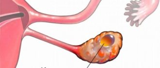
A multi-chamber ovarian cyst is a benign neoplasm on the female gonads. Anatomically, it is a cavity divided into chambers using thin partitions.
This pathology is formed from the epithelial tissue of the glands and nearby structures of the pelvic organs, and its contents become liquid of varying density.
Multilocular ovarian cysts can occur at any age, but most often they are found in women after 30 years of age. Late detection, refusal of timely treatment or delay in treatment threatens serious complications in the form of cancer, since the walls of the cyst and its septa often contain atypical cells.
What is an ovarian cyst with a septum: features of the neoplasm
Neoplasms in the ovarian area are considered one of the pathologies of the female reproductive system. In some cases, doctors make a diagnosis: multilocular cyst.
This formation is a wall of tissue or epithelium of the ovary filled with mucus or fluid.
Since such a pathology not only causes a lot of concern, but is also potentially dangerous, it is worth considering the reasons for the formation of the disease, its symptoms, as well as methods of treatment and prevention.
Causes of the disease
Having considered the definition of an ovarian cyst with a septum, what it is, it is important to determine why such a disorder appeared. Most often, the disease is diagnosed in women of childbearing or menopausal age. This may be caused by the following reasons:
- problems with the menstrual cycle, including severe delays;
- hormonal imbalance, which provokes the release of progesterone, which increases the risk of the formation of a corpus luteum cyst;
- inflammatory processes in the appendages;
- pathologies of the fetus leading to changes in the genital organs;
- sexual activity at an early age;
- forced termination of pregnancy by abortion;
- gynecological operations;
- disturbances in the functioning of the endocrine system;
- other diseases of the genital organs in an advanced state.
Important. The type and contents of the cyst largely depend on the factors that provoked its appearance. To determine them, a full gynecological examination is prescribed, based on the results of which treatment will be prescribed.
This problem should not be ignored. Firstly, in an advanced state, a benign cyst can develop into a malignant tumor with all the ensuing consequences. Secondly, a multilocular tumor is susceptible to rupture.
Then all its contents end up in the internal organs, which can cause infection. Thirdly, in the absence of proper treatment, pus may begin to appear inside the cyst, which externally manifests itself in the form of weakness, throbbing pain in the lower abdomen.
The most dangerous consequences are hemorrhage and tubo-ovarian abscess.
Types of cysts
Depending on their origin, there are several types of cysts. They differ in structure, symptoms and features of further development;
- A follicular cyst is a consequence of the onset of an inflammatory process. It is characterized by a large number of external signs. For its treatment, a course of anti-inflammatory drugs lasting 2-3 months is prescribed.
- A corpus luteum cyst suggests the presence of fluid with blood impurities. The disease goes away with virtually no external manifestations. And it can only be determined by ultrasound. Although in most cases there are no complications of this disease, in some situations cyst rupture is possible.
- The result of disturbances in the hormonal background of women is a paraovarian cyst. It differs from other types in that it is formed not on the basis of ovarian tissue, but with the capture of nearby organs. Accompanied by irritability, a constant feeling of fatigue and pain in the lower abdomen, this tumor tends to increase in size. The only solution to this problem is surgery.
Important. You can help your doctor correctly determine the type of cyst by explaining your feelings as fully as possible. This will help to recognize the alarming symptoms of the disease and make the correct diagnosis after conducting all the necessary tests.
If the disease develops during pregnancy
Often during pregnancy, a woman develops a special type of luteal cyst. Unlike other types of neoplasms, it appears for specific purposes:
- production of progesterone, vital for the developing fetus;
- installations of trophoblastic villi invasion;
- assistance in the formation of the placenta;
- ensuring proper growth of the embryo;
- stabilization of the immune system during gestation.
Therefore, the luteal cyst is observed, but not touched if it performs its functions normally. The decision to remove the tumor may be made if it begins to increase in size with a risk of rupture.
During a normal pregnancy, 1.5 months after birth, such a cyst resolves on its own. But if it remains and continues to grow, the doctor will take all measures to remove it.
How is diagnostics performed?
After listening to the patient’s complaints, the gynecologist needs to conduct a series of studies aimed at achieving the following goals. First, the doctor must make sure that the neoplasm itself is present.
Then it is necessary to understand the degree of complication, including the size of the cyst and its tendency to further growth.
Finally, a set of actions is determined to prevent it from developing into a malignant tumor.
At the first stage of research, the doctor has the following methods at his disposal:
- blood test for hormones and tumor markers;
- Ultrasound;
- examination of the vaginal condition;
- metrosalpingography;
Important. If the disease is already in an advanced state, the doctor may resort to CT or nuclear magnetic resonance. In this way, it is possible to obtain information that is not available by other research methods.
Treatment methods
When making a diagnosis, including establishing the size, origin and degree of activity of the tumor, the course of therapy is determined. It may additionally be influenced by the following factors: the patient’s age, ultrasound results, CA-125 level. Depending on this, treatment may be:
- conservative;
- puncture;
- operating procedures performed using laparoscopy or laparotopy.
In the presence of a follicular cyst or neoplasm of the corpus luteum, intervention in internal processes is not always required. With proper care, such formations can disappear on their own. If this does not happen, the doctor will prescribe hormonal medications that will bring the condition of the ovaries back to normal within three months.
Some types of cysts require a combination of traditional therapy with surgery. The first set of tools allows you to reduce the tumor in size. Then the subsequent operation will be less extensive and destructive to healthy tissue in the immediate vicinity of the ovaries.
Some types of cysts are treated surgically in combination with puncture therapy. This combination allows you to achieve a long-term effect with a low probability of relapse. But if the formation of a cyst poses a risk of it becoming a malignant tumor, the doctor will try to prescribe surgery as soon as possible.
In this case, the laparoscopic method of surgical intervention is most often used. To do this, several small incisions are made in the abdomen, through which instruments and a camera are delivered to the affected organ to control the process.
The role of removing the formation is taken on by the electrocoagulator. They not only manage to effectively eliminate damaged tissue, but also cauterize blood vessels.
Since the intervention takes place with minimal blood loss, this allows for rapid healing of the organs that were manipulated.
Prevention measures
Since the fight against diseases of the genital organs is an unpleasant and time-consuming task, it is worth taking care of a number of simple rules that can protect against this disease:
- use of hormonal contraceptives;
- compliance with hygiene standards, which will protect yourself from the onset of inflammation;
- Hygiene is especially important during menstruation;
- If signs of problems in gynecology appear, it is worth taking measures to eliminate them in a timely manner.
Since timely detection of an illness is often the key to maintaining health, regular visits to the doctor are necessary. In the case of a gynecologist, the gold standard is an examination once a year.
Let's sum it up
A multi-chamber ovarian cyst is a benign formation that occurs as a result of a number of disorders in the genital organs. In the absence of proper treatment, there is a risk of rupture of the cyst, or its transition to a malignant state.
A doctor can make a diagnosis based on the external symptoms of the disease, as well as a number of diagnostic techniques. Depending on the type of cyst, therapy is prescribed, from simple observation of changes in the condition to surgery. But the main guarantee of health will be the use of preventive measures and timely visit to the doctor.
Source: https://kistayaichnika.ru/simptomy/kista-yaichnika-s-peregorodkoj-chto-eto.html
Classification
Any true ovarian cysts have a tendency to form several cavities, regardless of the histological composition of their walls and contents. In this case, the partitions are formed either initially, from the moment the tumor appears, or during its growth.
Follicular cyst
It is the most common type of cystic neoplasm of the female reproductive system. It is formed from the tissues of the dominant follicle as a result of a violation of the ovulatory stage, that is, in the absence of an egg release.
Subsequently, the resulting cavity is filled with the contents of blood, lymphatic vessels or secretory fluid, which is secreted by the epithelial cells of the follicle. Despite the fact that this pathology most often turns out to be single-chamber, in the advanced stage, if left untreated, internal septa form.
Corpus luteum cyst or luteal cyst
It is a functional ovarian cyst. Appears in place of a non-regressing corpus luteum due to the accumulation of fluids that nourish it.
Paraovarian cyst
Formed from the tissues of the supraovarian appendages. Diagnosis is complicated by the fact that the neoplasm is similar in structure to the structures that form it.
Endometrioid cyst
Its walls are formed by the epithelial tissue of the ovary, and the contents become menstrual blood. It is a manifestation of endometriosis - abnormal growth of glandular tissue of the uterus.
Dermoid cyst
It refers to congenital pathologies of the female reproductive system, since its occurrence is facilitated by a violation of the intrauterine differentiation of the tissues of the embryonic germ layers of the fetus.
As the child grows, the cavity is filled with a jelly-like mass with inclusions of derivatives of the ectoderm, endoderm and mesoderm - hair, sebaceous and sweat glands, particles of skin, bones and adipose tissue.
The treatment tactics for a multi-chamber cyst are selected individually, based on its physiology and histological composition, since, unlike single-chamber cysts, they are difficult to treat without surgery.
This is explained by the fact that they are formed by several cavities at once, prone to the secretion of the corresponding fluid. Therefore, surgical intervention is considered the only sure way to prevent the development of complications.
If the disease develops during pregnancy
Often during pregnancy, a woman develops a special type of luteal cyst. Unlike other types of neoplasms, it appears for specific purposes:
- production of progesterone, vital for the developing fetus;
- installations of trophoblastic villi invasion;
- assistance in the formation of the placenta;
- ensuring proper growth of the embryo;
- stabilization of the immune system during gestation.
Therefore, the luteal cyst is observed, but not touched if it performs its functions normally. The decision to remove the tumor may be made if it begins to increase in size with a risk of rupture.
During a normal pregnancy, 1.5 months after birth, such a cyst resolves on its own. But if it remains and continues to grow, the doctor will take all measures to remove it.
Can it develop into cancer?
In the absence of timely diagnosis and treatment, a multilocular ovarian cyst can transform into a malignant neoplasm.
This is explained by the fact that its walls and septa almost always contain atypical cells, which, in the presence of provoking factors, can transform into malignant ones. That is, provoke the development of cancer of the cyst itself, and then, through metastases, of the remaining organs of the reproductive system.
Therefore, during the diagnosis of pathology, additional blood tests for tumor markers are often performed. If there is a positive result, the material obtained during the operation to remove the cyst is sent for histology, based on the results of which further treatment of the patient will be carried out.
Causes
There must be good reasons for the formation of a multi-chamber cyst in both the right and left ovaries.
On this topic
- Female reproductive system
Differences between cytology and colposcopy
- Natalya Gennadievna Butsyk
- December 6, 2021
Usually they themselves are the result of problems with the functioning of the pelvic organs, poor lifestyle and abuse of medications that affect the level of hormones in the blood. For this reason, pathology is most often diagnosed during puberty or menopause, when the patient’s hormonal background and reproductive system are completely rebuilt.
The main factors provoking the development of multilocular ovarian cysts are:
- anomalies of the organs of the reproductive system;
- problems with the functioning of the endocrine glands;
- early sexual life;
- sexual infections;
- surgical abortion;
- pregnancy;
- surgical interventions and further rehabilitation therapy with hormonal drugs.
According to statistics, two-chamber functional cysts most often form on the right ovary, since this part of the reproductive system is physiologically much more active than the left part.
Symptoms
The symptoms of the development of a multi-chamber ovarian cyst do not differ from the signs of the development of a similar single-chamber neoplasm in this area.
It is indicated by nagging pain in the lower abdomen, which is caused by the gradual growth of the tumor and its pressure on surrounding organs. Sharp localized pain appears only during gynecological palpation or sexual intercourse; the rest of the time it periodically occurs in the sacrum or lumbar region.
If the tumor is large in size, it changes the circumference of a woman’s abdomen and can cause shortness of breath, since in this case additional pressure occurs in the abdominal cavity.
The main reason for the development of a multi-chamber cyst is a change in hormonal levels. Subsequently, in a short period of time, the patient develops signs of hyperandrogenemia (male-type hair growth): an increase in the amount of hair on the chin, nasolabial fold and along the abdominal line.
On this topic
- Female reproductive system
Echosigns of adenomyosis
- Olga Vladimirovna Khazova
- December 4, 2021
Also signs of the development of pathology are a violation of the duration of the menstrual cycle, its complete absence or the appearance of scanty, irregular brown discharge.
The worsening of the situation, namely the rupture of the neoplasm and the opening of intra-abdominal bleeding, is expressed in a sharp increase in body temperature, weakness, dizziness, even loss of consciousness. This condition is life-threatening for the patient, so immediate hospitalization is required.
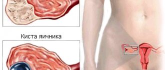
Two-chamber ovarian cyst (left or right): main symptoms, diagnosis and treatment methods
A two-chamber ovarian cyst is a phenomenon in gynecology that many women of reproductive age encounter. A benign growth, hollow inside and filled with liquid, is divided by a septum into separate cells.
The formation is more typical for the left ovary than for the right. A woman should know what it is, the reasons for the appearance of a cyst and be aware of the possible health consequences.
If the formation is diagnosed late or the woman refuses to undergo the prescribed treatment, internal bleeding or the development of peritonitis if the cyst bursts cannot be ruled out.
Symptoms that appear
A multi-chamber cyst, like other types, is difficult to detect at an early stage. Almost always it is asymptomatic and does not bother the patient until its size increases.
When the compaction reaches 4–5 cm in diameter, it begins to put pressure on the internal organs and causes discomfort.
Possible symptoms include:
- painful sensations in the ovary where the compaction is localized. Sometimes the pain also radiates to the lower torso or back; Pain in the lower abdomen, on the side.
- during intimacy, the pain syndrome intensifies;
- disruptions occur in the menstrual cycle. It becomes shorter or, on the contrary, longer;
- the type of discharge changes, and spotting may appear in the intervals between menstruation;
- the abdominal cavity increases in size, a feeling of tightness occurs;
- intestinal function is disrupted, disorders or constipation may appear;
- With the pressure of a two-chamber ovarian cyst on the bladder, urination becomes more frequent.
In case of complications (spontaneous rupture of the membranes, twisting of the leg, infection), other signs are additionally added. The temperature rises, the pain changes from aching to acute, and bleeding appears.
Dimensions and location
Based on localization, multi-chamber ovarian cysts are divided into the following types::
- Follicular cyst - appears at the site of the follicle;
- Corpus luteum cyst - appears in place of the corpus luteum;
- Paraovarian cyst - located between the layers of the broad ligament of the uterus.
If a woman suffers from ovarian endometriosis (the appearance of endometrioid tissue in the ovaries), then an endometrioid cyst may also appear in place of the endometrium.
Such a cyst will be called endometrioid.
Multilocular cysts can have different sizes.
Cyst sizes up to 10 cm are considered small . Accordingly, those cysts that are larger than 10 cm are considered large.
Why does it occur
The development of multi-chamber formation occurs in women of any age. Factors influencing the formation of the disease are different. More often these are embryonic disorders, when the cyst is a congenital pathology and is considered a developmental anomaly. This happens if during pregnancy the development of the embryo was seriously affected by external negative factors:
- tobacco products;
- alcohol;
- stress;
- use of medications;
- prolonged exposure to the sun's rays.
Also, a hereditary predisposition at the genetic level cannot be ruled out. Quite often, this pathology occurs in women who have suffered from inflammatory diseases of the genitourinary system, especially in the acute or chronic stages.
Factors of etiological origin are considered quite common causes. These include any irregularities in the menstrual cycle, such as length or regularity. Every third woman with a normal cycle, and every second woman with dysmenorrhea, is diagnosed with cystic formations.
A functional neoplasm can appear during pregnancy, as well as after surgery, such as abortion. Also, the development of pathology is influenced by the early onset of sexual activity, past or advanced sexually transmitted diseases.
Types of formations
Benign tumors have several classifications. One of them is related to the nature of the appearance. There are paraovarian and dermoid seals (congenital), follicular cyst (occurs due to hormonal disorders), serous, mucinous. In addition, pathology has a classification related to the location.
Source: https://PlastikaPlus.ru/opuholi/trehkamernaya-kista-yaichnika.html
Diagnostics
A woman can independently suspect the presence of a multilocular cyst based on the occurrence and intensification of the above symptoms. In this case, she needs to visit a gynecologist as soon as possible, where during an in-person appointment she needs to voice the relevant complaints.
He, in turn, using a bimanual examination method, must assess the condition of the uterus, appendages and ovaries for enlargement and pain of the latter. If suspicions are confirmed, the doctor prescribes a number of additional examinations.
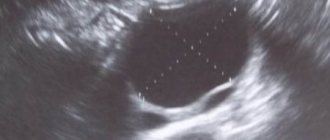
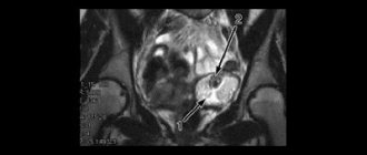
First of all, an ultrasound of the pelvic organs is performed. During its implementation, the exact location of the neoplasm, its size, structural features and structure are determined. If necessary, Doppler ultrasound is used to evaluate the circulatory system of the cyst.
On this topic
- Female reproductive system
Do I need to remove an ovarian cyst if it doesn’t bother me?
- Olga Vladimirovna Khazova
- December 4, 2021
If there is a suspicion that the neoplasm has reorganized into a cancerous tumor, the patient's blood is examined for the presence of tumor markers of the following series: CA-125, CA-15-3, HE-4, ROMA index. If all is well, then these indicators should be below or at the border of the normal table values.
In emergency situations, namely when a cyst ruptures or its base is torsioned, a puncture of the posterior vaginal fornix is performed to check for intra-abdominal bleeding.
Treatment
The treatment tactics for a patient with a multilocular ovarian cyst depend on the main characteristics of the neoplasm: its type, the number of internal cavities, their size and contents.
For example, in the presence of a small two-chamber functional neoplasm, preference is given to conservative treatment. With it, the patient, in addition to taking appropriate medications, will have to undergo regular gynecological examinations and have an ultrasound of the pelvic organs.
In this case, surgery is prescribed if an increased number of tumor markers is detected in the patient’s blood or when the cyst does not respond to conservative treatment for a long time.
On this topic
- Female reproductive system
Can there be a delay and a positive test for an ovarian cyst?
- Natalya Gennadievna Butsyk
- December 4, 2021
If an ultrasound revealed an ovarian dermoid cyst, it is surgically removed.
Women with endometrial cysts are treated in several stages, combining conservative methods of therapy with surgical ones.
The order of medical interventions is influenced by the size of the tumor, the patient’s condition and the presence of concomitant diseases. Usually, at the first stage, the cause of the ovarian cyst is eliminated, for example, inflammation or hormonal imbalance, and then, if the tumor does not resolve, it is removed surgically.
In practice, surgery is the most reliable way to get rid of a multilocular ovarian cyst. The choice of method is influenced by a large number of factors, including the severity of the pathology, its stage and size.
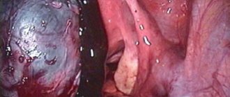
Laparoscopy
Laparoscopic surgery to remove cystic multilocular tumors is performed through small punctures in the navel and ovaries.
Through them, carbon dioxide is pumped into the abdominal cavity using a Veress needle until normal visibility of the area being examined is ensured. Then a laporoscope is inserted through them - a medical rigid endoscope equipped with a lens system and a camera.
The diagnosed cyst is removed with a loop of electrocoagulator, and its vessels are cauterized. The extracted material is sent for histological examination. In practice, laparoscopic surgery is considered the most accurate and least traumatic way to remove cysts of any nature.
Laporotomy
Open abdominal surgery to excise a multi-chamber ovarian cyst is rarely performed when other methods of removing it are impossible. For example, if the cyst is recurrent, large in size (more than 10 cm in diameter), fused with ovarian tissue, or complications arise in the form of twisting of the pedicle.
In this case, it is removed along with part of the gland through a surgical incision under the navel. Typically, this treatment method is used in women of menopausal age.
Types, features and treatment of multilocular ovarian cyst in women
A benign formation, called an ovarian cyst, has a number of features regarding structure and structure. It may consist of different cellular forms, be filled with serous, hemorrhagic or purulent fluid, and be divided into cavities. It's called a multilocular tumor.
Types of two-chamber and three-chamber cysts
Ovarian cysts are divided according to their structure into two-chamber, three-chamber and multi-chamber. This is due to the number of partitions inside the benign cavity. The septal wall can consist of a connective tissue structure or epithelial cells.
Multi-chamber cysts of the left and right ovary are classified by location:
- right-sided;
- left-sided;
- bilateral.
Example of a multi-chamber formation
Often one side is affected in young women (from 20 years to 45 years). Bilateral formations may be related to polycystic disease.
According to the course of the disease, a two-chamber cyst of the right ovary can be uncomplicated or complicated. It has also been established that the number of complications associated with torsion of the pedicle, rupture of the capsule and apoplexy of the appendage is much higher than that of single-chamber cysts.
A two-chamber ovarian formation can be attached by means of an anatomical or surgical pedicle, (More about this is described: torsion of the pedicle of an ovarian cyst.), it happens on the surface or inside the gonad.
Multi-chamber ovarian cysts are more characteristic of true formations than functional ones. This statement explains the frequent formation of oncology in this location.
According to histological structure, they are distinguished:
- Follicular;
- Luteal;
- Serous, serous-papillary and mucinous cystadenoma;
- Mature and immature dermoid (teratoma);
- Paraovarian.
An ovarian cyst with a septum can be present in the body of a woman of reproductive age for a long time and not make itself felt. Pathological symptoms occur with active growth, compression of adjacent structures and the development of complications.
The structure of multi-chamber ovarian formations
Functional cysts:
Almost always single-chamber, consisting of ovarian stroma and connective tissue membrane, the increase in size occurs due to the accumulation of intracapsular contents, and not cell proliferation as in cystomas. The follicular and luteal cyst will resolve on its own after 1–3 months.
Cystadenoma - according to its cytological structure can be a serous cyst, serous-papillary and mucinous:
- A serous cyst of the left or right ovary can be either single-chamber or double-chamber. The inside is lined with simple epithelium.
- Serous-papillary - consists of a capsule and papillary epithelial tissue. The rough papillary lining resembles a malignant tumor.
- Mucinous - lined with mucus-like epithelial tissue, filled with jelly-like contents.
Teratoma:
An underdeveloped remnant of the germ layers. A single-chamber structure is noted, but may have one or more septa. The remains of the ectoderm and endoderm are actively dividing, due to which it reaches large sizes. A multi-chamber ovarian cyst contains inside hair follicles, cartilage and bone tissue, teeth, sweat and sebaceous glands.
Paraovarian ovarian cyst:
It is a tumor-like formation against the background of impaired embryogenesis of gonadal tubules. The smooth-walled structure is single-chamber or two-chamber, in rare cases separated by a large number of partition walls. The parietal cells are unable to divide, and the increase in volume occurs due to the accumulation of secretory fluid. No cases of malignancy were registered.
Diagnosis of a two-chamber cyst
The diagnostic procedure begins with an examination on a gynecological chair; for medium and large sizes, the enlarged appendage on the affected side is palpated. The formation is moderately dense, mobile, and may be painful.
Taking into account the preliminary diagnosis and personal characteristics, instrumental techniques are prescribed:
- Ultrasound examination of the internal genital organs, preferably intravaginal ultrasound;
- Computer and magnetic resonance imaging determines the type, structural features, location if a malignant process is suspected;
- Fine-needle biopsy - a puncture is performed with a biopsy sample taken for cytological examination;
- Diagnostic laparoscopy;
- Puncture of the pouch of Douglas to exclude fluid in the pelvis.
Together with hardware methods, laboratory diagnostics are performed, general blood tests, urine tests, and biochemical blood tests are prescribed. Tests for tumor markers, human chorionic gonadotropin, progesterone, and estrogen are indicative. A pregnancy test is required.
Treatment of a two-chamber cyst
Based on the examination results and symptoms of the disease, a decision is made on further treatment tactics for the patient. As a rule, two-chamber ovarian cysts must be radically removed.
Is it possible to do without surgery?
Small tumors are treated without surgery. The effectiveness of therapy is high for functional formations (follicular, corpus luteum), due to the ability to involute and completely disappear after 1–3 months. Cystoms can initially be treated with hormonal and anti-inflammatory drugs, but often require surgical excision.
Elective patients with small and medium-sized lesions are subject to laparoscopy. After which a course of antibacterial, anti-inflammatory, and, if necessary, hormonal immunomodulatory agents is prescribed. In case of complications with bleeding, rupture of the capsule or ovary, emergency laparotomy and treatment in a gynecological or intensive care unit are indicated.
Removal is performed by enucleation or wedge-shaped excision; if it is large in size and damages most of the ovarian tissue - oophorectomy (removal of one or both ovaries).
The disease in pregnant women is observed in dynamics, with a tendency to growth or suspected malignancy - removal in the second trimester.
Why is multilocular cystosis dangerous?
The danger lies in the sudden rupture of an overfilled capsule after any type of physical activity. Severe pain is complicated by traumatic shock and peritonitis. Hemorrhagic syndrome occurs with ovarian apoplexy, the woman loses consciousness, the skin is pale, the pulse is poorly determined, blood pressure is below 100/60 mm Hg. Art.
When a secondary bacterial infection occurs, suppuration and abscess formation develop. A cavity with purulent exudate breaks through and pours into the pelvis, resulting in severe peritonitis and sepsis.
Cystic formations on the pedicle are prone to torsion with subsequent necrosis of the connection. Untreated or untimely diagnosed cystomas become malignant. The process goes unnoticed and is diagnosed in the later stages.
A two-chamber ovarian cyst is subject to careful diagnosis and further competent therapy. Individual selection of treatment is important in order to completely eliminate the disease. If you suspect a gynecological pathology, it is recommended to immediately contact a gynecologist.
Source: https://prokst.ru/mnogokamernaya-kista-yaichnika/
Complications
Despite the fact that multilocular ovarian cysts are benign neoplasms, their presence poses a danger to the patient’s life, even in the absence of the likelihood of developing cancer.
Leg torsion
Reaching large sizes, multi-chamber cysts can move in the abdominal cavity, including around their axis, during increased physical activity, sports or intimacy. The consequence of this development of events is the cessation of blood supply to the neoplasm and, as a consequence, tissue necrosis.
Suppuration
The cavities of multilocular ovarian cysts are isolated and often filled with blood. As a result, there is a high probability of their suppuration due to infection.
Shell rupture
May occur due to increased physical activity. The acute course of the pathology is dangerous for the patient’s life, since in this case intra-abdominal bleeding occurs.
Infertility
Functional multi-chamber ovarian cysts can produce excessive amounts of sex hormones, which, in turn, will interfere with the normal course of the ovulatory process and conception.
Since multi-chamber cysts usually reach large sizes, they can cause compression of nearby organs: the rectum and bladder, causing their dysfunction.
Multilocular ovarian cyst: treatment, surgery, large size
Multilocular ovarian cyst is one of the most dangerous types of benign tumors. In a short time it can increase significantly in size. The most dangerous complications of this neoplasm are rupture of the membranes and transformation of healthy cells into cancerous ones.
The tumor can grow in size very quickly.
What is it?
Contains a large number of partitions and chambers.
Multilocular cystic formation is a compaction that occurs in the tissues of the ovary. It is a cavity containing liquid contents. It may include purulent or bloody spots.
The difference from a two-chamber cyst is that this type of thickening contains more septa and chambers. First, individual bubbles appear on the internal organ, and then unite into one large tumor.
Features of the pathology
A multi-chamber ovarian cyst has features that are unique to it. The main difference is the large size of the tumor. Often its diameter is more than 10 cm. The significant growth of the capsule is due to the fact that it contains several chambers at once, each of which can be large.
Features of a three-chamber (or more) cyst include:
- seals are present in the body for a long time. At first there are several bubbles and only in the absence of therapy do they merge into one capsule;
- in most cases, pathology develops on the right side, since this ovary has more blood vessels;
- degeneration into a malignant tumor often occurs precisely in connection with multilocular neoplasms;
- have a tendency to complications (twisting of the leg, rupture of the membranes, infection);
- cannot resolve spontaneously, and also respond poorly to conservative treatment methods.
Complications
It is these types of cystic formations that often cause numerous consequences. If left untreated, multilocular ovarian tumors provoke the following complications:
- twisting of the leg on which the capsule is attached. This usually occurs during increased physical activity or during sex;
- spontaneous rupture of the membranes leads to the contents entering the abdominal cavity. This causes peritonitis, sepsis, etc.; Spontaneous rupture of the tumor causes peritonitis, sepsis.
- infection gets inside. Because of it, an inflammatory focus develops, tissue suppuration appears;
- large thickenings put pressure on other abdominal organs. This usually affects the activity of the gastrointestinal tract and bladder;
- prolonged absence of therapy leads to infertility. The proper functioning of the genital organs is disrupted, the menstrual cycle is disrupted; Prolonged lack of therapy leads to infertility.
- Benign cells often degenerate into malignant ones, which triggers the process of cancer development.
Symptoms of a multilocular ovarian cyst
At an early stage, it is difficult to understand that a multi-chamber seal has formed on the ovary. As it reaches large sizes, the following symptoms appear:
Nagging pain in the side and lower abdomen.
- disruptions in the menstrual cycle (increasing or decreasing);
- nagging pain in the side where the cyst is located. They can radiate to the lower part of the abdominal cavity, to the lower back;
- bleeding or spotting between cycles;
- increase in the size of the abdominal cavity;
- weight loss may occur;
- increased body hair growth (due to increased production of male sex hormones);
- the patient cannot become pregnant.
When an inflammatory focus develops, signs of general intoxication also appear. These include fever, paroxysmal pain, nausea or vomiting, headache, etc.
Types of education
Multiseptate tumors, like other varieties, are one of the following types:
- follicular cyst. Usually this variety increases slightly in size and is treated with oral contraceptives. Appears on the ovary when the follicle has not completed the entire maturation cycle. The liquid inside the capsule contains estrogens. Cystic thickening causes disruptions in the menstrual cycle, heavy bleeding at the beginning of the cycle, as well as aching pain in the abdominal cavity; It appears when the follicle has not completed the entire maturation cycle.
- Corpus luteum cyst is a similar type of lump that develops mid-cycle. Where the corpus luteum should form, a cavity appears and fills with contents. Unpleasant symptoms appear when ovulation occurs;
- endometrioid. It occurs as a consequence of excessive growth of epithelial tissue. With this type, menstruation is heavy and painful. A woman will not be able to become pregnant until the tumor is removed; It occurs due to excessive growth of epithelial tissue.
- cystadenoma. This type of benign neoplasm usually has one chamber. As it grows, thin septa form, and pain appears in the lower abdomen. Cystadenoma, according to the type of fluid contained in the cavity, can be serous, mucinous or papillary (the most dangerous type, which provokes the development of cancer cells).
Diagnosis and treatment methods
Typically, a woman consults her doctor when the first symptoms appear. But sometimes a neoplasm can be detected during a routine examination. By palpation, the gynecologist will determine that there is an extraneous thickening on the ovary. In the future, it will be necessary to clarify the diagnosis using modern examination methods.
By palpation, the gynecologist will determine that there is an extraneous thickening on the ovary.
The doctor will prescribe the following diagnostic procedures:
Using the device, the exact location of the capsule attachment and dimensions are determined.
- Ultrasound. Using the device, the exact location of the capsule’s attachment is determined, its dimensions are determined, as well as the number of chambers;
- if the ultrasound examination turns out to be insufficiently informative, an MRI or CT scan may be required;
- since there is a high risk of transformation of a benign tumor into a malignant one, a biopsy is required. Histological examination will determine whether cancer cells are present;
- Blood and urine tests are necessary to determine whether an inflammatory focus is present in the body. In addition, a hormonal blood test is performed;
- smears are taken to determine concomitant infections of the pelvic organs.
After receiving a complete picture of the pathology, the doctor prescribes treatment. It will depend on the type of thickening and its size, on the number of chambers, etc. At an early stage of development, a decision is often made to choose conservative treatment. In this case, hormonal drugs and anti-inflammatory drugs are used. They can be effective in cases where the tumor is small in size, there are no cancer cells, or other complications.
If the cyst does not resolve within 3 to 4 months, a decision is usually made to surgically remove it.
Surgery
There are several types of surgery that are used in the presence of a multi-chamber capsule. The most gentle method is considered to be laparoscopy, which involves making pinpoint incisions and removing the tumor through them. But this method can only be used for small cavities.
Laparoscopy involves making pinpoint incisions and removing the tumor through them.
Classic surgical excision of tissue is used most often.
During the operation, the capsule is removed along with its contents to eliminate the risk of relapse. Recovery is long and difficult. If the lump is very large, then the entire ovary may be removed along with it.
This occurs in cases where the capsule interferes with its proper functioning or tissue necrosis occurs.
Danger during pregnancy
It is observed during pregnancy and removal occurs after childbirth.
It is advisable to identify and remove the cyst before planning, since it often causes miscarriages, missed abortions and other complications. Doctors avoid performing surgeries on pregnant women, as this also leads to miscarriage or developmental abnormalities in the fetus.
In most cases, they take a wait-and-see approach and monitor any changes that occur. After childbirth, removal of the multi-chamber capsule is indicated. In rare cases, when there is a direct threat to the life or health of the patient, surgical intervention during pregnancy is prescribed.
An ovarian cyst, consisting of several capsules, poses a danger to a woman at any age. Once identified, it must be immediately removed surgically. The exception is small tumors, which are tried to be cured using conservative methods.
Source: https://kistateka.ru/yaichniki/mnogokamernaya