Thanks to fibrogastroscopy, the endoscopist can examine all parts of the upper segments of the gastrointestinal tract. In particular, using this technique, proliferation of the integumentary pituitary epithelium of the stomach can be detected. To identify it, a biopsy is used, which involves taking histological material using a special brush or forceps. In this case, pieces of tissue are taken and a histological examination is carried out on their basis, which makes it possible to detect various morphological changes. This method can detect proliferation of the integumentary epithelium.
Today, various modern techniques are used to diagnose this pathological process, which were specifically designed to detect tumors inside the abdominal cavity. In particular, proliferation of the integumentary epithelium is diagnosed using fine-needle aspiration biopsy, which can be performed by fiberoscopy or during ultrasound examination. If you place the sensor close to the area being analyzed, you can get the most accurate results. Therefore, the doctor will be able to detect even relatively unexpressed proliferation of the integumentary pitted epithelium.
In addition, proliferation of the gastric integumentary epithelium can be detected using immunocytochemical tests. To carry out these studies, the material is placed in a liquid medium. This technique involves the use of disposable brushes. After the examination is completed, the tips of these brushes are placed in a liquid with a special composition. Immunocytochemical testing is used to confirm various cancers, such as lymphoma or sarcoma. If the patient is expected to have proliferation of the surface epithelium, then it is possible to conduct a cytological study, in which less blood impurities are obtained.
What is proliferation
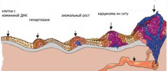
Cell proliferation is a normal process necessary for the body to renew itself. In other words, this is the physiological reproduction of cells with the result in the form of an increase in tissue volume. This process is controlled by hormones. Proliferation occurs in any organ and tissue. It controls the state of the immune system, leads to the destruction of tissue defects, regenerates them and restores the previous functioning of organs. Due to proliferation, connective tissue grows, new vessels are formed, burns are scarred, damage is eliminated and tissue is replaced with new cells or connective tissue. This applies to blood cells, epithelium and mucous membranes, cartilage, myocardial walls, etc. The most active process occurs in the gastric mucosa.
But proliferation under certain conditions can become pathological, that is, excessive. For example, with increased concentrations of somatropin, it leads to enlargement of organs and limbs. If cell differentiation is disrupted during the process of proliferation, this becomes the beginning of oncology with the appearance of atypical growths. This uncontrolled active (abnormal) cell division is called hyperproliferation or hyperplasia. This can also be explained by the predominance of proliferation over differentiation of mucosal cells. Then the number of narrowly differentiated cells decreases and the epithelium begins to lose its “legal properties” due to immature cells. Proliferation of the integumentary epithelium can and is often accompanied by processes of intestinal metaplasia of the stomach.
Marked proliferation of the foveal epithelium
The wall of the stomach consists of 4 layers: the inner mucosa, the submucosa, the muscular layer and the outer serosa.
The mucous membrane (MS) is normally folded, it has depressions - pits, flat areas (fields) and folds. The mucosal epithelium is highly prismatic (columnar) cells that line the entire stomach and all its folds in one layer. The cells of this epithelium secrete special mucus; it forms a layer up to 1-1.5 mm thick.
During the production of enzymes and hydrochloric acid, temporary channels are formed in this layer. Mucus protects the wall from digestion by stomach juice and various damage. The epithelium is renewed every 1-3 days.
What is proliferation
Cell proliferation is a normal process necessary for the body to renew itself. In other words, this is the physiological reproduction of cells with the result in the form of an increase in tissue volume. This process is controlled by hormones. Proliferation occurs in any organ and tissue.
It controls the state of the immune system, leads to the destruction of tissue defects, regenerates them and restores the previous functioning of organs. Due to proliferation, connective tissue grows, new vessels are formed, burns are scarred, damage is eliminated and tissue is replaced with new cells or connective tissue.
This applies to blood cells, epithelium and mucous membranes, cartilage, myocardial walls, etc. The most active process occurs in the gastric mucosa.
But proliferation under certain conditions can become pathological, that is, excessive. For example, with increased concentrations of somatropin, it leads to enlargement of organs and limbs. If cell differentiation is disrupted during the process of proliferation, this becomes the beginning of oncology with the appearance of atypical growths.
This uncontrolled active (abnormal) cell division is called hyperproliferation or hyperplasia. This can also be explained by the predominance of proliferation over differentiation of mucosal cells. Then the number of narrowly differentiated cells decreases and the epithelium begins to lose its “legal properties” due to immature cells.
Proliferation of the integumentary epithelium can and is often accompanied by processes of intestinal metaplasia of the stomach.
Hyperplasia
Proliferation of the integumentary pitted epithelium of the stomach is one of the subtypes of mucosal proliferation. The name is given because as a result of tissue growth, corkscrew-shaped pits are formed.
This proliferation means hyperplasia of mucus-producing cells (goblet cells), which play a protective role. The integumentary epithelium undergoes mutation, the content of mucin in the cells increases, pushing the nuclei to the base of the cell, pathological cells begin to replace healthy ones.
Pits form in the mucosa, which are shaped like a corkscrew pin. The process is a harbinger of oncology.
Most often, proliferation of the integumentary pitted epithelium occurs with hyperplastic gastritis, peptic ulcer disease, and ulcer-like cancer. Clinically and morphologically, they are identical, so a biopsy is required.
Cytological examination can reveal pronounced proliferation of the integumentary pitted epithelium of the stomach.
In fact, this is a precancerous condition, and such patients should be registered with an oncologist and undergo a systematic examination.
Reasons for development
the cause is chronic or long-term damage and irritation of the mucous membrane, which causes the appearance of wounds and ulcers. In addition to the above pathologies, the cause may be:
- hereditary predisposition;
- hormonal imbalances;
- the presence of Helicobacter pylori - its metabolic products contaminate the entire body, which suppresses the immune system, destroys the mucous membrane and causes proliferation of the integumentary epithelium; this bacterium leads to inflammation of the gastric mucosa and the development of gastritis in 90% of cases;
- nutritional disorders, which contain a lot of E additives, preservatives, alcohol;
- taking NSAIDs;
- stress;
- changes in the functioning of the duodenum (it provokes excess gastrin, which irritates the gastric mucosa; backflow of bile during reflux) and the pancreas.
Classification
Several types of gastric hyperplasia have been identified. They differ in type and areas affected. The main ones include the following:
- antral;
- focal;
- foveal;
- glandular;
- polypoid;
- lymphofolicular;
- proliferation of the integumentary pitted epithelium of the stomach;
- lymphoid.
Briefly about the types
There are the following types of pathology:
- Foveal hyperplasia of the stomach. In this case, the folds and gastric pits are deformed, they are elongated and curved. The most common and harmless type. Develops when taking NSAIDs.
- Antral - tissue grows at the junction of the gastric mucosa and duodenum. The cause is malnutrition. These changes look like multiple small growths.
- Lymphofollicular - the mucous membrane thickens and grows due to the accumulation of lymphocytes that form follicles. Possible malignancy. With lymphoid hyperplasia, the antrum of the stomach is most often affected, when the number of lymphocytes in the lymph nodes increases. With an increase in lymphocytes, the mucous membrane thickens and hyperplasias. Moreover, the enlargement of lymph nodes in this case is not inflammatory in nature. Causes: peptic ulcer and infectious diseases of the digestive tract.
- Glandular is the rarest form, with the proliferation of glandular cells and the formation of round and oval polyps. Occurs when the stomach stretches.
- Polypoid – the appearance of multiple polyps in any part of the stomach. The reason is unknown.
- Focal - “wart” - the appearance of additional tissue in one or several places. The process is observed in the initial stages of hyperplasia and is benign.
That is, we can say that the proliferation of cells of the integumentary pit epithelium is the proliferative growth of cells that produce mucus.
Degrees of proliferation
Today, hyperplasia is divided into 3 stages:
- First degree – mucus production decreases, hyperchromatosis is noted. The process is initial, it is reversible with proper treatment.
- The second degree is moderate proliferation of the integumentary pitted epithelium. The process is already more obvious, cell division accelerates and becomes more frequent, the number of Paneth and goblet cells decreases. The process can still be stopped with timely and modern treatment.
- The third degree is pronounced proliferation of cells of the integumentary pit epithelium of the stomach. Large areas proliferate, mucus is not secreted, there is no protection, the process becomes precancerous.
Symptoms and signs
For a long time, a person does not realize that he has a pathology, he does not have any illness, which is why early diagnosis is so late. Then, as the process progresses, first after eating there is severe pain in the upper abdomen, nausea, loss of appetite, sour belching, and hiccups.
In advanced stages, vomiting, general weakness, bloating, and flatulence are observed. The pain can be cramping in nature - at first it is stabbing or in the form of a burning sensation, then it becomes aching. The stool is disturbed and prone to diarrhea.
Signs of intoxication appear - pallor, headache, dizziness, fever, feeling of tense muscles. Symptoms are nonspecific and therefore often go unnoticed.
Early diagnosis is of paramount importance: it is necessary to promptly identify and prescribe effective treatment for proliferation of the integumentary pitted epithelium of the stomach. It is dangerous, but in the early stages it is amenable to therapy.
Diagnostic measures
The main diagnostic method is gastroscopy. With such an examination, it is possible to perform a biopsy followed by histology. The gastric mucosa is thoroughly examined and the specialist can see all changes in it immediately.
During the examination, the doctor can see neoplasms, abnormalities of tissues and membranes.
Barium X-ray will reveal polyps, their shape and the size of their legs. When establishing the etiology, an analysis is performed for the presence of Helicobacter pylori. This includes a urea breath test and a blood test for antibodies.
If a diagnosis of “pronounced proliferation of the integumentary epithelium hp” with a certain number of crosses is made, then this indicates the presence of Helicobacter pylori in the mucosa.
A biopsy can play a significant role; it is performed during gastroscopy. The study makes it possible to establish the presence of the disease and its subtype.
It must be said that the combination of proliferation of the integumentary epithelium and Helicobacter is not such a rare phenomenon.
Principles of treatment
Therapy for the disease is multicomponent and aims to improve gastric motility and normalize the production of hydrochloric acid.
Treatment of proliferation of the integumentary pit epithelium, i.e. hyperplastic gastritis, depends on the etiology, subtype of proliferation, hypo- or hyperacidity, severity of the clinic and concomitant diseases. Self-prescription of drugs is strictly prohibited.
How to treat proliferation of the integumentary pitted epithelium of the stomach? The following groups of drugs are prescribed for therapy:
- Antispasmodics - “No-shpa”, “Papaverine”, “Spazgan”, “Drotaverine”. Relieves spasm and pain.
- Antibiotics - Amoxicillin, Tetracycline, Clarithromycin, etc.
- Antimicrobial agent "Metronidazole" and bismuth preparations. Used to eradicate (remove) Helicobacter bacteria if present.
- Enzyme preparations - “Festal”, “Creon”, “Mezim”, “Panzinorm”, etc. They are used to improve digestion.
- Antacids - “Rennie”, “Almagel”, “Maalox”, etc. Relieve heartburn, pain, and protect the mucous membrane.
- Amino acids - “Methionine”, “BCAA”, etc. – are necessary to compensate for protein deficiency.
- Multivitamins - B vitamins, vit. S and R.
- For hypoacid conditions, gastric juice preparations are prescribed - Pepsin, hydrochloric acid.
- To reduce increased secretion - proton pump inhibitors. They are rarely prescribed and are effective in the initial stages. In recent years, hyperbaric oxygen therapy has been used for conservative therapy.
Traditional methods of treatment
As for traditional methods of treatment, they can help in the initial stages. Herbal medicine is used only in consultation with the attending physician and is symptomatic. Decoctions of herbs with an anti-inflammatory effect are used.
Chamomile is considered very useful - tea made from it has antispasmodic and anti-inflammatory effects. Ginger has antibacterial properties. Tea with mint, infusion of parsley or fireweed will relieve nausea and heartburn.
When is surgery needed?
Surgical treatment of hypertrophic gastritis is carried out when conservative therapy is ineffective. Indications for surgery are as follows:
- stomach bleeding;
- stomach polyps;
- presence or suspicion of malignancy of the process;
- severe course of the pathology.
Endoscopic removal of polyps (polypectomy) and gastric resection (complete or partial) are performed. Polyps are formed in all types of hyperplasia and can also be hereditary. The latter are more often malignant
Polyps larger than 1 cm are removed endoscopically. Small formations are not removed unless there is a suspicion of malignancy, but their development is monitored - examined once every six months.
Special diet
Nutrition should be balanced. You should eat food in small portions (fractionally), preferably at the same time, every 3 hours.
It is advisable to completely avoid alcohol and any products that irritate the mucous membranes: pickles, smoked meats, marinades, preserves, fermented foods, fast foods, soda and semi-finished products. Salt must be minimized; canned food and red meat are prohibited. All food should only be boiled, everything fried is prohibited.
Table No. 2 is recommended. This nutritional system is prescribed for intestinal diseases and gastritis. Its purpose is to stop the development of inflammation and normalize the functioning of the secretory apparatus of the stomach.
Forecast depends on the subtype of hyperplasia and its intensity, timeliness and modernity of diagnosis.
Preventive actions
There is no specific prevention of gastric hyperplasia, but general recommendations for improving health have not been canceled. The diet should contain sufficient vitamins and minerals, smoking and alcohol should be stopped, negative emotions and stress should be avoided. Try to choose products with the least amount of preservatives and carcinogens.
Get into the habit of drinking a glass of water before each meal so that your body receives at least 2 liters of clean water per day. Don't overuse anti-inflammatory drugs, even if they help you. An active lifestyle will also help get rid of many diseases. Don't forget to visit a gastroenterologist every six months.
If you really don’t like the EGD method, you can simply undergo ultrasound.
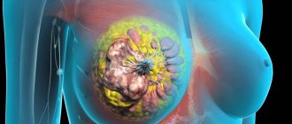
Proliferation of the integumentary pit epithelium is a dangerous pathology, which in advanced cases provokes cancer. Early diagnosis is important to stop lesions in the initial stages.
Source: https://worldwantedperfume.com/vyrazhennaja-proliferacija-foveoljarnogo-jepitelija/
Hyperplasia
Proliferation of the integumentary pitted epithelium of the stomach is one of the subtypes of mucosal proliferation. The name is given because as a result of tissue growth, corkscrew-shaped pits are formed. This proliferation means hyperplasia of mucus-producing cells (goblet cells), which play a protective role. The integumentary epithelium undergoes mutation, the content of mucin in the cells increases, pushing the nuclei to the base of the cell, pathological cells begin to replace healthy ones. Pits form in the mucosa, which are shaped like a corkscrew pin. The process is a harbinger of oncology.
Most often, proliferation of the integumentary pitted epithelium occurs with hyperplastic gastritis, peptic ulcer disease, and ulcer-like cancer. Clinically and morphologically, they are identical, so a biopsy is required. Cytological examination can reveal pronounced proliferation of the integumentary pitted epithelium of the stomach. In fact, this is a precancerous condition, and such patients should be registered with an oncologist and undergo a systematic examination.
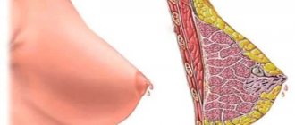
Breast
Breast changes are quite common, including among young girls and women. The organ constantly experiences the action of sex hormones, undergoes characteristic changes throughout the menstrual cycle, during pregnancy and lactation, and is therefore susceptible to various kinds of pathologies. According to statistics, up to 60% of women of reproductive age have signs of mastopathy.
Mastopathy is considered a benign process, but if it is present, the risk of malignancy increases several times. Proliferative variants are even more dangerous, they increase the likelihood of cancer by 25-30 times.
The presence or absence of proliferation is the most important sign when assessing the type of mastopathy. Non-proliferative forms are represented by foci of fibrous tissue, cystically altered ducts, the epithelium of which does not proliferate and is even atrophic. The risk of malignant transformation is relatively small.
Proliferative mastopathy is much more dangerous, because the cells of the ducts and lobules of the gland are actively dividing, creating the threat of cancerous degeneration. The more such foci of proliferation in the gland tissue and the more active the proliferation of the epithelial component, the more likely breast cancer is in the future, therefore all forms of proliferating mastopathy are considered precancerous conditions.
According to the severity of proliferation, several degrees of mastopathy are distinguished. In the first degree, proliferation is not detected, in the second, it is there, but the cells do not show signs of atypia, the third degree of mastopathy is manifested by active proliferation of epithelial cells with atypia.
Thus, proliferation of mammary gland cells is not only a criterion for the form of mastopathy, but also an indicator of the likelihood of cancer, therefore excessive division of the epithelium always attracts the attention of specialists. Diagnosis of this change is carried out by cytological examination of tissue obtained during puncture.
Reasons for development
The main reason is chronic or long-term damage and irritation of the mucous membrane, which causes wounds and ulcers. In addition to the above pathologies, the cause may be:
- hereditary predisposition;
- hormonal imbalances;
- the presence of Helicobacter pylori - its waste products contaminate the entire body, which suppresses the immune system, destroys the mucous membrane and causes proliferation of the integumentary pitted epithelium; this bacterium leads to inflammation of the gastric mucosa and the development of gastritis in 90% of cases;
- nutritional disorders, which contain a lot of E additives, preservatives, alcohol;
- taking NSAIDs;
- stress;
- changes in the functioning of the duodenum (it provokes excess gastrin, which irritates the gastric mucosa; backflow of bile during reflux) and the pancreas.
The main factors against which proliferation and hyperplasia develop
The main cause of hyperplastic and proliferative processes is hormonal dysfunction. Therefore, proliferation of columnar epithelial cells most often develops with impaired ovarian function or long-term uncontrolled use of hormonal contraceptives. Metabolic disorders such as obesity and diabetes mellitus play an important role in the development of proliferation of the columnar epithelium of the cervix.
The main types of cervical proliferation:
- Glandular proliferation of the uterus is the proliferation of glandular structures in the vaginal segment of the uterine cervix. Often, proliferation of the cervix of a glandular nature is mistaken for erosive changes. As a result, cryodestruction is performed, which is absolutely not indicated for this pathological condition.
- Glandular-cystic hyperplasia is the proliferation of glandular tissues with the formation of cystic formations.
- Microglandular hyperplasia is a proliferation of the cervix involving the glands of the cervical canal.
- Adenomatous hyperplasia (atypical) is a proliferation of columnar epithelium of the cervix, which quite often gives rise to malignant degeneration. In this case, columnar epithelium with signs of proliferation is detected.
Thus, the main differences between these types of hyperplasia are determined by the level of development of the process and its neglect. The proliferation of cylindrical cells is considered the most aggressive in terms of the possibility of cancerous degeneration. Therefore, if this pathology is present, patients should undergo regular examinations by a gynecologist to prevent the occurrence of malignant neoplasms.
Briefly about the types
There are the following types of pathology:
- Foveal hyperplasia of the stomach. In this case, the folds and gastric pits are deformed, they are elongated and curved. The most common and harmless type. Develops when taking NSAIDs.
- Antral - tissue grows at the junction of the gastric mucosa and duodenum. The cause is malnutrition. These changes look like multiple small growths.
- Lymphofollicular - the mucous membrane thickens and grows due to the accumulation of lymphocytes that form follicles. Possible malignancy. With lymphoid hyperplasia, the antrum of the stomach is most often affected, when the number of lymphocytes in the lymph nodes increases. With an increase in lymphocytes, the mucous membrane thickens and hyperplasias. Moreover, the enlargement of lymph nodes in this case is not inflammatory in nature. Causes: peptic ulcer and infectious diseases of the digestive tract.
- Glandular is the rarest form, with the proliferation of glandular cells and the formation of round and oval polyps. Occurs when the stomach stretches.
- Polypoid – the appearance of multiple polyps in any part of the stomach. The reason is unknown.
- Focal – “wart” – the appearance of additional tissue in one or several places. The process is observed in the initial stages of hyperplasia and is benign.
That is, we can say that the proliferation of cells of the integumentary pit epithelium is the proliferative growth of cells that produce mucus.
The main factors against which proliferation and hyperplasia develop
The main cause of hyperplastic and proliferative processes is hormonal dysfunction. Therefore, proliferation of columnar epithelial cells most often develops with impaired ovarian function or long-term uncontrolled use of hormonal contraceptives. Metabolic disorders such as obesity and diabetes mellitus play an important role in the development of proliferation of the columnar epithelium of the cervix.
The main types of cervical proliferation:
- Glandular proliferation of the uterus is the proliferation of glandular structures in the vaginal segment of the uterine cervix. Often, proliferation of the cervix of a glandular nature is mistaken for erosive changes. As a result, cryodestruction is performed, which is absolutely not indicated for this pathological condition.
- Glandular-cystic hyperplasia is the proliferation of glandular tissues with the formation of cystic formations.
- Microglandular hyperplasia is a proliferation of the cervix involving the glands of the cervical canal.
- Adenomatous hyperplasia (atypical) is a proliferation of columnar epithelium of the cervix, which quite often gives rise to malignant degeneration. In this case, columnar epithelium with signs of proliferation is detected.
Thus, the main differences between these types of hyperplasia are determined by the level of development of the process and its neglect. The proliferation of cylindrical cells is considered the most aggressive in terms of the possibility of cancerous degeneration. Therefore, if this pathology is present, patients should undergo regular examinations by a gynecologist to prevent the occurrence of malignant neoplasms.
Degrees of proliferation
Today, hyperplasia is divided into 3 stages:
- First degree – mucus production decreases, hyperchromatosis is noted. The process is initial, it is reversible with proper treatment.
- The second degree is moderate proliferation of the integumentary pitted epithelium. The process is already more obvious, cell division accelerates and becomes more frequent, the number of Paneth and goblet cells decreases. The process can still be stopped with timely and modern treatment.
- The third degree is pronounced proliferation of cells of the integumentary pit epithelium of the stomach. Large areas proliferate, mucus is not secreted, there is no protection, the process becomes precancerous.
Cervical dysplasia
Cervical dysplasia is a precancerous condition, which is based on pronounced proliferation of the columnar epithelium of the cervix without transition to stromal cells and surface epithelium. Such proliferation of the uterus is characterized by a serious disruption of cellular structures, which is manifested by nuclear polymorphism, an increase in the nuclear-cytoplasmic ratio and pathological vacuolization. In this case, the proliferation of the cervix is initially localized in the basal layer, and subsequently moves to higher layers. As a rule, columnar epithelium with signs of proliferation is found in any areas of dysplasia characteristic of polyps, pseudoerosions and leukoplakias.
Symptoms and signs
For a long time, a person does not realize that he has a pathology, he does not have any illness, which is why early diagnosis is so late. Then, as the process progresses, first after eating there is severe pain in the upper abdomen, nausea, loss of appetite, sour belching, and hiccups.
In advanced stages, vomiting, general weakness, bloating, and flatulence are observed. The pain can be cramping in nature - at first it is stabbing or in the form of a burning sensation, then it becomes aching. The stool is disturbed and prone to diarrhea. Signs of intoxication appear - pallor, headache, dizziness, fever, feeling of tense muscles. Symptoms are nonspecific and therefore often go unnoticed. Early diagnosis is of paramount importance: it is necessary to promptly identify and prescribe effective treatment for proliferation of the integumentary pitted epithelium of the stomach. It is dangerous, but in the early stages it is amenable to therapy.
Ointments with hormonal agents
Products containing glucocorticoid hormones have gained quite controversial popularity. Treatment of sprains is usually carried out with hydrocortisone and prednisolone ointments.
Indeed, such gels are an ambulance for the most unbearable pain. In addition, they are able to quickly stop the inflammatory process. Ointments from this group cope well with such tasks, and patients note a significant improvement in the condition of the injured leg.
Therefore, having tried such therapy once, a person refuses other treatment. But it is difficult to predict what the future consequences of such therapy will be.
All hormonal agents included in the ointments are synthesized from cortisol. Long-term use of a stress hormone can provoke not only a number of negative consequences, but also addiction, as well as withdrawal syndrome. The skin and muscle tissue are at risk. Bruising, tissue thinning, and muscle damage are often observed.
Diagnostic measures
The main diagnostic method is gastroscopy. With such an examination, it is possible to perform a biopsy followed by histology. The gastric mucosa is thoroughly examined and the specialist can see all changes in it immediately.
During the examination, the doctor can see neoplasms, abnormalities of tissues and membranes.
Barium X-ray will reveal polyps, their shape and the size of their legs. When establishing the etiology, an analysis is performed for the presence of Helicobacter pylori. This includes a urea breath test and a blood test for antibodies.
If a diagnosis of “pronounced proliferation of the integumentary epithelium hp” with a certain number of crosses is made, then this indicates the presence of Helicobacter pylori in the mucosa. A biopsy can play a significant role; it is performed during gastroscopy. The study makes it possible to establish the presence of the disease and its subtype. It must be said that the combination of proliferation of the integumentary epithelium and Helicobacter is not such a rare phenomenon.
Diagnostic methods for detecting proliferation and hyperplasia
Hyperplasia, which is based on proliferation of the cervix, is characterized by the following clinical symptoms:
- Intense, prolonged menstrual bleeding.
- Intermenstrual bleeding.
- Regular cycles without the onset of ovulation, which makes it impossible to adequately plan pregnancy.
In some cases, proliferation of columnar epithelium may be asymptomatic.
To identify columnar epithelium with signs of proliferation, it is necessary to conduct the following studies:
- Hysterocopy;
- Colposcopy;
- Targeted biopsy sampling from the area of the pathological focus;
- Ultrasound of the pelvic organs.
If the study revealed columnar epithelium with signs of proliferation, the doctor uses additional laboratory tests. They involve taking an analysis for luteinizing hormone, follicle-stimulating hormone, estradiol and progesterone. For a cytological examination, a smear is taken, which reveals columnar epithelium with signs of proliferation. In parallel with this, to exclude malignant degeneration, it is recommended to undergo oncocytology. Since cervical proliferation can develop against the background of inflammatory diseases, all these patients are recommended to undergo bacteriological or bacterioscopic studies to identify pathogenic pathogens of a specific and nonspecific nature, such as Escherichia coli, cocci, chlamydia, Gardnerella and Trichomonas. If a patient exhibits proliferation of columnar epithelial cells, PCR diagnostics can be used to determine its etiology. It can also help identify herpes viruses and human papillomavirus, which can contribute to the development of uterine proliferation.
Principles of treatment
Therapy for the disease is multicomponent and aims to improve gastric motility and normalize the production of hydrochloric acid.
Treatment of proliferation of the integumentary pit epithelium, i.e. hyperplastic gastritis, depends on the etiology, subtype of proliferation, hypo- or hyperacidity, severity of the clinic and concomitant diseases. Self-prescription of drugs is strictly prohibited.
How to treat proliferation of the integumentary pitted epithelium of the stomach? The following groups of drugs are prescribed for therapy:
- Antispasmodics – “No-shpa”, “Papaverine”, “Spazgan”, “Drotaverine”. Relieves spasm and pain.
- Antibiotics – “Amoxicillin”, “Tetracycline”, “Clarithromycin”, etc.
- Antimicrobial agent “Metronidazole” and bismuth preparations. Used to eradicate (remove) Helicobacter bacteria if present.
- Enzyme preparations - “Festal”, “Creon”, “Mezim”, “Panzinorm”, etc. are used to improve digestion.
- Antacids – “Rennie”, “Almagel”, “Maalox”, etc. Relieve heartburn, pain, and protect the mucous membrane.
- Amino acids – “Methionine”, “BCAA”, etc. Necessary for replenishing protein deficiency.
- Multivitamins – B vitamins, vit. S and R.
- For hypoacid conditions, gastric juice preparations are prescribed - “Pepsin”, hydrochloric acid.
- To reduce increased secretion - proton pump inhibitors. They are rarely prescribed and are effective in the initial stages. In recent years, hyperbaric oxygen therapy has been used for conservative therapy.
Medicines for sprains
The doctor will prescribe the following groups of medications:
- non-steroidal anti-inflammatory drugs - cooling ointments and creams. They help quickly relieve pain in the affected area. Common medications from this group are Voltaren, Ketoprofen;
- anticoagulants - necessary to improve blood flow. They prevent blood clots from forming in blood vessels and also prevent the development of vascular diseases.
- cooling - creams or ointments with menthol and camphor oil that help relax muscles and eliminate cramps.
In some cases, your doctor may recommend using ointments based on steroid hormones. They suppress inflammation and also reduce swelling. This effect is achieved by reducing the permeability of the walls of blood vessels. Warming ointments may also help.
Additionally read:
- Ankle sprain: ICD-10 code, treatment
If the pain does not go away for a long time, it is very important to consult a doctor and stop self-medication. The reason may lie deeper, and incorrectly selected pharmaceuticals will only aggravate the situation.
Remedies for sprains prices in the pharmacy
At the pharmacy you can buy various painkillers, as well as drugs that relieve swelling and speed up the recovery process. You can give your pharmacist a list of medications from your doctor and find out their costs, then compare offers at other pharmacies. Or enter the name on the Apteka24 website and quickly place an order without wasting time. We have the best offers and best prices!
When is surgery needed?
Surgical treatment of hypertrophic gastritis is carried out when conservative therapy is ineffective. Indications for surgery are as follows:
- stomach bleeding;
- stomach polyps;
- presence or suspicion of malignancy of the process;
- severe course of the pathology.
Endoscopic removal of polyps (polypectomy) and gastric resection (complete or partial) are performed. Polyps are formed in all types of hyperplasia and can also be hereditary. The latter are more often malignant
Polyps larger than 1 cm are removed endoscopically. Small formations are not removed unless there is a suspicion of malignancy, but their development is monitored - examined once every six months.
Cervical dysplasia
Cervical dysplasia is a precancerous condition, which is based on pronounced proliferation of the columnar epithelium of the cervix without transition to stromal cells and surface epithelium. Such proliferation of the uterus is characterized by a serious disruption of cellular structures, which is manifested by nuclear polymorphism, an increase in the nuclear-cytoplasmic ratio and pathological vacuolization. In this case, the proliferation of the cervix is initially localized in the basal layer, and subsequently moves to higher layers. As a rule, columnar epithelium with signs of proliferation is found in any areas of dysplasia characteristic of polyps, pseudoerosions and leukoplakias.
Special diet
Nutrition should be balanced. You should eat food in small portions (fractionally), preferably at the same time, every 3 hours.
It is advisable to completely avoid alcohol and any products that irritate the mucous membranes: pickles, smoked meats, marinades, preserves, fermented foods, fast foods, soda and semi-finished products. Salt must be minimized; canned food and red meat are prohibited. All food should only be boiled, everything fried is prohibited.
Table No. 2 is recommended. This nutritional system is prescribed for intestinal diseases and gastritis. Its purpose is to stop the development of inflammation and normalize the functioning of the secretory apparatus of the stomach.
Forecast depends on the subtype of hyperplasia and its intensity, timeliness and modernity of diagnosis.
Diagnostic methods for detecting proliferation and hyperplasia
Hyperplasia, which is based on proliferation of the cervix, is characterized by the following clinical symptoms:
- Intense, prolonged menstrual bleeding.
- Intermenstrual bleeding.
- Regular cycles without the onset of ovulation, which makes it impossible to adequately plan pregnancy.
In some cases, proliferation of columnar epithelium may be asymptomatic.
To identify columnar epithelium with signs of proliferation, it is necessary to conduct the following studies:
- Hysterocopy;
- Colposcopy;
- Targeted biopsy sampling from the area of the pathological focus;
- Ultrasound of the pelvic organs.
If the study revealed columnar epithelium with signs of proliferation, the doctor uses additional laboratory tests. They involve taking an analysis for luteinizing hormone, follicle-stimulating hormone, estradiol and progesterone. For a cytological examination, a smear is taken, which reveals columnar epithelium with signs of proliferation. In parallel with this, to exclude malignant degeneration, it is recommended to undergo oncocytology. Since cervical proliferation can develop against the background of inflammatory diseases, all these patients are recommended to undergo bacteriological or bacterioscopic studies to identify pathogenic pathogens of a specific and nonspecific nature, such as Escherichia coli, cocci, chlamydia, Gardnerella and Trichomonas. If a patient exhibits proliferation of columnar epithelial cells, PCR diagnostics can be used to determine its etiology. It can also help identify herpes viruses and human papillomavirus, which can contribute to the development of uterine proliferation.
Preventive actions
There is no specific prevention of gastric hyperplasia, but general recommendations for improving health have not been canceled. The diet should contain sufficient vitamins and minerals, smoking and alcohol should be stopped, negative emotions and stress should be avoided. Try to choose products with the least amount of preservatives and carcinogens. Get into the habit of drinking a glass of water before each meal so that your body receives at least 2 liters of clean water per day. Don't overuse anti-inflammatory drugs, even if they help you. An active lifestyle will also help get rid of many diseases. Don't forget to visit a gastroenterologist every six months. If you really don’t like the EGD method, you can simply undergo ultrasound.
Treatment of cervical proliferation and hyperplasia
The main treatment method that can eliminate cervical proliferation is the use of hormonal drugs. If cylindrical epithelium with signs of proliferation is observed against the background of inflammatory and infectious diseases of the uterus and vagina, then these patients are recommended to receive full anti-inflammatory and antibacterial therapy. However, if the proliferation of columnar cells is in an advanced state, surgical removal of the affected areas is recommended for these patients. The choice of a specific technique depends on how pronounced the proliferation of cylindrical cells is.
Follicular problem
Follicular hyperplasia of the stomach is also disguised as other diseases in the early stages. But this type of disease is insidious in that it can unnoticeably develop into a malignant form. It should be noted that follicular gastritis is quite rare - one in a hundred. What it is is when white blood cells accumulated at the site of mucosal damage transform into follicles.
The cells grow gradually without causing problems. And only when there are a lot of these cells does the patient experience painful and unpleasant sensations. The following symptoms should alert you:
seemingly causeless increase in body temperature,
weakness in the body,
occasional pain in the digestive organ.
When a person has another disease, for example, gastritis, the patient will feel symptoms characteristic of it. The presence of follicles can only be determined if you go to the clinic about disturbing problems.
Important! If follicular hyperplasia begins to bother you with frequent, severe pain, there is a possibility that the disease has turned into a malignant form.
Starts from the hearth
The focus of hyperplasia in the stomach is a polyp in its early development, which has a benign course and is located in one specific place, the focus. Hence the name.
This disease is determined by the endoscopic method and using special dyes. The lesion is immediately colored. It can be large, small, multiple or single. It is usually formed as a result of erosion of the gastric mucosa.
Erosion of the mucous membrane, as a rule, provokes quite noticeable pain. Therefore, those suffering from this disease may experience cramping pain. They recur periodically, often arising as a reaction to provoking foods.
In addition, another sign of the presence of focal hyperplasia is developing anemia. Its symptoms may appear from time to time. And this is also a reason to contact a gastroenterologist.
In the treatment of focal hyperplasia, medications are used, as well as dietary nutrition that completely excludes fats. Medicines are selected depending on the root cause of the disease. In most cases these are hormonal drugs.
Treatment of cervical proliferation and hyperplasia
The main treatment method that can eliminate cervical proliferation is the use of hormonal drugs. If cylindrical epithelium with signs of proliferation is observed against the background of inflammatory and infectious diseases of the uterus and vagina, then these patients are recommended to receive full anti-inflammatory and antibacterial therapy. However, if the proliferation of columnar cells is in an advanced state, surgical removal of the affected areas is recommended for these patients. The choice of a specific technique depends on how pronounced the proliferation of cylindrical cells is.