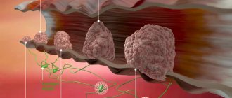
The most common cancer in women is breast cancer (BC). Among the many types of breast cancer, the safest is non-invasive cancer in situ (in situ)
, which is characterized by minimal aggressiveness and does not form metastases.
It is classified as stage zero of breast cancer and is often called “ precancer
.”
Translated from Latin, in situ sounds like “ cancer in place.”
" The size of the tumor remains unchanged because cells die at a rate equal to the rate of cell formation.
Recently, the number of women diagnosed with non-invasive breast cancer has increased significantly. But this situation is explained not by an increase in incidence, but by an increase in diagnosability - more frequent mammography.
What is carcinoma in situ of the cervix?
Cervical carcinoma (cancer) is a malignant neoplasm formed in the lower part of the uterus, that is, in the cervix. Currently it ranks third in detecting cancer in women. The in situ species is distinguished from the group of this disease. This is a type of oncology that has an initial stage. What is another name for carcinoma in situ?
- zero degree;
- latent (hidden) carcinoma;
- First stage;
- possible carcinoma;
- preinvasive cervical cancer;
- intraepithelial cancer.
Cervical in situ develops in the cervical epithelium, without spreading to other tissues. That is, without invasive cancer. It begins its development in the squamous epithelium at the junction with the columnar epithelium. Hence the concept of in situ: it develops in itself (in the epithelium) without affecting all other organs. It is interesting that this type affects women over 40 years old. And the time it takes for it to develop into invasive cancer ranges from a year to 17 years.
Who is at risk of getting this disease?
- those who have been diagnosed with a dangerous type of HPV;
- those who have low immunity;
- those who smoke;
- those who have sexual intercourse before the age of 18;
- those who become pregnant before the age of 16;
- those who have a large number of sex partners;
- those who suffer from sexually transmitted diseases.
The above factors do not inevitably lead to carcinoma of the female cervix, but greatly increase the risk of the disease.
Different types of non-invasive cervical carcinoma
The structure of the epithelial tissue of the cervix is heterogeneous. The glandular layer or columnar epithelium covers the cervical canal and uterus. Multilayered squamous epithelium is located on the outer portion of the cervix.
Cylindrical (single-layer) is an integral part of the cervical canal. The tumor in this part is called carcinoma in situ of the cervix.
Pathology in the area of the squamous epithelium of the cervix is called in situ cervical cancer.
Clear cell carcinoma of the cervix
Adenocarcinoma is more common in older people, is characterized by rapid development and exposes the ovary to metastases. The clear cell variant occurs in 4% of carcinoma cases (a rare variant). Most often, the mesonephroid type of neoplasm manifests itself in postmenopausal patients who have taken the drug diethylstilbestrol (used in the treatment of cancer).
The neoplasm is microscopically comparable to clear cell tumors found in other organs. Macroscopically it can manifest itself in the form of small ulcers to large variants. The cells resemble the shape of shoe nail heads, with large nuclei.
85% of cases of this disease are diagnosed at stages 1-11 of development. In the first stage, the patient has a 90% chance of survival.
Infiltrative carcinoma of the cervix
The preinvasive form, after penetrating into deeper layers, gradually acquires the infiltrating stage of neoplasm development. The decisive factor in this issue is invasion (penetration) into other organ tissues. With invasion up to 3 mm. The cancer is classified as microcarcinoma (microinvasive variant). Next, the malignant tissue penetrates further into the cervix, forming the first stage of the tumor. The following stages develop lymphogenously.
- The patient, having an infiltrative variant of cervical carcinoma, suffers from excessive and unusual discharge (Leucorrhoea). Their presence depends on disorders of the lymphatic system;
- Also, if infiltration is diagnosed, there is bloody discharge from the genitals of an acyclic nature, regardless of the achievement of menopause. At first, such a sign appears at the slightest examination, but later the symptom appears more often;
- A putrid smell of discharge appears, provoked by the presence of a concomitant infection;
- The late stage is manifested by pain, indicating compression of the nerve pathways by a cancerous tumor.
For timely diagnosis, a cervical biopsy, as well as vaginal and rectal examination, are necessary.
Forms of cancer in situ
Preinvasive cervical cancer is usually divided into three forms.
- Squamous cell carcinoma in situ. This configuration is found in the vaginal lobe of the cervix. It originates from stratified squamous epithelium. With this form, keratinization of some cells or an entire group of cells is observed. Free from form, cell atypia and a large number of pathological mitoses are observed.
- Reserve gene. The name refers to the source of cancer development. These are reserve cells. It is in them that it begins to ripen in situ. These cells are located in the endocervix. This is a cancer of heterogeneous structure, which, very often, is difficult to distinguish from squamous cell carcinoma, as it can appear in the squamous epithelium. Still, there are certain distinctive features. The cells have the shape of bundles with elongated nuclei. With this configuration of cancer in situ, mucous manifestations are found in the cytoplasm.
- Glandular carcinoma in situ. Quite a rare form. They are noticed mainly due to adenomatosis. Appears in the prismatic epithelium. The cells of this cancer are round, elongated and often very ugly in appearance.
All these forms have a common feature: preservation of the basement membrane.
Treatment methods
The treatment method for in situ cancer is selected depending on the type, size and location of the tumor, the presence of other diseases and the individual characteristics of the body.
If lobular carcinoma is detected, the pathological area is surgically removed.
When diagnosing hormone receptor-positive breast carcinoma, the doctor selects hormonal medications that block hormones, preventing the process from reoccurring.
For ductal carcinoma, surgical treatment is also recommended. In most cases, radiotherapy is performed in addition to surgery. Targeted treatment is also possible - drugs are used that have a targeted effect on malignant cells without causing harm to healthy ones.
In most cases, organ-preserving operations are performed - lumpectomy or quadrantectomy, during which the affected area is removed.
The need for a total mastectomy (complete removal of the mammary gland) arises if:
- several tumors were identified;
- despite a previous operation, relapse occurs.
- there is a high hereditary predisposition to breast cancer;
- intraductal carcinoma will grow beyond the duct.
After a mastectomy, breast reconstruction is recommended, restoring the shape of the breast and the aesthetic appearance, which relieves the woman of the feeling of inferiority.
Morphological characteristics of cancer in situ
Morphology is a science that studies the shape, appearance, color, structure of a particular organism or neoplasm. How is the morphological characteristic of cervical in situ?
At the first stage, changes are observed in the cells of the epithelial layer. These cells are located incorrectly, have a deformed structure and strive to take the place of healthy cells. Cells lose mitoses, and the nucleus itself changes (especially its shape and size). This altered epithelium is embedded in the glandular layer. But at the in situ stage, it will not pass into the basement membrane.
Histology
The time between the appearance of latent cancer and its detection is difficult to determine. It may not be detected for a long time, and it, in turn, may not develop. Cancer in situ has the ability to disappear on its own. How to detect it in time?
Factors in the development of the disease
Viral etiology plays a major role as the cause of atypical processes in the cervical canal area. The main destructive factor is called 34 possible types of HPV (human papillomavirus).
The high and medium risk zone includes the HPV stamp 16, 18, 31,32,35,39,48, 51, 58. These types of pathogens infect stem cells at the junction of different epithelial layers. The work of the cell is disrupted after failures in the genetic code, which is a prerequisite for oncological tumors many times over.
The following factors also influence directly and indirectly:
- Inappropriate pH level, which is regulated by the activity of lactobacilli, affects the functioning of epidermal cells;
- Sexually transmitted diseases cause various inflammatory processes. The endometrial system of the cervix suffers. Such phenomena increase cell atypia (deviation from the norm), facilitate the process of degeneration of a functional cell into a malignant one;
- Various injuries that occur during medical operations can cause pathological formations;
- Inappropriate levels of hormones can lead to negative changes in the process of cell division; hormonal levels can also increase tissue inflammation.
Indirect factors influencing the transition of dysplasia to the stage of in situ carcinoma:
- Early experience of sexual activity;
- Hereditary predisposition;
- Frequent change of sexual partner;
- Violation of the body's protective functions;
- Bad habits (smoking, alcohol);
- Systematic use of contraceptive pharmaceuticals.
It is difficult to say the exact cause of the development of the disease due to the frequent absence of symptoms of the pathology and accidental detection during examination.
Carcinoma in situ of the cervix: treatment
Before making a diagnosis, invasive studies are necessary.
Marker in the treatment of cervical carcinoma. Tumor marker analysis is used to study predisposition to tumors and identify the most effective methods for treatment. A tumor marker appears in the form of a substance that is produced by pathological neoplasms. Specific marker molecules are secreted by cancer cells. The use of such means makes it possible to establish the presence of the disease even in the early stages of penetration. Elevated levels of substances indicate the dynamics of tumor development. The marker can be determined in a patient’s blood test.
The marker is used in the following cases:
- Determination of the source of the tumor.
- Monitoring the patient's treatment.
- Dynamics of the disease.
- Carcinoma in situ of the cervix relapse.
When researching, you should not rely on only one marker value. In some women, the cancer marker is always present in the body in quantities.
Cervical carcinoma is cured using various methods. In cases of age under 50 years, high amputation of the cervix using the Sturmdorff method is practiced. The removed tissue is taken as a preparation for histological studies. If traces of tumor invasion are detected in a series of sections, an extirpation operation is performed using the Wertheim treatment method. Radiation therapy is also prescribed. If the patient's age exceeds 50 years, the doctor performs extirpation of the appendages. The woman's uterus is also removed without the use of radiation therapy.
If carcinoma is detected in a young patient or in a pregnant girl, a conservative treatment method is started.
If hormonal imbalance is indicated as the main factor, hormonal therapy is prescribed. The course of treatment lasts 3-6 months, based on individual dynamics.
In the case of papilloma virus, cytological, chemical, and immunorestorative drugs are prescribed.
The diagnosis of carcinoma at the in situ stage can be successfully treated in 90% of cases, guaranteeing a prognosis without the appearance of relapses.
The highest degree of spread of metastases, if the uterine variant of cancer is taken into account, occurs through the circumuterine and iliac lymph nodes. The external lymph node is also involved. If a cancer patient begins to be treated for metastases, the case becomes severe. If you have any doubts, consult different doctors. Remember that the disease can progress quickly, so the reaction of doctors and the patient must be lightning fast. Women regularly need to undergo a medical examination by a gynecologist.
Video: cancer in situ of the cervix
We can cure cancer in situ
Diagnostics
During the initial examination, pre-invasive cervical cancer is almost impossible to detect. Additional diagnostic methods are required.
- A cytology test is performed on all women who come with any complaints or changes. With its help, cancer processes are successfully detected. This synthesis shows the slightest changes in cells.
- Colposcopy. Examination of the cervix using a special apparatus. The doctor, as if through a microscope, examines the cervix and determines whether there are any changes in it.
- Biopsy. A piece of the altered epithelium is taken to examine it for histology. This test shows whether cancer is actually developing and, if so, by how much.
- HPV. Analysis for the presence of pathogenic strains of human papillomavirus. It leads to cancer.
- Tumor markers are special substances that are detected in a woman’s blood. They indicate exactly whether there is oncology in the body. This also applies to carcinoma in situ.
- Other diagnostic methods. Additionally, a woman may be prescribed an MRI, ultrasound, x-ray, and additional blood and urine tests.
Causes of pathology
The exact causes of non-invasive cancer are unknown.
It is believed that they increase the risk of developing cancer:
- hereditary predisposition, the presence of genes for predisposition to breast cancer - BRCA1
,
BRCA2
and
CHEK2
; - hormonal disorders;
- ionizing radiation;
- smoking;
- prolonged contact with carcinogenic substances;
- long-term (more than 5 years) hormone therapy;
- taking oral contraceptives;
- age over 50 years;
- late pregnancy (after 30 years);
- late menopause (after 55 years)
- having many children and being childless;
Treatment
After successful diagnosis and diagnosis, treatment begins. Carcinoma in situ is a little easier to treat than other stages of cancer. How is she treated?
If the woman is under 50 years of age, the affected area of the cervix is removed. This prevents the development of cancer in the future. This process is called conization. Up to 1 cm of the cervix is removed. The patient is warned about all the consequences of the operation, after which she consents to the procedure.
If the patient is over 50 years old, then the uterus and appendages are completely removed without the use of radiation.
But what if carcinoma in situ was found in a young woman, or even a pregnant woman? In this case, conservative treatment methods are used. Determine the cause of this phenomenon:
- Hormonal disbalance;
- HPV virus.
Treatment is aimed at eliminating the cause.
In the first case, hormonal therapy is prescribed using an OK regimen. The medications are prescribed for 3-6 months, based on individual characteristics.
If the cause in situ is HPV, the following drugs are prescribed:
- Cytological: Podophyllin, Condilin.
- Chemical: Feresol, Solcoderm.
- Preparations for restoring immunity: Interferon, Alfaferon, Cycloferon.
After eliminating the cause, the effect (latent carcinoma) goes away.
Conization of the cervix in carcinoma, consequences after surgery
Conization is the process of an advanced method of studying tissues by intravital sampling of cells or tissues from the body. This form of research is also called a “conization biopsy” because tissue is taken from the cervix in a cone-shaped area. The doctor removes tissue from the cervix in the area of the cervical canal with a small part of normal cells.
Using this research method, the treating doctor is able to obtain the required area of tissue for analysis for carcinoma. Diagnostics also often uses the method.
Attention! Conization allows for the physical removal of all abnormal cells from the cervix. If the operation is successful, only a smear of the cervix is required for cytological analysis (examination of the structure of cellular elements).
Modern implementation of the method is carried out using:
- Carbon dioxide laser;
- Electrosurgical loop excision;
- Classic scalpel.
Canonization takes place under regional or local anesthesia. During the operation, the doctor uses vaginal speculum to visually access the cervix. After preparation, the process of conization of the tissue occurs.
After the operation, the woman is in the recovery room under the supervision of specialists. If no deviations are observed, the patient is free after 3-4 hours.
After conization and complete recovery, the necessary functions of the cervix are fully restored.
Evidence of the negative impact of removing part of the cervix has also been documented. If part of the reproductive organ is completely removed, there is a risk of miscarriage in the later months of pregnancy.
Scar marks on the cervical canal can lead to secondary and primary infertility. This will serve as an additional obstacle for sperm cells.
During gestation at 16-18 weeks, the development of isthmic-cervical insufficiency is possible, when the cervix can open under the weight of the child.
Prognosis for recovery
If you have been diagnosed with this, don’t panic! Remember, in situ stage cervical carcinoma is treatable. In 90% of all cases, this type of cancer is detected in the early stages and is completely cured without subsequent relapse. This figure is significantly underestimated if a woman delays receiving professional treatment or does not consent to surgery. Remember the main thing:
- visit your gynecologist regularly;
- avoid bad habits;
- be selective in choosing a sexual partner.
Clinical manifestations
In the initial stages, breast carcinoma does not reveal itself in any way.
Later symptoms of ductal carcinoma include:
- soreness in the mammary glands;
- puffiness and swelling;
- discharge mixed with blood.
Lobular carcinoma is usually asymptomatic and in advanced stages. Therefore, it is discovered by chance when a diagnosis is made for another disease.