The myometrium is an important constituent layer of the reproductive organ. Its task is to ensure normal distensibility of the uterus during pregnancy and the required contractile activity during and after childbirth. The condition of the myometrium reflects the disturbed hormonal sphere of a woman - if there is an imbalance in the production of hormones, diffusely heterogeneous structures, foci of endometriosis, fibroids, cysts and malignant tumors may appear in the muscle layer. It should be noted that diffuse changes in the myometrium are often visualized by gynecologists on ultrasound in women who have achieved their reproductive function. Such changes are caused by the vascular component and are normal.
The myometrium changes its condition throughout a woman’s life, which can be seen during an ultrasound examination by its heterogeneous structure. In young nulliparous women, the state of diffuse changes in the myometrium is not determined. However, this is precisely the conclusion that women often hear in the ultrasound room.
Causes of diffuse changes
A diffusely heterogeneous structure of the myometrial layer appears for a number of reasons:
- endometriosis or adenomyosis;
- blood diseases, including anemia;
- exposure to stress and regular emotional shocks;
- caesarean section or other operations performed on the uterine cavity;
- abortions, curettage or mechanical cleanings;
- hormonal disorders;
- infectious and inflammatory processes.
It is possible to determine what the true cause of diffuse changes in the myometrium is only after a comprehensive examination. An ultrasound examination alone will not be enough. Childbirth also becomes an indirect cause of the formation of structural changes and heterogeneous foci, however, this natural process does not significantly affect the condition of the muscular layer of the reproductive organ. A woman will have to give birth at least 2 times for diffuse deformation of the muscle layer to begin.
There are also physiological reasons for diffuse changes in the structure of the middle muscular layer - the uterine myometrium. They do not cause persistent pathology. Natural causes provoke the appearance of a temporary heterogeneous structure. These include cyclical changes in hormonal levels, pregnancy and the postpartum period. Diffuse changes and distortion of the structure of the uterine layers are diagnosed during reproductive age and menopause. The pathological process can remain unattended for a long time. The absence of complaints does not guarantee a woman that diffuse disorders do not occur in the uterus.
How to treat
When myometrial heterogeneity is treated, the treating doctor must select an individual course based on the patient’s characteristics: general health, degree of uterine damage, age, as well as past previous diseases.
There are three main ways to treat this disease:
- Drug treatment;
- Surgical intervention during which the uterus is preserved;
- Removal of the uterus.
Drug treatment involves taking a certain type of drug that reduces the growth of endometrial cell mass. In order to eliminate pain, a specialist can prescribe tablets with antispasmodic and analgesic spectrums of action, and to treat the disease itself, hormonal medications are prescribed.
When the drug treatment method does not give the desired effect, then it is worth resorting to a method such as hysteroscopy, which involves curettage of the uterus. Sometimes after this, relapses occur, and the meometrium again becomes heterogeneous.
Cauterization of the uterine walls is a subsequent method of aligning the myometrium. The way the doctor straightens the myometrium is called coagulation. The chances of a relapse are extremely low; moreover, this process is completely reliable and harmless.
In the event that this disease is advanced and the heterogeneous myometrium provokes a host of other diseases, complete removal of the uterus is possible. After such an operation, the patient will have a chance to continue her normal life without problems with women’s health.
It is worth remembering that the absence of abortions, regular consultations with a gynecologist, as well as timely prevention of infection - all this is the prevention of this disease.
Signs
There are many known signs of structural and diffuse changes in the uterine myometrium, however, all of them are indirect. Knowing about the complaints, the gynecologist cannot make a reliable diagnosis - heterogeneous structure of the myometrium. The doctor only has assumptions. Additional diagnostics will confirm your suspicions. The reason to consult a doctor in women with structural and diffuse changes in the myometrium is:
- large blood loss during menstruation;
- painful monthly uterine contractions;
- discomfort during urination;
- discomfort during sexual intercourse;
- painful ovulation;
- brown spotting in the middle or second half of the cycle;
- long absence of pregnancy, primary or secondary infertility.
Most often, diffuse heterogeneous changes in the myometrium are asymptomatic for women.
Diagnostics
It is possible to determine that the myometrium lining the median wall of the uterine cavity is diffusely heterogeneous or has an altered structure using ultrasound scanning. During the procedure, the sonologist evaluates the echogenicity, thickness and homogeneity of this area. If there are any non-compliances with the standards, they are described in detail in the ultrasound report. It should be noted that there are no standards for structural changes. If diffuse distortions are detected, then this already indicates pathology.
To differentiate the diagnosis, the gynecologist may prescribe a series of tests, including blood tests for tumor markers and sex hormones.
If diffuse heterogeneous changes are accompanied by pathological signs in the endometrium, diagnostic curettage is prescribed, which itself becomes a provocateur of the formation of heterogeneous myometrium and its diffuse changes with structural disturbances. If the doctor recommends starting the examination with this manipulation if the endometrium is normal and excludes other methods, it makes sense to consult another specialist.
A clearer picture of what is happening in the uterine cavity is given by endoscopic examination: hysteroscopy or laparoscopy. The procedure is prescribed to patients with a questionable ultrasound result and can go from diagnostic to therapeutic.
Endometriosis
The finding of diffusely uneven myometrial structure suggests that this condition is caused by uterine endometriosis. The disease is characterized by heterogeneous growth of the mucous layer of the reproductive organ and its penetration into the muscular layer. During an ultrasound examination, the sonologist may discover that there are peculiar cellular zones - foci - in the myometrium. Symptoms of the pathology include irregular menstruation and constant pain in the pelvic area.
Often, a woman turns to a gynecologist with a complaint about the lack of pregnancy, and during diagnosis she is diagnosed with endometriosis of the uterus with structural changes in the muscle layer. The disease has three forms:
- genital (uterus, ovaries, fallopian tubes and peritoneum are affected);
- extragenital (adjacent organs are affected);
- mixed (combines the previous two).
If diffusely heterogeneous myometrium is detected, it should be understood that this condition cannot be eliminated in one day. To normalize the function of the muscle layer, the cause of the structural changes must be eliminated.
Endometriosis is corrected with hormonal drugs and minimally invasive interventions. It is also necessary to try to find the exact cause of the development of uterine endometriosis, which is not always possible.
Why does this happen?
The main causes of heterogeneous myometrium:
- Endometriosis and its internal genital type, also called uterine adenomyosis.
- Myometritis. In the vast majority of cases, it is a consequence of complicated endometritis, so in fact we are talking about endomyometritis.
- Uterine fibroids. It can lead to local nodular changes or almost total thickening of the myometrium (in the diffuse form of the disease).
Each of these diseases not only has special symptoms, but also leads to characteristic changes in the myometrium. They can be differentiated using ultrasound; in the protocol of the study, the specialist describes the echographic picture of the detected pathology and indicates its type.
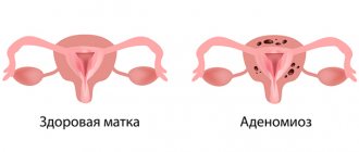
Adenomyosis is the most common cause of myometrial changes
Endometriosis is the pathological growth of endometrial cells outside the lining of the uterus. When the myometrium is predominantly affected, we speak of adenomyosis, which can be of diffuse and nodular type, and of varying degrees of severity.
This disease is classified as benign hyperplasia of a hormonal-dependent nature. So it is typical for women of reproductive age, and in the postmenopausal period and against the background of adequately selected hormonal therapy, the activity of the process subsides.
With adenomyosis, growths of endometrioid tissue appear in the myometrium. They can be of two types:
- In the form of blind branched deepening pockets communicating with the endometrial layer of the uterus. In such cases, the ultrasound report usually indicates that the echostructure of the myometrium is heterogeneous and cellular. In severe degrees of the disease, germination of the entire thickness of the myometrium is noted, which is accompanied by the formation of fistula-like formations between the uterine cavity and other structures of the small pelvis.
- In the form of nodes - closed round lesions with an uneven central cavity filled with blood or a chocolate-colored liquid mass. They are usually multiple, of varying sizes, with an uneven distribution in the uterine wall. With this variant of the disease, the conclusion of an echographic examination usually notes that the myometrium is heterogeneous with signs of adenomyosis.
Any endometriotic formations undergo repeated changes in accordance with a woman’s ovarian-menstrual cycle and lead to an inflammatory process. Under the influence of sex hormones, cells of the abnormally located endometrium grow and are rejected in the same way as in the uterine mucosa. This leads to the appearance of clinical symptoms of the disease.
Adenomyosis is characterized by cyclic uterine bleeding, which is more profuse and painful compared to normal menstruation. And the emptying of stagnant cavities and endometrioid pockets leads to the appearance of chocolate-colored discharge from the genital tract. They are actually menstrual blood that has accumulated and undergone incomplete decomposition.
Adenomyosis
Diffuse changes in the myometrium can be caused by adenomyosis. With this disease, the structure of the muscle tissue becomes heterogeneous, and foci of the endometrium are identified in it.
The main difference between adenomyosis and endometriosis is the localization of germination of the mucous layer. With endometriosis, the lesions are more extensive. Adenomyosis is characterized by ingrowth of the endometrium only into the myometrium and is manifested by diffuse changes in its structure.
Often adenomyosis occurs without obvious signs. With the same degree of probability, a woman may be bothered by abdominal pain and discomfort during palpation. The causes of adenomyosis include trauma to the uterine mucosa and muscle layer. Damage can occur during diagnostic curettage, abortion, or during inflammation.
Treatment of adenomyosis begins with drug correction. The patient is prescribed hormonal drugs that regulate the functioning of the ovaries, anti-inflammatory and painkillers. It is often necessary to introduce an artificial hormonal menopause into a woman. If this technique is ineffective, surgical treatment is performed, which involves removing the reproductive organ. The appointment of surgical intervention is prescribed to patients with mandatory consideration of age and desire for subsequent birth of children.
Treatment
The disease is cured strictly under the supervision of a doctor. Mostly, hormonal drugs are prescribed. Diagnostic curettage is used to determine the nature of the pathogen and the degree of changes. In some cases, antibiotics are also prescribed.
Antibiotics help defeat the inflammatory process, if present. Hormone therapy normalizes hormonal balance. Sometimes it is also necessary to cauterize the altered diffuse area of the myometrium. The disease is not severe; the prognosis is usually favorable.
Anemia
Changes in the structure of the myometrium and the formation of heterogeneous foci can be caused by anemia. Lack of oxygen impedes blood supply to organs. As a result, the nutrition of the uterine layers is disrupted, and it becomes heterogeneous. Often, getting rid of anemia does not restore proper functioning of the uterus. In this case, they talk about mixed pathology, which is accompanied by several causes. Anemia is treated with medications that normalize blood counts, as well as diet and a healthy lifestyle. Doctors use iron supplements and B vitamins in treatment, as well as combination products that include several microelements that normalize the structure of the myometrium.
Consequences and complications
Timely detection and treatment of the heterogeneous structure of the myometrium or foci of diffuse changes has a favorable prognosis. Women of reproductive age, with a competent approach, can quite easily get rid of the existing problem. If you do not look for the causes of diffuse changes, then the formation of a heterogeneous structure of the muscle layer will continue. The result of this may be:
- infertility;
- progression of fibroid growth;
- severe adenomyosis;
- malignant neoplasms of the muscle layer;
- lack of sex life (due to pain);
- menstrual irregularities;
- bleeding leading to fainting and other problems.
It will not be possible to independently detect the diffusely heterogeneous structure of the myometrium. A woman may suspect the presence of a problem based on her symptoms. If you are concerned, you should definitely get examined, since adenomyosis and endometriosis are progressive pathologies. The more extensive the damage to the myometrial structure, the more difficult it will be to get rid of the problem.
Myometrial structure
The muscular wall of the uterus has a complex structure. It consists of several layers:
- Circular (vascular). Consists of rings of pipes. There are many vessels included. The strongest layer of the uterus.
- Longitudinal (subserosal). Includes longitudinal and circular muscle fibers.
- Submucosal. Consists of longitudinal homogeneous fibers. The most fragile of all layers.
This structure helps the uterus to contract well during childbirth and push out fluid during menstruation. The frequency of contractions is regulated by female sex hormones - oxytocin and estrogen. The structure of the muscular uterine wall is examined with ultrasound, but if this does not show a clear picture of the state of the uterine cavity, then the method of hysteroscopy can be used.
Articles on the topic
- Omnik - instructions and mechanism of action, contraindications, side effects, dosage regimen and analogues
- Warfarin - instructions for use, dosage, active substance and contraindications
- Routes of transmission of hepatitis: symptoms and types of diseases