Anatomical and physiological features of the organ
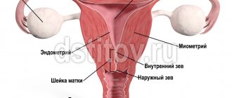
The uterine, or fallopian, tubes are considered a hollow and somewhat elongated paired organ, which is an important part of the female reproductive system. They are located almost horizontally and start from the fundus of the uterus on both sides.
The main functions of the fallopian tubes:
- transportation of eggs and sperm;
- creating favorable conditions for fertilization, development of the zygote and its movement into the uterine cavity.
Normally, each tube begins with the uterine (or interstitial) section, which is enclosed in the thickness of the myometrium and has a length of about 15-30 mm. Outside the uterus, the organ passes into the isthmus (isthmic section) - a thin horizontal canal in the leaves of the broad ligament, having a length of 27-40 mm. The isthmus is followed by the ampulla. It occupies about 50% of the entire length of the pipe and ends in a kind of funnel, on which there are narrow and long villi - fimbriae. The latter capture the egg released into the pelvic cavity as a result of ovulation and transfer it to the tube.
In women of reproductive age, the average length of the fallopian tube ranges from 100-120 mm, and its thickness is 5 mm. At the same time, according to statistics, the left tube is slightly shorter than the right one.
Causes of disorders in the reproductive system
The main reasons that can provoke genital dysfunction include:
- termination of ectopic pregnancy
- development of inflammatory processes in the appendages: salpingoophoritis, adnexitis, oophoritis
- progression of endometritis - an inflammatory process that affects the inner lining of the uterus and occurs as a complication of abortion, curettage
- formation of adhesions caused by surgical interventions, peritonitis
- abnormal development of internal genital organs (bicornuate uterus)
To confirm the diagnosis, a comprehensive examination by a gynecologist and a number of diagnostic procedures are required: both instrumental (ultrasound) and laboratory. In order to fully examine the uterus and its appendages, the administration of contract drugs is required. The procedure is carried out only in case of objective indications; this is not a routine study recommended for all categories of women.
Bicornuate, vestigial uterus
A bicornuate uterus is a pathology of organ development, which is accompanied by the formation of two separate parts with cavities. In the lower part of the uterus, such “horns” unite, and a common canal opens into the vaginal area. Such a disorder occurs as a result of hereditary predisposition and exposure to external provoking factors. These can be toxic substances, viral infections, heavy metals, pathogenic microorganisms of bacterial origin.
There are no specific symptoms for this disorder. In some cases, menstrual irregularities, uterine bleeding, miscarriage, and difficulty conceiving may occur. In most cases, a bicornuate uterus is discovered by chance, during a routine examination by a gynecologist. In most cases, specific therapy is not required. Surgical correction is performed only if there are difficulties with conception.
Oophoritis
Oophoritis is an inflammatory process that affects the ovaries. Develops under the influence of infectious and inflammatory processes: endometritis, vulvovaginitis, adnexitis, sexually transmitted infections. Among the predisposing factors that can provoke pathology are impaired functioning of the immune system, regular hypothermia, a history of multiple abortions, surgical interventions, the use of an intrauterine device, and disruption of the natural microflora.
Why and how to do an ultrasound of the fallopian tubes
However, with a transvaginal examination, in a small percentage of patients, doctors are able to remove the organ against the background of peritoneal fluid that has leaked during ovulation.
Thus, if the study protocol states that the fallopian tubes are not detected on ultrasound, this means that they were not subject to pathological changes. Indications for echohysterosalpingography (EchoGSS), or hydrosonography (ultrasound examination of the uterine appendages):
- menstrual irregularities (irregularity, painful menstruation, changes in their duration);
- frequent inflammatory processes of the internal genital organs;
- suspected infertility (unsuccessful attempts to conceive a child within 12 months);
- pain in the lower lateral abdomen, suprapubic region;
- a history of sexually transmitted diseases;
- preparation for in vitro (artificial) fertilization.
If a transabdominal ultrasound , then the woman should exclude foods that increase gas formation in the intestines 2-3 before the procedure. On the day of the study, 1-1.5 hours before the procedure, you need to drink 800-1000 ml of liquid to fill the bladder. With the transvaginal technique, the bladder must be empty, that is, you need to urinate before the examination.
How is hydrosonography performed:
- The woman takes a comfortable position in a special gynecological chair.
- The doctor inserts a thin catheter into the uterine cavity through the cervix. After which, a sterile saline solution at a temperature of 37 degrees (or Echovist) is supplied to the organ.
- Under ultrasound control, the doctor monitors the process of filling the uterus and fallopian tubes with the solution.
- At the end of the study, the sensor is removed from the vagina, and the catheter is removed from the uterus.
With a regular scan, without the use of a contrast agent, this organ is not described in any way - this means that the fallopian tubes are not visualized. According to official statistics, complete or partial organ obstruction occurs in 42-80% of infertile women.
Advantages of undergoing sonohysterography at CELT
The multidisciplinary clinic CELT invites you to undergo the procedure effectively and obtain all the necessary data. Having been working in the market of paid medical services for more than a quarter of a century, we have everything necessary to carry out accurate diagnostics and effective treatment.
By contacting us, patients can count on comfortable conditions and the procedure being carried out in accordance with international standards. We employ experienced ultrasound specialists, doctors of the highest category, doctors of science. They have at their disposal innovative ultrasound devices produced by Electric Medical System (USA) and Philips (Holland).
They are equipped with high-resolution monitors providing high quality and clarity of images. The procedure can be carried out in various modes and allows you to identify any anomalies and pathologies at different stages of their development.
The price of ultrasound examination of fallopian tube patency depends on what access is used, contrast enhancement and anesthesia. You can view our prices by going to the “Services and Prices” tab in this section of our website. You can eliminate any misunderstandings and find out the exact numbers by contacting our operators: +7 (495) 788-33-88.
Why fallopian tubes are not visualized on ultrasound
Normally, these structures are so thin and small that the resolution of most devices simply does not allow them to be examined using ultrasound. In addition, the pipes constantly make floating movements.
With the transvaginal technique , provided that a sufficient amount of fluid accumulates in the pelvis, it is sometimes possible to “catch” this organ. Normal pipes have a uniform structure; the muscle layer and lumen are not determined. In some cases, the doctor may see the ampullary segment with fimbriae.
That is why, to assess the condition of the “appendages” of the uterus, hydrotubation, or hysterosalpingoscopy, is performed.
Visualization
Ultrasound examination allows you to quickly and accurately detect pathological changes in the reproductive system and determine the size and structure of the ovaries.
Sometimes a medical professional will tell a patient that her ovary cannot be visualized. To find out why the gonad is not visible on the monitor, you need to undergo additional research. But most often, the lack of visualization is a consequence of menopause, hormonal disorders, and pathologies of the pelvic organs. The ovaries may not be visible on the ultrasound machine monitor if:
- undergone surgery in the pelvic area;
- congenital absence of gonads;
- the onset of menopause, accompanied by drying out of the ovaries;
- flatulence and intestinal fullness with feces;
- displaced location of the pelvic organs as a result of congenital or previous pathologies;
- serious diseases of the uterus, accompanied by proliferation of uterine tissue.
The reason for the invisibility of the left ovary on the monitor is most often the empty state of the bladder. Many patients do not understand why it is necessary to come to an ultrasound with a full bladder. As a result, the doctor’s recommendation is ignored, which makes the study ineffective.
You need to understand that the structure of the organs of the pelvic region is such that the left ovary can be closed by intestinal loops. There may be gases and feces in the intestines that reflect ultrasonic waves; as a result, the ultrasound simply does not reach the reproductive gland. The fluid-filled bladder inflates, pushes aside the intestinal loops, and becomes a lumen through which ultrasonic vibrations pass.
The reasons for the lack of visualization of the right gonad may be different. The right ovary may not be visualized if it is covered by uterine tissue or areas of the pelvic bones.
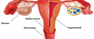
One of the most important organs in a woman's reproductive system is the fallopian tubes. It is with their help that you can count on increased chances of conceiving a baby and successfully consolidating the embryo.
Normally, the fallopian tubes are not visualized during an ultrasound examination. This means that they are not visible. They can be seen on an ultrasound if there is an accumulation of fluid in the lumen of the fallopian tubes. And this is a sign of an inflammatory process, that is, pathology.
In addition to inflammatory processes in the fallopian tubes, an adhesive process can also develop. In this case, if the lumen of the tube is completely closed, a sactosalpinx and hydrosalpinx are formed - a cavity filled with liquid. The nature of the fluid can be determined through further examination, and an ultrasound will determine the presence of an inflammatory process and its location (right and/or left tube).
When there is cause for concern
Due to the progression of the inflammatory process, activation of adhesive processes in the area of the fallopian tubes may be observed. This is accompanied by nagging pain, menstrual irregularities, and difficulties in conceiving a child. For any of the described or any other symptoms, it is recommended to refrain from self-medication and seek advice from an experienced gynecologist. The sooner specialists identify the cause of the disorder and direct efforts to eliminate it, the more favorable the prognosis for the woman.
How much does hysterosalpingoscopy cost?
An ultrasound scan of the fallopian tubes is prescribed to check the health of the female reproductive system. During the examination, the doctor uses a contrast liquid with ultrasound.
The price of a fallopian tube ultrasound is influenced by the region in which the woman lives. The cost of the examination ranges from 1500–5000 rubles. In some paid clinics in the Moscow region, the price reaches 10,000 rubles.
Have you undergone such a procedure? Tell us about it in the comments. Share the article with your friends on social networks. Be healthy.
Decoding the results
If the woman is healthy and there are no pathologies in the pelvic area, then the injected contrast liquid should completely remain in the recess of the abdominal cavity (more precisely, between the rectum and the uterus). If there is no fluid there, but it is found in the fallopian tubes and the uterus itself, it means that the patency of the tubular organs is seriously impaired.
Increased diameter
One of the possible pathologies detected during ultrasound of the fallopian tubes is their expansion (scientifically called hydrosalpinx).
Important! Often the disease is a consequence of fluid accumulation, the development of inflammation or impaired blood circulation.
What causes the fallopian tubes to become dilated? The reason may be:
- Hydrosalpinx, both simple and follicular. The first leads to an enlargement of the pipe in only one cavity and occurs in a mild form, but is dangerous with a high probability of complications. Therefore, it is so important to identify the disease in time and completely treat it.
- Salpingitis is an inflammation that occurs in the tubular organs of the uterus.
- Enlargement of the isthmus of the tube.
Ultrasound hysterosalpingoscopy is a fairly informative method that allows you to assess the condition of the uterine cavity and fallopian tubes. At the very beginning of the procedure, when filling with saline, the rate of its entry into the uterine cavity is assessed. At this stage, the presence of adhesions inside the uterus is visible, and polyps and myomatous nodes can also be visualized.
If there is obstruction of the fallopian tubes, then the solution injected through the catheter will not enter the abdominal cavity, but will accumulate. And then the specialist will be able to definitely say at which segment there is an obstacle to the movement of liquid.
It should be remembered that there are smooth muscle cells in the wall of the fallopian tubes, therefore, in response to the introduction of the solution, a spasm may occur, and then the flow of the substance into the abdominal cavity will stop. In this case, you may get an unreliable result.
Standard ultrasound scanning is not able to identify and localize the adhesive process, since during such an examination the lumen of the fallopian tubes, as a rule, is not visualized. As an alternative and informative diagnostic method, in which the woman does not experience pain, 4D ultrasound can be recommended.
Norm and decoding
During the manipulation, the uterus and fallopian tubes are examined, and ultrasound examination determines their patency. If the saline solution does not encounter any obstacles on its way, then gradually the entire volume of the injected substance accumulates in the vaginal-rectal pocket. This situation is perfectly visualized on the screen of an ultrasonic unit.
During the process of filling with saline solution, the structure and condition of the endometrium, the presence of pathological volumetric processes and uterine anomalies can be assessed.
Violation or slowdown in the filling of the uterus and fallopian tubes suggests the presence of any obstacles. During the manipulation, the specialist clearly determines the localization of the problem, which subsequently determines the management tactics for each woman.
Sometimes pregnancy occurs quickly after the procedure. The fact is that the passage of saline solution through the pipes cleanses their mucous membrane and destroys small adhesions in the lumen of the organs. And the liquid irritates the villi of the fallopian tubes, stimulating them to actively capture mature eggs.
Hysterosalpingoscopy shows any enlargement of the fallopian tubes that is interfering with the natural process of conception.
Fallopian tubes are not visualized on conventional pelvic ultrasound. Therefore, ultrasound hysterosalpingography uses a safe pigment that is injected through a thin catheter.
As a result, the sonologist can see hydrosalpinx, an accumulation of transudate on ultrasound with polyps and myomatous nodes. The listed pathologies are diagnosed if the solution from the catheter does not penetrate into the abdominal cavity, but accumulates. This is how the length of obstruction in the fallopian tubes is determined.
Normally, the saline solution from the catheter passes through the fallopian tubes. In some cases, after ultrasound hysterosalpingoscopy, a long-awaited pregnancy occurs.
This happens due to the fact that saline solution cleanses the mucous membrane of the fallopian tubes and irritates their villi. The latter, in turn, actively capture the mature egg and push it into the uterine cavity.
Indications and contraindications
Usually, if there is a suspicion of obstruction in the fallopian tubes, the woman is sent for an ultrasound. Other indications for the procedure include:
- Previously diagnosed infertility. Periodic ultrasonography is prescribed to monitor the condition of a woman’s reproductive system.
- An inflammatory process in the uterus and appendages, which began due to infection. Usually the disease becomes chronic.
- Frequent pain in the lower abdomen. The gynecologist directs the woman to check the pelvic organs with an ultrasound to rule out diseases associated with this part of the body.
- Irregularity of menstruation up to its complete absence, amenorrhea.
- Previous sexually transmitted infections.
We invite you to read: A child chooses a year
The main cause of fallopian tube obstruction is considered to be adhesions. Blockage of the tubes occurs due to long-term inflammatory diseases in the pelvic organs, previous abortions and other surgical interventions.
Contraindications for the procedure are:
- pregnancy that develops normally, without complications;
- presence of infections;
- bleeding in the uterus, no matter what etiology;
- exacerbation of chronic diseases;
- cervical diseases;
- development of the inflammatory process in the uterine cavity.
Ultrasound of the fallopian tubes is optimally done between the 5th and 20th day of the menstrual cycle. A more informative result is obtained if the study is carried out before the onset of ovulation, approximately 9–11 days after menstruation.
It is during this period that the cervix dilates to its maximum, which eliminates the appearance of cramps during an ultrasound. The endometrium becomes thinner after the next menstruation. This facilitates the research and guarantees the accuracy of the results obtained.