Proliferation refers to the rapid growth of cells or microorganisms. In multicellular organisms, it occurs on the basis of accelerated division of cells involved in the structure of the tissue, for example, in the healing phase of damage or inflammation. Cell hyperplasia is controlled by a number of factors.
The most important of these are hormones and growth factors, such as macrophage colony-stimulating factor, which influence cell metabolism.
The proliferation of glandular epithelium in gynecology is the quantitative growth of glandular structures in the epithelial layer of the cervix.
Since the structure of the uterine mucosa is heterogeneous, its function is also ambiguous.
There are 4 uterine cycles, during which the endometrium changes:
- The desquamation phase is manifested by menstruation.
- The regeneration phase involves the restoration of the functional layer due to the proliferation of the epithelium.
- In the proliferation phase, the glands of the mucous membrane and stroma grow. The layer becomes moderately wide, long glandular tubes directed towards the surface are visible.
- Secretion phase. The endometrial layer increases significantly and forms 2 separate layers.
Kinds
Proliferation in gynecology is a condition in which the proliferation of surface epithelial cells of varying degrees of severity occurs. Doctors classify endometrial hyperplasia based on the types of cellular changes in the mucous membrane.
WHO approved hyperplasia classification table.
| Endometrial hyperplasia | |||
| Proliferation of glands without cytological atypia | Proliferation of glands with signs of cytological atypia | ||
| Simple | Complex (adenomatous) | Simple | Complex (adenomatous) |
| Adenocarcinoma | |||
As hyperplastic processes of the endometrium develop, it is customary to distinguish:
- glandular hyperplasia, when there is an increase in the number of glandular cells;
- development of glandular-cystic cells, supplemented by cystic formations;
- atypical hyperplasia, which is a precursor to oncological pathology.
Proliferation without cytological atypia
Benign endometrial hyperplasia is a condition that occurs due to abnormally enlarged growth of the endometrial glands, resulting in an uncharacteristic thickening of the uterine lining.
Proliferation in gynecology
This usually occurs due to prolonged exposure to the hormone estrogen, which is not balanced by the hormone progesterone. Benign endometrial hyperplasia may cause symptoms such as menstrual irregularities and abnormal vaginal bleeding or discharge.
The adenomatous condition is characterized by a close arrangement of glandular cells throughout the entire organ or individual areas. The proliferation of glandular tissue is often accompanied by cysts.
This condition may improve without treatment. In some cases, hormone therapy helps.
Atypical hyperplasia
This precancerous condition is caused by excessive growth of abnormal cells. The atypical form of the endometrium develops from benign hyperplasia as a result of prolonged exposure to the hormone estrogen. Characteristic signs: abnormal vaginal bleeding, the presence of polyps in the endometrium.
Most atypical cases occur in postmenopausal women. Risk factors include a positive family history of the disease, obesity, polycystic ovary syndrome, and treatment for breast cancer with the drug tamoxifen.
The adenomatous condition portends a high risk of endometrial cancer (sometimes up to 45 times). Treatment options may include hormonal medications, endometrial ablation and, in some cases, hysterectomy. The prognosis depends on the underlying cause and the body's response to treatment.
Adenocarcinoma
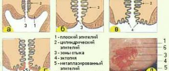
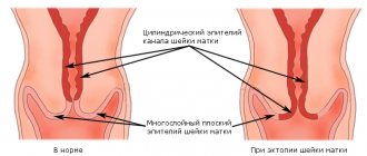
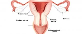
A woman's reproductive system includes the uterus, cervix, vulva, ovaries, vagina and fallopian tubes. The cervix has a cylindrical shape, its outer surface opens into the vagina, and the inner, cervical canal, faces the uterine cavity. The cervix is covered by 2 types of cells: glandular cells inside the cervical canal and squamous cells on the outer layer.
The glandular ones produce mucus and develop inside the uterus and cervix. Cervical cancer consists of 2 main types of cancer cells: adenocarcinoma and squamous cell carcinoma.
The latter accounts for 80 to 90% of cervical cancers and usually begins in the thin cells in the lower lining of the cervix. Adenocarcinoma cells make up 10 to 20% and begin within the glandular cells at the top of the cervix.
Cervical cancer grows slowly, but the disease caused by abnormal glandular cells is usually more aggressive. Not all abnormal glandular cells indicate cancer. Sometimes the cells are precancerous, which means they are abnormal and may become cancerous in the future. In other cases, they can be triggered by a virus and cause other complications of an inflammatory nature.
Gynecological pathologies
Pseudo-erosion itself, in which the cervix is partially covered with columnar epithelial cells, is not dangerous. But to avoid problems, doctors often prescribe chemical destruction or surgical treatment:
- laser coagulation;
- cryodestruction;
- radio wave coagulation;
- electrocautery.
With this therapy, the lesions are removed, and the cervix, after healing, is again covered with squamous epithelium.
Normally, it is acceptable for there to be moderate proliferation of squamous epithelium. This process is necessary to renew the cellular layer that covers the cervix. Do not panic if pregnant women exhibit proliferation of the columnar epithelial layer. This is possible due to serious hormonal changes taking place in the body of expectant mothers.
The proliferation process becomes more pronounced when:
- the addition of inflammation (cervicitis);
- the appearance of tumors (polyps, dysplasia, papillomas, cancer);
- violation of the integrity of the integument (ruptures during childbirth, removal of pathologically changed areas of the cervix, diagnostic curettage, abortion).
Depending on the reasons for its appearance, the following types of proliferation are distinguished:
- hyperplastic;
- post-traumatic;
- inflammatory.
If it is revealed that there is a process of increased division of squamous stratified epithelium, then there is no reason to panic. The doctor determines whether the patient requires treatment or should simply keep the situation under control. If atypically changed cells are detected against the background of proliferation, it is necessary to clarify the diagnosis and, depending on the results obtained, carry out treatment.
Stages and degrees
Proliferation of the endometrium occurs when the normal function of the uterine mucosa is disrupted. In gynecology, degrees of endometrial development are distinguished, which depend on the level of sex hormones in the body.
Simple glandular hyperplasia
At this stage, the endometrium is sharply thickened, the glands are elongated, tortuous or corkscrew-shaped. In most cases, there is an unclear distinction between the basal layer and the functional layer. The glandular epithelium is in a state of vigorous proliferation.
Endometrial cells grow to their largest possible size, but after all cellular resources are exhausted, endometrial tissue is rejected. In this case, uterine bleeding occurs outside the cycle.
This is the least harmful of all types of hyperplasia. The polyps formed in this case do not contain cancer cells. An endometrial polyp without atypia indicates a benign growth of the mucous membrane.
Simple glandular cystic endometrial hyperplasia
At the next stage of endometrial proliferation, cysts are formed from glandular cells of the uterine mucosa. They are small cavities filled with fluid containing a large amount of estrogen.
When the function of glandular cells is impaired, they are not able to absorb large amounts of hormones and estrogen is forced out of the cells into the space between them.
Focal glandular hyperplasia
The glandular form of hyperplasia is characterized by thickening of the endometrium with active division of glandular cells, the location of which is uneven (group accumulation is often observed).
Proliferation processes are detected primarily in those places that have thickenings in a normal healthy state. In these places, foci, polyps are formed, growths of the glandular covering along with the tissues that are located under them.
Focal anomaly in endometrial hyperplasia is a rare phenomenon. This type is very difficult to distinguish from endometrial polyps because its characteristics are more or less the same.
Proliferation in gastroenterology
Proliferation of the gastric epithelium occurs normally and continuously. The mucous membrane of this organ needs regular renewal, so the cells are actively dividing. At the same time, any damage causes an acceleration of cell division.
The main reasons causing increased proliferation of the gastric epithelium are gastritis, ulcers, polyps and tumors. In acute gastritis, against the background of a pronounced inflammatory component in the form of swelling of the mucous membrane, desquamation of the surface layer, hemorrhages, areas of proliferating integumentary epithelium are found, which reflects the process of regeneration of the mucous membrane.
Chronic gastritis, so common these days in all age groups, also occurs with increased proliferation of the epithelium. This process is especially evident in hyperplastic forms of the disease, when the mucous membrane thickens and polyp-like growths are possible.
When a stomach ulcer worsens, it causes damage to the wall of the organ, which the body seeks to eliminate. The result is proliferation of the epithelium, clearly expressed at the edges of the ulcerative defect. Hyperplastic polyps, often found at the edges of the ulcer, are associated with such proliferation.
Of particular danger is precancerous proliferation in the wall of the stomach, when dark elongated cells from the neck of the gastric glands begin to multiply. Initially, this process is regenerative in nature, but since cell differentiation is impaired, normal restoration of the mucosa does not occur. Instead, the glands are covered with cells with dark, elongated nuclei located at their base.
Precancerous proliferation may be limited or diffuse, but is always found in the upper parts of the glands, while the deeper layers remain little changed. Dark cells of dysplastic epithelium do not secrete mucus and, accordingly, do not perform their direct functions. Over time, they displace the normal glandular lining, completely replacing it.
Symptoms and signs
There are several noticeable symptoms that indicate the disease. Usually these signs are associated with changes in the menstrual cycle.
Some of the symptoms of hyperplasia:
- irregular or absent menstrual cycle;
- menorrhagia - increased bleeding during menstruation;
- the appearance of acne;
- vaginal dryness;
- bleeding between periods;
- pain during intercourse;
- sudden feeling of heat in the chest, neck, face;
- increased heart rate;
- excessive fatigue;
- sudden mood swings
- increased body hair growth;
- vaginal discharge;
- severe abdominal pain.
Causes
Proliferation in gynecology is a process induced by estrogen. The main cause of the disease is a lack of balance between two female hormones, estrogen and progesterone. Too much estrogen and lack of progesterone leads to excessive growth of endometrial cells, thickening its layer.
In addition to this reason, the disease can be caused by a number of chronic pathologies, such as diabetes, polycystic ovary syndrome and obesity. Some drugs affect hyperplasia, such as Tamofixen, which is used to treat breast cancer.
Diagnostics
The proliferation of the glandular epithelium of the cervix is diagnosed primarily in local inflammatory, dyshormonal or post-traumatic processes. Glandular proliferation is often associated with pseudo-erosion of the cervix. In this case, proliferating glandular epithelium can be detected in the area of the vaginal part of the cervix.
The proliferation condition is diagnosed using a number of methods:
- The procedure used is gynecological smear microscopy (analysis costs from 400 rubles).
- The hysteroscopy procedure allows for complete curettage and removal of pathologically altered endometrium (average price 5 thousand rubles).
- The endometrial scraping undergoes a histological examination procedure, during which the type of hyperplasia is determined and a morphological diagnosis is established (the cost of a smear is from 1 thousand 500 rubles).
- Ultrasound. Transvaginal ultrasound examination is performed to determine the level of thickness of the endometrial mucosa (cost from 500 rubles).
- Biopsy. Endometrial hyperplasia is easily detected by biopsy. The cellular material is sent to the laboratory for research (cost from 2 thousand rubles).
A biopsy of the uterine lining is the most effective test for diagnosing hyperplasia.
All diagnostic procedures are carried out in medical clinics, gynecological clinics, reproduction and family planning centers.
How to get tested for dysbiosis in a baby?
For the procedure under consideration, it is necessary to bring to the laboratory the freshest possible morning stool obtained no earlier than 2 hours before. How to get tested for dysbacteriosis in a child:
- For 4-7 days before collecting the material, do not introduce new foods into the baby’s diet.
- Temporarily avoid taking any medications, especially those affecting digestion. Do not administer suppositories or give enemas.
- Collect at least 8-10 g of feces.
- Place it in a special sterile container with a lid.
- Take the feces to the laboratory immediately. If it's hot outside, use a cool pack or bag.
Test for dysbiosis in infants - how to collect?
In this case, it is not advisable to donate feces from a disposable diaper. Pediatricians recommend performing an analysis for dysbacteriosis in infants using the most pure biological material without impurities. To do this, you will need certain equipment and the attentiveness of parents. How to properly test for dysbacteriosis in an infant:
- If the baby poops at a certain time, it is necessary to remove his diaper during this period and put him on a clean oilcloth. After bowel movement, collect feces.
- Massage, gymnastics (bending the legs towards the navel), and laying on the stomach will help speed up the process of defecation.
- When your baby has problems with bowel movements, you will have to stimulate him. It is necessary to place the child on a clean oilcloth and insert a sterile gas outlet tube 0.5-1 cm into the anus. The end of the device is lubricated with petroleum jelly. A bowel movement should occur within 3-5 minutes.
- The resulting biomaterial is collected with a spoon, which comes with the medical container.
Treatment methods
Proliferation in gynecology is a disease that involves a whole range of measures in its treatment. Conservative, radical, home methods are used, it all depends on the type of disease. The most dangerous form is atypical hyperplasia, which requires radical treatment.
If the disease develops without polyps and hyperplasia does not cause changes in the uterus, medications are used. It is possible to use folk remedies. In case of development of cysts or polyps, surgical intervention is used.
Drug therapy
Various groups of drugs are used for treatment, each of which is selected individually. This is done in order to prevent side effects such as weight gain and hair loss.
| Group of drugs | Name | Action |
| Synthetic progesterone analogues (gestagens) |
| Analogues of natural progesterone, promote the transition of the uterus from the proliferation phase to the secretory phase, used by women of any age, |
| Combined oral contraceptives |
| Estrogen-gestagen contraceptive drugs that restore cycle regularity and reduce the risk of developing endometrial cancer. Prescribed to women of reproductive age, young girls during puberty. The general course of treatment is 6 months. |
| Analogs of Riesling hormones of the hypothalamus |
| Hormonal drugs are considered the most effective treatment method, especially in the female age group over 35 years. The course of treatment is up to 6 months. The drugs are administered once every 28 days. |
In addition to medications, iron-containing drugs are prescribed, such as Ferlatum, Sorbifer, and B vitamins.
When prescribing physiotherapy, preference is given to electrophoresis and acupuncture.
Surgical treatment
Surgical methods involve conservative and radical treatment methods:
- Scraping The most common and productive treatment method, the procedure takes no more than 25-30 minutes. This eliminates hyperplastic endometrium and polyps. After the biopsy, the woman is selected, taking into account the results of the analysis, hormonal therapy.
- Laser or thermal ablation. During the operation, the affected areas of the mucous membrane are burned out. Subsequently, they are destroyed and independently leave the uterine cavity.
- Hysterectomy. In case of adenomatosis, the development of cancer cells, the uterus, ovaries, and fallopian tubes are completely removed. If there is no pathology in the ovaries, they are not removed. Most often, such operations are performed during menopause. In the postoperative period, hormonal drugs are prescribed.
Partial hysterectomy (removal of the uterus and cervix) is the method of choice for hyperplasia with atypia in patients who have completed childbearing. Removal of bilateral tubes and ovaries is performed in postmenopausal women.
Since 17 to 59% of cases of complex hyperplasia with atypia contribute to the development of cancer, removal of the ovaries is necessary. In premenopausal women, the decision to remove the ovaries during a hysterectomy is more difficult.
In 25% of uterine cancer cases in premenopausal women, the cancer cells are located in the ovaries. In some cases, it is advisable to remove only the uterus and wait for definitive pathology to determine whether there is cancer in the uterus. If pathology is detected, a repeat operation will be required to remove the ovaries.
Home Remedies
Relieving symptoms such as pain, fatigue, and cramps is important for women trying to cope with the disease, especially if a treatment plan has not yet been developed. There are many home remedies that can help relieve symptoms quickly.
- Heat can relax the pelvic muscles, which can reduce cramping and pain. You can use warm baths, hot water bottles, or heating pads applied to the abdominal area.
- Castor oil can be used at the very beginning, when cramping is first felt, to help the body rid itself of excess tissue. It is important that this method is used only before menstruation. A light massage with oil mixed with a few drops of essential oil, such as lavender, relaxes the pelvic muscles. You can apply an oil compress to the abdominal area.
What to do if there is proliferation?
If proliferation is detected anywhere, the specialist will first determine its cause and then draw up a management plan for the patient. There is no specific treatment for proliferation as such, because it is not an independent pathology, but a reflection of other diseases. If increased division is caused by inflammation, the doctor will prescribe anti-inflammatory therapy, supplementing it with antibacterial or antiviral agents if necessary.
Precancerous proliferation with atypia against the background of dysplasia may require more radical measures - excision of the affected area. In the case of proliferation against the background of carcinoma, treatment is carried out according to all principles of oncological care, up to organ removal.
Any proliferation indicating pathology serves as an alarming signal, so patients with such changes are always in the doctor’s field of vision. After treatment of the underlying disease, a control cytological examination or biopsy is usually performed to assess the effectiveness of therapy and the risk of tumor transformation in the future.
Author: oncologist, histologist Goldenshlyuger N.I. (OICR, Toronto, Canada)
Epithelial proliferation is the final stage of any inflammatory process, in which the focus of inflammation is separated from the surrounding tissue. At the same time, proliferation of epithelial cells can form at the beginning of the inflammation phase, immediately after the first release of mediators. This process is based on the proliferation of mesenchymal, cambial, endothelial and adventitial cells. In addition, blood cells such as T and B lymphocytes and monocytes also participate in inflammation. At the same time, pronounced proliferation of the epithelium is combined with the processes of cellular differentiation and transformation.
Possible consequences and complications
Proliferation in gynecology is the growth of the endometrium, which most often occurs in the form of active reproduction of glandular tissue. In advanced stages, a woman is often diagnosed with infertility. And in the future, the disease can lead to hormonal imbalances and cancer.
In most cases, it is not cancerous and responds well to treatment. It is very important to ensure that hyperplasia does not develop into atypical cells, causing one of the most common types of gynecological cancer - uterine cancer.
Many women have signs of the disease but are unaware of it and therefore do not seek medical attention for diagnosis and treatment. It should be remembered that hyperplasia can be treated if it is detected at an early stage, and thus avoid the risk of developing uterine cancer.
Older women, those diagnosed with polycystic ovary syndrome, and those with a high body mass index are at higher risk of the disease.
Proliferation in gynecology has several types, differing from each other in their course, nature, treatment methods and prognosis of recovery. Regular examinations by a gynecologist, timely treatment of gynecological diseases and a correct lifestyle are the key to women's health.
Breast
Breast changes are quite common, including among young girls and women. The organ constantly experiences the action of sex hormones, undergoes characteristic changes throughout the menstrual cycle, during pregnancy and lactation, and is therefore susceptible to various kinds of pathologies. According to statistics, up to 60% of women of reproductive age have signs of mastopathy.
Mastopathy is considered a benign process, but if it is present, the risk of malignancy increases several times. Proliferative variants are even more dangerous, they increase the likelihood of cancer by 25-30 times.
The presence or absence of proliferation is the most important sign when assessing the type of mastopathy. Non-proliferative forms are represented by foci of fibrous tissue, cystically altered ducts, the epithelium of which does not proliferate and is even atrophic. The risk of malignant transformation is relatively small.
According to the severity of proliferation, several degrees of mastopathy are distinguished. In the first degree, proliferation is not detected, in the second, it is there, but the cells do not show signs of atypia, the third degree of mastopathy is manifested by active proliferation of epithelial cells with atypia.