What are the signs of fibroids on the back wall of the uterus? About 20% of women of childbearing age experience a benign tumor from the muscle cells of the uterus. Myoma can be localized both on the anterior and posterior walls of the organ, which affects the clinical picture and approach to treatment of the pathology. As the tumor grows, it disrupts a woman’s normal functioning, causes infertility, chronic pain, and can sometimes become life-threatening.
General information
Uterine fibroids along the anterior wall of the uterus are detected in 5 patients per 100 clinical cases in which the presence of a benign tumor is suspected. The pathological condition is detected mainly in women of reproductive age. Often, fibroids affect the body of the uterus, but nodes can also be located on the cervix of the organ.
Myoma on the anterior wall of the uterus. Source: laroses.ru
Unfortunately, recently the disease has been increasingly diagnosed in young women, so it is imperative for them to undergo regular examinations by a gynecologist. If we talk about the tumor itself, then it is definitely benign, and it is a hormone-dependent formation, since with increased production of estrogen in the body, it begins to rapidly increase in size.
Causes
Fibroids along the anterior wall of the uterus, as is considered by most gynecologists, the main provoking factor in the occurrence and development of fibroids is the high level of estrogen, which is produced in the woman’s body.
However, there are other reasons why pathology may develop:
- Termination of pregnancy by artificial means, curettage;
- Mechanical damage to the uterine cavity, perforation, ruptures;
- The presence of gynecological diseases, in particular endometritis;
- Poor nutrition;
- Excess body weight;
- Constantly being in nervous states and stressful situations;
- The presence of pathologies of the cardiovascular system.
In addition, uterine fibroids of the anterior wall can develop due to genetic predisposition. Women who are at risk because they have the described diseases or conditions need to regularly visit a gynecologist and undergo timely treatment of pathologies.
How does fibroid on the back wall of the uterus affect pregnancy?
What are the signs of fibroids on the back wall of the uterus? About 20% of women of childbearing age experience a benign tumor from the muscle cells of the uterus.
Myoma can be localized both on the anterior and posterior walls of the organ, which affects the clinical picture and approach to treatment of the pathology.
As the tumor grows, it disrupts a woman’s normal functioning, causes infertility, chronic pain, and can sometimes become life-threatening.
Peculiarities
Myomatous nodes located on the posterior wall of the uterus are less common than tumors located along the anterior edge.
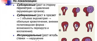
They can be located on the surface of the organ (subserous uterine fibroids), in the thickness of the myometrium, or interstitially, and also extend into the cavity (submucous fibroids).
Most often, tumors are found in the body of the organ (in 90% of cases); they can also grow in the lower segment and cervix, grow into the uterine ligaments and neighboring organs (parasitic fibroids).
The node consists of muscle and fibrous cells covered with a pseudocapsule.
As it grows, it acquires its own network of blood vessels, which are branches of the uterine arteries. On the surface of fibroids there are receptors for estrogen and progesterone, which makes it hormonally dependent.
Causes
The main reason for the occurrence and intensive growth of fibroids is considered to be increased synthesis of ovarian hormones. Changes in hormonal levels can be observed with:
- A woman entering menopause.
- Carrying a pregnancy.
- Hormonally active tumors of the ovaries and adrenal glands.
- Obesity (excess lipids go into the formation of estrogen).
- Various lesions of the hypothalamus and pituitary gland (tumors, cerebral hemorrhages, injuries, neurological disorders).
- Taking incorrectly selected hormonal contraceptives.
Active tumor development requires a decrease in the body's defenses. The immune system, being suppressed, will not be able to detect and destroy fibroid cells.
A decrease in immunity contributes to:
- Chronic stress and overwork.
- Physical inactivity.
- Bad habits.
- Hypothermia.
Symptoms
A benign neoplasm on the posterior wall of the uterus may not manifest itself clinically for a long time. When the tumor size is up to 4 cm, there are no pronounced symptoms, and pathology is usually detected only by ultrasound.
Nodes larger than 4-6 cm, located on the posterior wall of the uterus, often manifest themselves with the following symptoms:
- Bleeding: heavy and prolonged discharge during menstruation, bleeding not associated with the menstrual cycle.
- Dull, nagging pain, localized in the inguinal, iliac, sacral regions, and also radiating to the anus.
- When the node is large, compression of the intestine is observed, accompanied by constipation and a painful urge to defecate.
Important! A feature of this localization of fibroids is the absence of enlargement of the anterior abdominal wall. A tumor growing along the back wall of the uterus must be gigantic in size (over 20 weeks of pregnancy) in order to be detected by visual examination.
Pregnancy
The effect of fibroids on the back wall of the uterus on pregnancy is quite noticeable. The tumor, deviating the uterus posteriorly, can prevent sperm from entering the cavity of the fallopian tubes, which makes conception impossible. Also, menstrual irregularities that accompany fibroids cause infertility.
If fertilization does occur, there are three possible outcomes of the pregnancy:
- Early miscarriage.
- Complications in the form of low attachment or placenta previa, threatened miscarriage, chronic fetal hypoxia and premature birth.
- Safe pregnancy and delivery on time.
The outcome of pregnancy in patients with nodes on the posterior wall of the uterus depends on the size of the pathological formation and its location. Small tumors located subserosally rarely complicate the gestation period and allow a woman to give birth naturally.
Tumors in the uterine cavity or in the thickness of the myometrium with a diameter of more than 4-6 cm disrupt the normal attachment of the fertilized egg, which can cause miscarriage or a difficult pregnancy.
Complications of the birth process may include discoordinated contractions, weak pushing, impaired discharge of the placenta, and postpartum hemorrhage.
In order for the period of bearing a child to pass safely, women with uterine fibroids should plan a pregnancy. At the stage of preconceptional preparation, medicinal or surgical treatment of fibroids is carried out. After postoperative intervention, in the absence of complications, you can become pregnant within a year.
Hormonal therapy for fibroids during pregnancy is prohibited.
In emergency cases (bleeding, disruption of the blood supply to the node with subsequent necrosis), surgical excision of the tumor on the posterior wall of the uterus during pregnancy or simultaneously with a cesarean section is possible.
Complications
The main complications of fibroids located on the back wall of the uterus are:
- “Birth” of a submucosal node when it is torn off from the stalk, accompanied by cramping pain and large blood loss.
- Torsion of the base of the tumor with the development of necrosis and inflammation of the peritoneum (with subserous localization).
- Compression of the intestine with large sizes of the node with the occurrence of mechanical obstruction and fistulas.
Important! These conditions require urgent medical attention, because may be life-threatening.
Treatment
When a myomatous node is detected on the back wall of the uterus, there are three main tactics for further action: watchful waiting, drug treatment, and surgery.
Waiting tactics
If the tumor on the posterior wall of the uterus does not exceed 2-3 cm in diameter, does not manifest itself clinically and does not bother the patient, it can be left alone. In this case, the woman is recommended to visit a gynecologist once every six months and undergo an ultrasound examination of the uterus and adnexal organs once a year to monitor the growth of the tumor.
In order not to provoke active tumor growth, women with uterine fibroids are contraindicated:
- Visiting the bathhouse and sauna.
- Excessive insolation.
- Procedures involving heating or irradiation of the iliac, lumbar and gluteal regions.
- Taking hormonal contraceptives without consulting a gynecologist.
Drug treatment
Medium-sized tumors (up to 6 cm / 12 weeks of pregnancy) can be treated with hormonal drugs. For this purpose, substances are used that suppress the synthesis of ovarian hormones or block receptors for them on the myomatous node itself.
Such drugs include:
- Gestagens. They are used in the form of tablets, injections and hormonal uterine devices.
- Combined oral contraceptives that suppress ovarian function.
- Gonadotropin releasing hormone agonists. They disrupt the hypothalamic-pituitary-ovarian connection, reducing the synthesis of sex steroids.
- Antigestagens. I block the receptors for the sex hormones of the node itself, which leads to its reduction.
Hormonal therapy lasts at least 3-6 months, after which a control ultrasound is required. If symptoms persist and there is no positive dynamics, surgical removal of the node is used.
Surgery
Indications for surgical intervention for fibroids along the posterior walls of the uterus are:
- Dimensions of myomatous tumor from 12 weeks of gestation.
- Active growth of pathological tissues in postmenopause.
- Suspicion of myosarcoma.
- Infertility.
- Bleeding.
- Prolapse of the uterus.
- Ineffectiveness of conservative treatment.
The extent of the operation depends on the size of the node and its location, as well as the age of the patient.
Women of reproductive age with nodes up to 16 cm without signs of malignancy undergo myomectomy - excision of pathological tissue while preserving the uterus.
In cases of gigantic tumor size, suspicion of myosarcoma, multiple nodes, and postmenopausal women, the uterus is removed (hysterectomy).
Surgical intervention can be with a wide incision in the anterior abdominal wall (laparotomy) or through small punctures near the navel with the introduction of endoscopic instruments (laparoscopy). There are also minimally invasive and less traumatic methods for eliminating nodes along the posterior wall of the uterus, such as uterine artery embolization and FUS ablation.
Symptoms
The majority of patients who were diagnosed with uterine fibroids on the anterior wall of the uterus say that the disease did not show any symptoms for a long time.
But with all this, there are certain signs by which one can suspect the development of pathology:
- Menstrual irregularities;
- Presence of pain in the lower abdomen;
- Discomfort during intimacy;
- Problems with urination;
- Impossible to conceive a child;
- Pain in the lumbar spine.
Depending on the form in which the pathological process progresses, the node will have certain characteristics. For example, with submucous fibroids, the neoplasm will be located under the mucous membrane of the reproductive organ. But in the case of the interstitial type, it will be localized in the muscle layer.
With the progression of the subserous type of the disease, myomatous nodes can simultaneously be located in different places. In most cases, the lump is on the outer surface of the uterus and under the mucous membrane. Myoma can also be pedunculated, which is a specific sign of a tumor. It can be localized anywhere, but it will always have a narrow base.
In most cases, treatment is carried out with medication, but if a formation is detected on the leg, it will be necessary to remove it. Doctors also note that they often detect not one, but several nodes, which indicates the progression of multiple fibroids.
Cervical
Cervical fibroids can be located on the lateral or posterior wall of the cervix, between the lobes of the broad ligament, in the anterior or posterior part, in the intercervical tissue. Regardless of location, small cervical nodes do not manifest themselves in any way and can only be detected during routine examinations.
As the isthmus grows, symptoms appear:
- increase in the duration and intensity of bleeding on critical days,
- bleeding or spotting between menstrual periods,
- pain in the lower third of the abdomen, perineum, lower back,
- difficulty urinating, constipation, frequent urge to go to the toilet.
Peculiarities
If a node on the anterior wall of the uterus grows inside the organ, then the subserous type of the disease is diagnosed. A characteristic feature of this pathology is that the neoplasm can be on a thick or thin stalk. In the first case, the tumor will very quickly increase in size and also create negative pressure on nearby internal organs.
Thin-pedunculated fibroids have a high likelihood of torsion. In this case, the nutrition of the neoplasm stops, which provokes the development of necrosis, which subsequently causes serious complications.
In most cases, fibroids of this type develop over a fairly long period of time. Moreover, during the progression of the pathological process, a woman almost always has no characteristic symptoms. The disease can be detected already at an advanced stage, when complications begin to develop, or during a gynecological examination.
Diagnostics
Uterine tumors located on the posterior wall of the organ are difficult to detect during gynecological examination and palpation. Such nodes are palpable only when they reach large sizes. The doctor pays attention to the patient’s characteristic complaints (menstrual irregularities, bleeding, abdominal pain, constipation and discomfort during bowel movements). To clarify the diagnosis, a number of examinations are required.
Additional methods for examining a node on the posterior wall of the uterus are:
- Ultrasound of the uterus and appendages with Dopplerography.
- Hysteroscopy is a transvaginal examination of the uterine cavity with an endoscope.
- Laparoscopy is an examination of the pelvic cavity by introducing endoscopic instruments through the anterior wall of the abdomen.
- CT or MRI.
- Separate diagnostic curettage - removal of the upper layer of the uterine epithelium followed by histological examination.
- Taking smears from the vagina and cervical canal for microflora and cellular composition.
Based on the results of these studies, the gynecologist prescribes the required amount of treatment.
Complications
The likelihood of developing complications depends on how quickly the tumor process progresses, as well as where it is localized. When the myomatous node is located on the anterior wall of the uterus, there is a possibility of the following conditions:
- A gradual, but constantly increasing, compression of the bladder will begin;
- Women will have problems with urination;
- The functioning of the kidneys is impaired;
- Pyelonephritis progresses;
- There are problems with the ureters.
When fibroids form on a pedicle, torsion cannot be ruled out. In this case, the woman will feel severe pain, and the treatment process will involve surgical removal of the tumor. The doctor can confirm the diagnosis using ultrasound examination of the pelvic organs, hysteroscopy, magnetic resonance imaging, and laparoscopy.
Treatment
If a single myomatous node or multiple tumors have been identified, it is imperative to begin treatment for this pathological process. The woman will need to adhere to a certain diet. It is imperative to exclude spicy, salty foods, as well as smoked foods from your diet. Particular attention should be paid to controlling the level of carbohydrates and fats consumed.
During the entire therapy, you should avoid any procedures associated with increased exposure to heat. Therefore, girls are not recommended to sunbathe on the beach, visit a bathhouse or sauna, or go to a solarium. Some physiotherapy procedures are also contraindicated, as they can cause tumor enlargement.
You definitely need to start taking multivitamin complexes, and ideally you should undergo a course of treatment with medications of this type. Doctors advise taking folic acid and B vitamins in the first phase of the menstrual cycle. After this period, it is better to take vitamin C and E.
In addition, if hormonal medications have been prescribed, you must take them. Doctors often develop a therapy complex that contains monophasic estrogens or hormonal contraceptives. Thanks to this effect, it is possible to prevent the increase in myomatous formations, as well as their degeneration into malignant tumors.
In most cases, when a woman is prescribed the correct drug therapy, the tumor thickens, as well as a decrease in blood circulation processes in it. This leads to the fact that the size of the tumor either decreases or stops increasing. If the fibroids were small, then medications will help relieve unpleasant symptoms.
If the tumor is large, the doctor will recommend that the woman have it removed. In this situation, medications may also be prescribed. However, their use is necessary in order to reduce the likelihood of large blood loss during the operation. They are also drunk in order to prevent complications from developing and in preparation for intervention.
Operation
Myomatous nodes can be quite large. Therefore, if a doctor detects such a tumor, it is possible that it may threaten the health and life of the patient. In this situation, urgent surgical removal of the tumor is required.
First of all, the specialist must clearly determine not only the type of fibroid, but also in what method it will be removed. To do this, he will have to take into account many factors, including the woman's age and physical condition. If it is possible, then preference is given to operations that are minimally invasive, which allows maintaining the full functioning of the reproductive organ.
In most cases, gynecologists detect a subserous type of tumor. If treatment is not carried out in a timely manner, the woman will subsequently have problems with the bladder, since the tumor grows inside the uterus.
Although pathological nodes are diagnosed quite often, in most clinical cases conservative therapy is sufficient for their treatment. That is why women are advised to visit a gynecologist in a timely manner, who can identify the disease at an early stage.
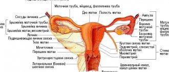
Interligamentous leiomyoma
To maintain normal anatomical conditions in the pelvis, the reproductive organs are held in place with the help of a ligamentous apparatus that maintains the correct position of the uterus, tubes, ovaries and bladder. On the sides of the uterus there are broad ligaments. A benign node growing interligamentously changes the anatomical relationships of organs and can cause unpleasant complications. This usually happens when fibroids form in the lower part of the uterus, closer to the isthmus. The ligamentous apparatus limits the mobility of the node, so the intraligamentous location of leiomyoma becomes a negative feature of the course of the disease, requiring surgical treatment.