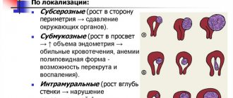
- Dimensions of uterine fibroids
Uterine fibroids
is one of the most common female diseases. According to medical statistics, among all gynecological diseases it accounts for 27%. Every fourth woman in the world suffers from it. In Russia, 80% of operations end in uterine amputation. So this tumor, although benign, is not at all as harmless as it seems at first glance. Therefore, identifying fibroids in the early stages in order to rid a woman of them while maintaining reproductive function is the main task of a diagnostician.
Most often, fibroids develop in women in the late reproductive period and before the onset of menopause, but sometimes it also occurs in younger representatives of the fair sex. The choice of treatment method for this disease depends on the size and location of the tumor in the walls of the uterus.
How to determine the size of uterine fibroids in weeks?
However, clinically, most often it does not manifest itself in any way, and only during a routine examination by a doctor can one suspect the presence of fibroids due to an increase in the size of the uterus, and with the help of an ultrasound examination can confirm the presence of nodes, their size and location. And also dynamically determine the presence or absence of their growth.
Uterine fibroids: sizes in mm are varied and, depending on their indicators, all tumors are divided into small, medium and large. How many weeks are uterine fibroids 2 cm? Small benign tumors include nodes up to 20 mm in diameter, which corresponds to 4-6 weeks of pregnancy. What is the treatment for uterine fibroids for 5 weeks? In this case, surgical treatment is carried out only in case of torsion of the tumor stalk, if any, or in case of heavy bleeding leading to the development of anemia, as well as infertility, which is caused by a myomatous node. Only in these cases should small fibroids be removed. Therefore, if the question is about how many weeks uterine fibroids are 11 mm, then it is approximately 2-3 weeks. And if the uterine fibroids are 6 mm, then this is completely equal to 1-2 weeks.
What to do with 4 cm uterine fibroids? Medium-sized fibroids - 40-60mm correspond to 10-11 weeks of pregnancy; they are not always subject to surgical treatment, but only if the nodes are not actively growing or there are no pronounced symptoms. If the size of the node exceeds 60 mm in diameter, which corresponds to 12-15 weeks of pregnancy, then the fibroid can only be treated surgically.
Complications
Multiple large fibroids can be especially dangerous. In this case, the gynecologist has to monitor several nodes at once, sometimes there are more than a dozen of them. If a woman is pregnant, but the placenta is affected by a myomatous node, this greatly increases the risk of miscarriage.
Large nodes put pressure on internal organs and can interfere with the normal functioning of the kidneys, promote the formation of stones and the development of infectious and inflammatory diseases. The functioning of the digestive system may also be disrupted: constipation develops.
The rate of growth is also important: it is considered accelerated if during the course of a year the tumor has increased by the gestation period corresponding to 5 weeks.
Impaired blood supply to fibroids can cause necrosis, and torsion of the tumor stalk is also possible. In some cases, the formation may prolapse from the uterus into the vagina, which is accompanied by severe pain and bleeding. In approximately 2% of cases it can develop into cancer.
Uterine fibroids 2 cm
What to do in such a situation and how to treat it correctly? Many women turn to gynecologists with this question, because the risk of its increase is quite high, which can cause not only the appearance of clinical symptoms, but also degeneration into a malignant tumor. Most often it is asymptomatic, but there may be an increase in the volume of menstruation, a shortening of the menstrual cycle, increased pain during menstruation, the appearance of spotting brown discharge, as well as discomfort in the lower abdomen. As a rule, treatment of such fibroids is associated with the normalization of a woman’s hormonal levels by reducing ovarian activity. For this purpose, antigonadotropins and gonadotropin-releasing hormone agonists are used, which are able to suppress the growth of gonadotropins in a woman’s body by acting on the hypothalamus. All this leads to regression of the tumor or to stopping its development. The dosage of the drugs is as follows: 3.75 mg of the drug from the 1st to the 5th day of the cycle, 28 days break and repeat the injection again. And so on for 3 to 6 months, and after 3 or 4 procedures the size of the tumor is reduced by half.
Pregnancy and tumor size
Of course, all women who do not yet have children are interested in the question of whether it is possible to get pregnant with uterine fibroids. If it is small in size, up to 3 cm as at 5 weeks, then it is quite possible to conceive a child, there are no obstacles to this. It’s another matter if education is large and growing very rapidly. In such a situation, the sperm simply will not be able to enter the cavity of the reproductive organ, therefore, it will not be possible to get pregnant.
Even if a girl manages to conceive a child, the tumor can negatively affect the development of the fetus. Miscarriages often occur, and quite late, from the fourteenth week. In most cases, pathology prevents the body from conducting a normal natural birth. The baby will not be able to pass through the birth canal due to the neoplasm. Therefore, children are born to patients via caesarean section.
In addition, it is worth remembering that during pregnancy, women's hormonal levels change, which is a very good environment for the occurrence of fibroids. In this regard, it is necessary to constantly be monitored by a gynecologist. The doctor is obliged to monitor the condition of the mother and baby throughout the entire period of pregnancy.
Uterine fibroids 5 cm
Refers to medium-sized fibroids and corresponds to 11-12 weeks of pregnancy. At the same time, a woman can often suspect that she has it if her stomach increases in size despite the fact that the pregnancy test is negative, pain syndrome, menstruation irregularities, hemorrhagic syndrome, as well as dysfunction of adjacent organs - problems with urination and intestines. In the absence of the above symptoms, treatment of a benign tumor can be conservative, and if they are present, then only surgical treatment is indicated.
Uterine fibroids at 5 weeks correspond to the small size of the node and are subject only to observation and hormonal treatment, because if complications arise, the likelihood of surgical treatment increases.
Clinical picture
When the uterine tumor is still small in size, for example, 7 mm, 9 mm or 10 mm, it does not manifest itself in any way. Therefore, a woman does not even realize that a disease is developing inside her, which can grow rapidly.
As soon as the fibroids begin to reach a higher size, starting at about eight weeks, the patient notices that her periods are intense, and they are accompanied by pain. The pain can be very severe, which even medications cannot eliminate.
Fibroids at 14 weeks cause the uterus to enlarge and the lower abdomen to swell. At the same time, the woman’s body weight does not increase. If the patient has a tumor on the leg, then torsion is possible, which causes very severe pain in the abdominal area.
Large fibroids, 10-20 weeks old, can put pressure on nearby organs, and also lead to problems with bowel movements and bladder, pain in the lumbar region, heart and legs. In addition, numbness of the lower extremities may occur.
If the tumor grows in the outer layer of the reproductive organ, then adhesions may form with the tissues and organs located near it. This process during physical activity causes pain, which can manifest itself in any part of the body.
Uterine fibroids 5 6 weeks
What to do and how to deal with it? This size of fibroids also requires dynamic observation by a specialist and a course of drug treatment, because in the case of rapid growth of such a node, surgical treatment will be required after hysteroscopy. Rapid growth is characterized by an increase in the size of the node by 5 weeks or 4 cm throughout the year, which is a leading signal for changing the direction of treatment and emergency surgery.
Uterine fibroids 5 weeks: treatment is carried out only hormonally and in each specific case the doctor selects a group of drugs that are the most effective, because the woman’s age, concomitant pathology, parity and a number of other factors influence not only the choice of drugs for treatment, but also the outcome of it .
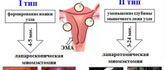
Uterine fibroids 6 weeks - what to do if the node is twisted? Here it is only possible to perform a myomectomy operation, and in rare cases, removal of the uterus.
Diagnosis of the tumor and its size
A female disease can be detected using various diagnostic methods, which differ in their capabilities. But before prescribing one or another method of detecting a tumor, the gynecologist first conducts an examination.
At the same time, he feels that the uterus is enlarged, he can say approximately its size. As already mentioned, doctors compare it with a certain time of pregnancy, for example, uterine fibroids at 8 weeks coincide with the volume of the uterus at the eighth week of an interesting position. But this is approximately, so the specialist recommends undergoing additional examination to determine the exact size.
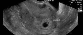
First of all, the patient is sent for an ultrasound examination. It can be carried out in two ways. The first is to diagnose through the anterior wall of the abdominal cavity. This method is considered the most preferable for patients because it does not cause discomfort, but it provides doctors with little information.
Most of all, experts like to do it in the second way, that is, through the vagina. It's much more informative. True, women have to endure some discomfort during the procedure. Ultrasound examination helps determine the presence of a tumor and its size.
If difficulties arise in diagnosing a uterine tumor, doctors prescribe hysterography. The specialist injects a contrast agent into the cavity of the female organ and takes an X-ray. Hysteroscopy may also be prescribed, which detects the disease using a special device that allows you to examine the cavity of the reproductive organ.
If there is a suspicion that the tumors are very small or located in hard-to-reach places, then a computed tomography scan or laparoscopy may be performed.
Uterine fibroids 6 7 weeks
What is this? This is a concept adopted among obstetricians and gynecologists around the world to assess the degree of development of fibroids depending on the size of the uterus, because normally the uterus increases in size only during pregnancy and these sizes are almost always stable. Therefore, if the doctor says that uterine fibroids are 6-7 weeks old, then this corresponds to the size of the uterus at 6-7 weeks of pregnancy. Here it should be understood that this is not the size of the node or tumor, because only ultrasound will answer this question.
In most cases, fibroids of this size are subject to conservative therapy, where hormonal treatment is combined with homeopathic remedies and traditional medicine. But, when the first symptoms appear indicating trouble in the body - pain or bleeding, you should immediately inform your doctor, because this may be the reason for its growth or the development of complications requiring immediate surgery.
What sizes can be dangerous?
Any neoplasm is always an additional health risk. Large fibroids begin at 12 weeks (about 60 millimeters). If you allow the disease to develop and the levels are already higher, it can be life-threatening.
Fibroids do not appear unexpectedly. If a woman is regularly examined by a gynecologist, it will not be difficult to miss the beginning of her growth and prevent the development of the disease.
Monitor your health, visit your gynecologist regularly and, if necessary, start treatment at the earliest stages of the disease. Large uterine fibroids are always dangerous and risky.
Myoma and spiral: development risks
It is known that the use of intrauterine devices increases the risk of developing fibroids in women, but this theory is not always confirmed by practice. In general, the contraceptive has no effect on the progress of the tumor. The risk of developing a tumor increases due to the incompetence of the gynecologist or the choice of a low-quality device. The main reason for the development of fibroids in women using a coil is its incorrect installation, which leads to excessive trauma to the mucous membranes.
It is impossible to refute the possibility of a therapeutic effect from the spiral when it is installed for the treatment of fibroids. The effect is achieved by restoring a woman’s hormonal levels. The process of tumor development stops, and then a decrease in its size is observed. If it is small, there is a chance of its complete disappearance. This is why doctors often recommend installing a spiral and thus treating fibroids.
A gynecologist should select a high-quality contraceptive. When using an IUD that releases synthetic estrogen, the fibroids will grow. The substance provokes active growth and division of cells in the uterus.
How does pregnancy proceed with fibroids with centripetal growth of the node?
A difficult task in obstetrics is the combination of fibroids and pregnancy. Many opinions of patients agree on the normal state during the period of illness and pregnancy. But this does not exclude serious risks, both during pregnancy and during childbirth. Complications can be of two types:
- At the site of tumor formation;
- According to the factors that influenced education.
Pregnancy during the disease is possible, but this is influenced by the location of the source of the disease, the number of nodules and size. A normal pregnancy is guaranteed by a small accumulation of tumor cells that occurs deep in the wall of the uterus. Large nodes, which especially occur at the isthmus and at the mouths of the fallopian tubes, cause difficulties when trying to get pregnant. If the fetus has time to develop, there is a risk of complications.
Risk factors
The growth of a fibroid node indicates the following risk factors during pregnancy:
- Birth of a child with pathology or miscarriage;
- Scars on the uterus that formed after operations;
- The appearance of more nodes;
- Location of the placenta near the node;
- Swelling of the “nodal” place;
- Uterine cells lose their ability to stretch and contract;
- Deformation of the organ shell;
- The size of the sore spot increases to 8 cm;
- Presence of an additional disease.
A small likelihood of risk is observed in young girls whose tumor is only at a developing stage. The early stage is characterized by a minimal number of nodes that do not exceed 8 cm. Moreover, the girl does not suffer from another disease of this organ. Pregnancy will proceed normally if the placenta of the embryo is fixed away from the node. Uterine fibroids during pregnancy, which are characterized as small in size, do not progress. The fetus develops normally, and the baby will be completely healthy at birth.
If at least one of the risk factors is observed during pregnancy, a complication may occur. In such cases, you should consult a doctor. It is possible that the woman will be prescribed an abortion.
Symptoms
The most alarming symptom of uterine fibroids is heavy, untimely bleeding. When the bleeding continues and does not stop, you should urgently seek medical help, because anemia may develop. This happens during prolonged menstruation.
The location and severity of pain is also an irreversible symptom of fibroids. A woman is often bothered by pain in the pelvic area. Many girls describe pain defects as a bursting, incomprehensible and sharp heaviness in the abdominal area. This happens with a constant increase in the volume of fibroids. The tumor exerts a burden on adjacent organs.
The pain may resemble contractions. This occurs when the uterine muscles contract.
Blood supply may be impaired due to acute pain attacks.
A woman can independently detect fibroids if there are no sharp pains and the nodes have grown sufficiently.
An indirect cause of the growth of nodules may be impaired functioning of the cardiovascular system. Developed anemia, which is expressed in a decrease in hemoglobin levels, provokes the formation of tumor nodes. Other symptoms are also present: chronic fatigue, depressed mood and decreased performance.
A woman must promptly notice changes in her body that indicate uterine fibroids, because a benign tumor can turn into a huge problem in an instant. For any uterine bleeding, you should contact a specialist.
Video: Intramural fibroids. Centripetal node growth
Intramural fibroids. Centripetal growth of the node.
Submucosal growth of fibroids
Women's worries about long periods and their excessive abundance are not groundless. Often the culprit of the disease is submucous fibroid, which, through its growth, causes uterine bleeding.
If a woman does not see a doctor on time, the fibroids continue to grow and cause a number of complications:
- Lack of oxygen to internal organs, which is caused by anemia;
- Inflammation and infection of the myomatous nodule, which is detected when pus is discharged along with blood;
- Decreased immunity, which causes septic disorders;
- Premature birth or miscarriage;
- The appearance of a mechanical obstacle during childbirth;