Types of myomatous node necrosis
Types of necrosis of the myomatous node are distinguished by morphological characteristics.
- Coagulation (dry) necrosis of the myomatous node. The dead zones of the neoplasm shrink and form cavernous cavities, where fragments of necrotic tissue accumulate.
- Wet necrosis of myomatous node. Dead tissue softens and forms cystic cavities.
- Hemorrhagic infarction (red necrosis of the myomatous node). The tissues of the node acquire a soft consistency and a red-brown color. Accompanied by dilation of tumor veins and thrombosis. This type of disease is more common in pregnant or postpartum women.
- Aseptic necrosis of myomatous node. Necrosis of areas of the myoma node is accompanied by infectious inflammation of a hematogenous or lymphogenous nature. It can be caused by pathogens such as E. coli, staphylococcus or streptococcus. This type carries the maximum risk of peritonitis and sepsis.
Sometimes aseptic necrosis of the myomatous node is interpreted as a separate type of complication of uterine fibroids.
Reasons for the development of necrosis of the myomatous node
Myoma increasing in size can provoke deformation of the vessels that feed it, or compress them. This happens most often for the following reasons:
- torsion of the leg of the myomatous node;
- bending of the tumor stalk;
- node ischemia;
- formation of blood clots in the myomatous node.
It is worth noting that twisting of the stalk is more typical for subserous fibroids, which for the most part have thin stalks. Intramural tumors that are under the strong influence of contractions of the muscular layer of the uterus are more often subject to ischemia. These reductions, in turn, can be caused by:
- the use of drugs that affect the uterine muscles;
- pregnancy;
- childbirth.
In general, necrosis of the myomatous node is caused by impaired blood flow in it.
Why is fibroid torsion dangerous?
Torsion of the fibroid stalk leads to compression of the veins, while the arteries continue to function for some time due to the elasticity of the walls. The outflow of blood from the tumor is disrupted, and it stagnates in the uterus. The node increases in size, becomes soft, its capsule stretches. At the same time, the peritoneum covering it is stretched, and foci of hemorrhage appear.
Against the background of impaired blood flow, fibroid necrosis develops quite quickly. Tissues deprived of nutrition die. Often a secondary infection occurs, and then the necrosis becomes septic. First, pelvic (local) and then diffuse peritonitis develops. This condition is life-threatening and requires emergency medical attention.
Symptoms of myomatous node necrosis
A disease such as necrosis of a myomatous node exhibits the same symptoms, regardless of the type. They differ, rather, in the intensity of manifestation depending on the nature of their occurrence:
- in case of blood supply disturbance - gradually;
- when twisting the leg - sharply.
In general, the symptoms of myomatous node necrosis are as follows:
- nagging or cramping pain in the lower abdomen;
- tension of the anterior abdominal wall;
- temperature rise to 37.5 degrees;
- chills;
- tachycardia;
- constipation, increased gas formation;
- dry mouth;
- nausea and vomiting;
- soreness of the node or the entire uterus during gynecological palpation.
Against the background of painful attacks, the development of low-grade fever is possible - a condition characterized by a constantly elevated body temperature about a degree above normal.
Impaired blood supply to the fibroid node
Definition
Impaired blood supply to a fibroid node can occur due to torsion of the pedicle of a subserous myomatous node, ischemia of a large interstitial node, or its rapid growth.
Pathogenesis
When the blood supply to the fibroid node is disrupted, degenerative processes occur in it: edema, necrosis, hemorrhage, hyaline degeneration, degeneration. There are dry, wet and red necrosis of fibroids. With dry tumor necrosis, gradual shrinkage of areas of necrotic tissue occurs with the formation of a cavernous cavity with remnants of dead tissue; when wet - softening and wet necrosis of tissue with subsequent formation of cystic cavities. Myomas located intramurally are more often exposed to red necrosis, while macroscopically the tumor nodes are colored red or brown-red, have a soft consistency, and microscopically reveal pronounced dilatation of the veins and their thrombosis.
Aseptic necrosis is often accompanied by an infection that penetrates the node by hematogenous or lymphogenous route. The causative agents of infection are staphylococcus, streptococcus, and E. coli. Infection of myomatous nodes poses a great danger due to the possibility of developing diffuse peritonitis and generalized infection (sepsis).
Clinic
With the acute development of the disease, pain in the lower abdomen and low-grade fever are noted; vaginal examination reveals an enlarged and painful uterus. When the leg of a subserous myomatous node is torsed, symptoms of an acute abdomen occur: pain in the lower abdomen, nausea, vomiting, increased body temperature.
Myoma necrosis is usually accompanied by acute abdominal pain, tension in the anterior abdominal wall, increased body temperature, nausea, vomiting, disturbances in stool and urination; During vaginal examination, the presence of myomatous nodes in the uterus is determined, one of which is sharply painful on palpation.
The diagnosis is established on the basis of typical complaints, medical history, objective examination, results of laboratory and additional research methods (ultrasound with Doppler ultrasound, laparoscopy).
Treatment is surgical. The scope of surgical intervention depends on the patient’s age, reproductive function, size, location, and number of myomatous nodes. In case of necrosis of the myomatous node, amputation or extirpation of the uterus and tubes is performed. Conservative myomectomy is not indicated, since severe complications may occur in the postoperative period: dehiscence of the sutures on the uterus, suppuration, peritonitis. It can be performed only in exceptional cases in young nulliparous women under conditions of intensive antibiotic therapy.
Abortion
Definition
Abortion is termination of pregnancy.
Stages of abortion: threatened, begun, abortion in progress (incomplete, complete).
Etiology
Reasons for spontaneous abortion: sexually transmitted infections; hormonal disorders; isthmic-cervical insufficiency; infectious diseases; malformations of the uterus, tumors of the uterus and ovaries; diseases of the kidneys, cardiovascular and other body systems that experience additional stress during pregnancy; bad habits (smoking, alcoholism, drug addiction) that affect the formation and development of the fertilized egg; excessive physical and nervous tension; negative environmental factors (radiation, gas pollution, high concentrations of chemicals, prolonged work at the computer).
Clinical picture.
A threatened abortion is manifested by nagging pain in the lower abdomen and lower back. Scanty bleeding from the genital tract is possible. The tone of the uterus is increased, the cervix is not shortened, the internal os is closed, the body of the uterus corresponds to the period of pregnancy. Ultrasound records the fetal heartbeat.
When an abortion begins, pain and bloody discharge from the vagina are more pronounced, the cervical canal is slightly open.
During an abortion, regular contractive contractions of the myometrium are detected. The size of the uterus is less than the expected gestational age. In later stages of pregnancy, amniotic fluid may leak. The internal and external pharynx are open, the elements of the fertilized egg are in the cervical canal or in the vagina. Bloody discharge can be of varying intensity, often abundant. There are incomplete and complete abortions:
Incomplete abortion is a condition associated with retention of fertilized egg elements in the uterine cavity. The lack of full contraction of the uterus and closure of its cavity leads to ongoing bleeding, which in some cases causes large blood loss and hypovolemic shock. More often, incomplete abortion is observed after 12 weeks of pregnancy in cases where the miscarriage begins with the rupture of amniotic fluid. On bimanual examination, the uterus is smaller than the expected gestational age. Bloody discharge from the cervical canal is abundant. Using ultrasound, the remains of the fertilized egg are determined in the uterine cavity, and in the second trimester - the remains of the placental tissue.
Complete abortion - the fertilized egg is completely released from the uterine cavity. The uterus contracts and bleeding stops. During bimanual examination, the uterus is well contoured, its size is smaller than the gestational age, and the cervical canal can be closed. In case of a complete miscarriage, the closed uterine cavity is determined using ultrasound. There may be slight bleeding.
During a general clinical examination, a general blood test may show a decrease in hemoglobin, hematocrit, slight leukocytosis without a shift of the formula to the left, and a slightly accelerated ESR.
Differential diagnosis is carried out with a polyp of the cervical canal, a myomatous node.
Treatment
The goal of treating threatened miscarriage is to relax the uterus, stop bleeding and prolong pregnancy if there is a viable embryo or fetus in the uterus.
They recommend physical and sexual rest, a nutritious diet, gestagens, methylxanthines (pentoxifylline), and as symptomatic treatment - antispasmodic drugs (drotaverine, suppositories with papaverine for severe pain), herbal sedatives (motherwort decoction, valerian).
Prescribing drugs that affect hemostasis (etamsylate, vikasol, tranexamic acid, aminocaproic acid and other drugs) to pregnant women for bloody discharge has no basis and no proven clinical effects because bleeding during miscarriages is caused by chorionic detachment of the early placenta), and not by coagulation disorders.
In case of incomplete abortion, emergency care is necessary - immediate instrumental removal of the remaining fetal egg and curettage of the walls of the uterine cavity. During the operation and after it, it is advisable to administer intravenously an isotonic solution of sodium chloride with oxytocin; carry out antibacterial therapy. If necessary, treatment of posthemorrhagic anemia. In Rh-negative women (during pregnancy from a Rh-positive partner), after vacuum aspiration or curettage during pregnancy for more than 7 weeks and in the absence of Rh antibodies, rhesus immunization is prevented by administering anti-Rhesus immunoglobulin.
In case of complete abortion in less than 14–16 weeks, it is advisable to perform an ultrasound and, if necessary, curettage of the uterine walls, since there is a high probability of finding parts of the fertilized egg and decidual tissue in the uterine cavity. At a later date, when the uterus has contracted well, curettage is not performed. Antibacterial therapy is prescribed, anemia is treated according to indications, and anti-Rhesus immunoglobulin is administered to women with Rh-negative blood.
Barren marriage
Definition
According to the WHO definition (1993), a marriage is considered infertile in which a woman of childbearing age does not become pregnant within a year of regular sexual activity without the use of contraceptives. The cause of infertility can be disorders of the reproductive system in one or both spouses. At the same time, the female factor causes infertility in marriage in 45% of cases, the male factor - in 40%, the combined factor - in 15% of cases.
Classification of infertility:
· according to the presence of pregnancies in the anamnesis - primary and secondary;
· the possibility of pregnancy - absolute and relative;
· according to the mechanism of development - congenital and acquired;
· by duration - temporary, permanent, physiological;
· according to etiopathogenesis:
o endocrine - anovulation, luteal phase deficiency: dysfunction of the hypothalamic-pituitary system, hyperandrogenism, hyperprolactinemia, chronic inflammatory processes of the uterine appendages, hypo- or hyperthyroidism, luteinization syndrome of unovulated follicle;
o tubal and peritoneal - dysfunction of the fallopian tubes, organic damage to the fallopian tubes, peritoneal form of infertility;
o gynecological diseases with a violation of the anatomical and functional state of the endometrium, not accompanied by anovulation and obstruction of the fallopian tubes (internal endometriosis, submucosal uterine fibroids, endometrial polyps, endometrial hyperplasia, external endometriosis with the formation of antiendometrial antibodies, repeated diagnostic curettage of the uterine mucosa, postpartum and postoperative complications , the effect of chemicals and cauterizing substances, endometritis of various etiologies);
o immunological - formation of antisperm antibodies;
o psychogenic;
o infertility of unknown origin.
Causes of female infertility: inflammatory diseases of the genital organs, diseases of the endocrine glands, underdevelopment and abnormal positions of the genital organs, endometriosis of the uterus, tubes and ovaries, tumors of the genital organs, extragenital diseases, inadequate nutrition in quantitative and qualitative terms, immunological factors.
The main causes of male infertility: pretesticular disorders (pathology of the hypothalamus, pituitary gland); congenital defect of GnRH secretion (Kallmann, Prader-Willi syndrome); acquired defect in GnRH secretion, hypopituitarism (tumors, trauma, ischemia, radiation); ZPR, isolated LH deficiency (Pasqualini syndrome); hyperprolactinemia; dysfunction of other endocrine glands; taking hormonal medications; testicular disorders; chromosomal abnormalities (Klinefelter syndrome); congenital and acquired anorchism, isolated aplasia of the spermatogenic epithelium (Sertoli cell, or Del Castillo syndrome); cryptorchidism; varicocele; testicular damage (trauma, torsion, orchitis); disorders caused by systemic diseases or exogenous factors; androgen deficiency or resistance; posttesticular disorders; obstruction of the vas deferens (congenital, acquired); hypospadias; impaired function or motility of sperm (autoimmune disorders, infections of the accessory sex glands).
Diagnosis of necrosis of myomatous node
The first stage of diagnosis for suspected necrosis of a myomatous node involves taking an anamnesis and physical examination of the patient. The doctor pays attention to the following:
- abdominal condition: the woman complains of bloating, positive peritoneal symptoms are observed in the lower parts, as well as pain.
- skin condition: pale in color.
- condition of the tongue: covered with a whitish coating;
- state of the cardiovascular system: blood pressure is normal, but the patient complains of tachycardia.
A blood test contains the following signs indicating necrosis of the myomatous node:
- increase in ESR;
- increase in leukocytes;
- shift to the left of the leukocyte formula.
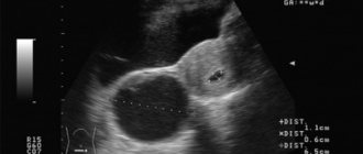
Ultrasound of the pelvic organs is aimed at identifying the following parameters:
- increase in the size of the uterus;
- the appearance of cystic cavities in the node;
- decreased node density;
- heterogeneity of the structure of the myomatous node;
- changing the contours of the node.
In some cases, an increase in the size of the uterus is determined during a gynecological examination.
Laparoscopy is often used to clarify the diagnosis. It allows you to establish the nuances of the course of the disease that cannot be detected by other methods, for example, hemorrhage or swelling in the node. In addition, diagnostic laparoscopy prepares access for subsequent surgery.
Treatment of necrosis of myomatous node
There is only one method of treating myomatous node necrosis - surgery. As soon as the symptoms of this disorder have been identified, the woman should immediately go to the hospital. Necrosis of a myomatous node cannot be cured on an outpatient basis. The choice of operation option will depend on:
- patient's age;
- the presence of her successfully completed childbirth;
- number of myomatous nodes;
- the size of each myomatous node;
- node locations;
- threat of peritonitis.
If a woman is young, of reproductive age, has not yet given birth, or is currently pregnant, she may be prescribed a conservative myomectomy. This operation involves removing fibroids while preserving the uterus. After such a surgical intervention, the patient will be able to give birth to a child.
If a woman is on the verge of menopause and her reproductive function has already been exhausted, there is no goal of preserving the organ. She can be removed:
- body of the uterus with preservation of the cervix;
- body and cervix;
- body and cervix, appendages and ovaries.
Depending on the complexity of the situation, the operation can be performed using laparoscopic or vaginal access.
It is possible to postpone the operation for a couple of days only in case of ischemia of the myomatous node. However, during these hours the patient will need infusion therapy, which will normalize the water-electrolyte balance and alleviate the degree of intoxication.
Prevention and prognosis
With timely surgical treatment, the prognosis is favorable. Preservation of reproductive function and pregnancy depends on the clinical situation. Prevention measures include regular visits to a gynecologist and an ultrasound scan of the pelvic organs, planning pregnancy in the presence of uterine fibroids, planned tumor removal if indicated, and medical examination of women with fibroids.
Sozinova Anna Vladimirovna, obstetrician-gynecologist
1, total, today
( 54 votes, average: 4.78 out of 5)
Types of endometriosis. Endometriosis and pregnancy
Uterine bleeding: causes, symptoms and first aid
Related Posts
Prognosis for necrosis of myomatous node
Modern medicine gives a fairly favorable prognosis if necrosis of the myomatous node was promptly identified and eliminated. In addition, doctors try to preserve reproductive function. However, after the operation, the patient must carefully monitor her condition.
The prognosis worsens if the pathology progresses. This could be peritonitis or blood poisoning, which without proper medical care can lead to disastrous consequences, including death.