From this article you will learn about what this medical term means, how uterine fibroids can grow, as well as the classification of nodes depending on their location in the wall of the organ.
Uterine fibroids can increase in size. Its growth according to the results of two or three ultrasound examinations performed over time is one of the indications for treatment. But nodes can grow in different ways. To indicate the nature of their growth, doctors use special terms:
- Centripetal growth - inward, towards the endometrium (the mucous membrane lining the uterine cavity). Such nodes eventually lead to deformation of the organ cavity.
- Centrifugal growth - outward, towards the abdominal cavity. Such fibroids are less likely to cause symptoms and usually make themselves known when they reach a large size.
What are the reasons for the emergence and growth?
In addition to a woman’s genetic predisposition, doctors identify a number of factors such as:
- Stress, depression, overexertion, nervous conditions;
- Trauma to female organs;
- Excessively long wearing of the intrauterine device (or incorrect installation);
- Hormonal imbalance: due to excess estrogen and progesterone;
- Inflammation in the urinary system, pelvis: infection;
- Physical overexertion;
- Metabolic disease;
- Heredity;
- Too early or late onset of sexual activity;
- Lack of full sexual activity: dissatisfaction;
- Frequent abortions;
- Hypertension;
- Bad habits: alcohol, cigarettes, drugs.
Despite the fact that the causes are varied, specialists often cannot identify a specific cause in their patients. Basically, the causes of pathology are multifactorial in nature.
Important! About half of women aged 30 to 40 experience this disease.
Causes of uterine fibroids with centripetal growth
The main reason for the development of uterine fibroids is hormonal imbalance in the body. When a woman produces more estrogen, the endometrium of the reproductive organ begins to actively grow, forming a tumor.
In addition to hormonal imbalances, there are a number of factors that can lead to the development of neoplasms:
- Weakening of the immune system due to pathologies, stressful situations, fatigue;
- Infectious diseases that develop in the uterine cavity;
- Damage to the uterus as a result of abortion, installation of contraceptive devices, operations.
Most often, uterine fibroids are detected in patients aged 30-40 years. It affects both parous and nulliparous women.
Symptoms
Undoubtedly, every woman is required to undergo an examination by her gynecologist at least once a year. But if this does not happen, then you should pay attention to the following symptoms:
- Severe pain premenstrually or during menstruation;
- Bleeding between periods;
- Heavy discharge during menstruation (as a rule, blood comes out in clots);
- The pain is localized in the lower abdomen and lower back;
- Abnormal bowel movements, frequent urination, intestinal disorders;
- Disruptions in the menstrual cycle (absence, reduction or increase in menstruation)
- Pain during intercourse, accompanied by bloody discharge.
It is worth noting that this symptomatology is characteristic not only of the centripetal type of fibroids.
Also, depending on the stage of development of the pathology, three types of pain can be distinguished:
- With a small size of fibroids , pain occurs only during menstruation (this is rare for such an early stage, due to the relatively small size of the tumor).
- At the beginning of the growth of fibroid nodes there is a constant cutting pain.
- The decomposition phase of the nodes is a sharp and severe pain, causing nausea and vomiting. The patient's temperature also increases.
Signs of uterine fibroids with centripetal growth of the node
Fibroids are characterized by vivid clinical manifestations. A woman may be bothered by the following symptoms:
- Menstrual irregularities. For some, menstruation becomes longer and more abundant, for others, on the contrary, it becomes shorter and sparser;
- Uterine bleeding. They occur regardless of menstruation, differ in intensity, and can lead to anemia;
- Pain in the lower abdomen and lower back;
- Frequent urination, stool upset. Occurs with a significant increase in neoplasm, which compresses neighboring organs and causes disruption of their functioning;
- Blood impurities in the discharge after sexual intercourse;
- Deterioration of general condition.
An e-mail consultation will help you learn more about the manifestations of the disease. A woman can ask questions to the best gynecologists and receive quick and constructive answers without leaving home.
Complications caused by interstitial fibroids during pregnancy
Basically, the main danger for a woman is complications associated with the pregnancy process. Therefore, it is very important to be checked for the presence of such a tumor before conception.
If the fibroid is already large enough and compresses the tubes, then it blocks the access of sperm to the uterus, and therefore prevents pregnancy.
However, if the size of the fibroid nodes is not large enough, then conception can be successful. But the growth of the tumor will pose a danger to the fetus and the pregnancy process may end in miscarriage.
Diagnosis of the disease
If you have the symptoms described above, a visit to a gynecologist is mandatory. With a full examination, the disease can be easily detected. To do this, the doctor will make an initial diagnosis and prescribe a series of tests that the patient needs to undergo.
The first and mandatory step will be taking tests to the laboratory. Among them:
- Complete blood count, blood test for cancer cells;
- Hormone analysis;
- Analysis of the microflora of female genital organs;
- Analysis of urine;
However, laboratory results alone will not be enough. After or before all tests are completed, the patient is referred for ultrasound or x-ray examinations.
Ultrasound diagnostics can determine the growth rate of fibroids, size, location, and type.
Rarely are MRI or CT scans used to clarify the diagnosis.
Treatment
As soon as the presence of tumor-like formations is detected, the patient is admitted to the hospital under medical supervision. This practice is carried out in case the growth of fibroids accelerates and urgent surgical intervention is necessary.
However, despite the severity of interstitial fibroids of the uterine body, depending on the stage of its development, drug treatment can also be used. The stage is considered early if the growth time of the node does not exceed eight weeks.
This treatment is based on taking hormonal drugs:
- Antiprogestogens (Triptorelin, Goserelin, Duphaston);
- Agonists (Mifepristone);
- Androgenic steroids.
- Combined oral contraceptives;
- Gestagens.
Additionally, homeopathic remedies may be prescribed.
These medications control the growth of fibroids, stop them and can help shrink tumor nodes.
Treatment of uterine fibroids with centripetal growth using uterine artery embolization
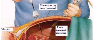
Doctors at fibroid treatment clinics treat the disease in a unique way - uterine artery embolization (UAE). The procedure is performed by endovascular surgeons, as knowledge in the field of radiology and vascular surgery is required. The expert council of fibroid treatment clinics consists of D.M. Lubnin. and Bobrova B.Yu., who are the best specialists involved in the management of patients treated with the UAE method.
The procedure is performed under local anesthesia and is absolutely safe and painless. The doctor performs a puncture of the right common femoral artery, after which a thin catheter is inserted into the artery, which, under the control of X-ray television, is passed directly into the uterine arteries. An embolic drug is then injected, which cuts off the blood supply to the tumor.
Immediately after embolization of the uterine arteries, menstrual bleeding is normalized, its duration and volume are reduced. After a few weeks or months, symptoms of compression of organs located near the uterus decrease and disappear. Within 4-6 months, myoma nodes growing centripetally decrease. After embolization of the uterine arteries, the disease does not recur.
Unlike other methods of treating fibroids, uterine artery embolization has the following advantages:
- The procedure is safe as it is performed under local anesthesia;
- High efficiency is 98.5%;
- Reduction or complete disappearance of symptoms after the procedure;
- No relapses;
- Short hospital stay (1 day);
- UAE is an organ-sparing operation, so a woman can subsequently become pregnant and give birth.
To obtain detailed information about uterine artery embolization, you should make an appointment with a doctor and visit the clinic at a time convenient for you.
A woman's reproductive organs are susceptible to various types of tumors. One of the most common pathologies is fibroids. The neoplasm is a benign tumor. Depending on the form of development and stage, treatment may involve therapeutic intervention or surgical intervention. Uterine fibroids with centripetal growth are the most difficult to treat.
Surgery
If treatment with medication is not possible, surgical intervention is performed. This determines a number of indications:
- Excessively large size of tumor-like nodes;
- Infertility;
- Inflammatory process in the body;
- Rapid growth of fibroids (at least 4 weeks per year);
- Heavy bleeding.
- Necrosis of the node;
- Formation of a subcumcosal node.
- Multiple nodes.
As soon as the doctor notices these symptoms, the patient is immediately taken to the surgical bay and begins her preparation for surgery.
The following methods are available for performing the operation:
- Ultrasound or FUS ablation (the least traumatic method, which involves rapid rehabilitation of the patient);
- Laparotomy (surgery in the abdomen);
- Laparoscopy (incision only 0.5 - 1.5 cm);
- Hysteroscopy (intravaginal tumor removal).
In more severe cases, not only the nodes are removed, but also the organ affected by the tumor. As a rule, a woman knows about the need for such an operation.
Doctors can resort to such radical measures only if the life of their patient is threatened.
The least dangerous and gentlest for the patient’s health are laparoscopy and gastroscopy. These operations minimize the risk of losing the uterus and the woman’s reproductive function.
Also, after these operations, the rehabilitation time is significantly reduced, as is the risk of a similar pathology occurring a second time.
Methods of combating the disease
The growth of fibroids must be monitored in order to understand how active the growth is and whether it requires gentle or radical therapeutic methods of elimination. Fibroids of any of the uterine layers can spread inside the hollow organ. Sometimes tumors have a base, sometimes they have a stalk. Progressive fibroids should be reduced or stopped hormonally (if the size is not large). If the node is large, you should think about other ways to eliminate the affected area:
- Laparoscopic myomectomy. Treatment involves penetration through several incisions (3-4 mm) necessary for the introduction of instruments.
- Hysteroscopic myomectomy. The method involves inserting an elongated instrument with an optical device at the end through the external genitalia. He can remove the fibroid and perform a diagnosis.
- Laparotomy. Treatment is carried out by cutting the uterus through the abdominal cavity. Only the fibroid is removed, the patient’s reproductive function is not affected.
- Removal without surgery. Using ultrasound, only the abnormal growth is crushed, which then comes out naturally.
- Removal of the entire organ.
For no more than 30 years, a method such as uterine artery embolization (UAE) has been used in gynecology.
In fact, this is not really a treatment. Its essence is to de-energize the blood channels that feed the fibroma. This technique has its disadvantages. The tumor remains in place, complicating the process of implantation of the embryo and, therefore, reducing the possibility of a successful pregnancy. Tissue necrosis negatively affects the patient’s general condition (pain) and reproductive function (uterine tissue is affected). Hormone-dependent disease requires further monitoring after successful surgical treatment.
In addition, immediate treatment can save the life and health of the patient, since heavy bleeding leads to anemia, a drop in hemoglobin and blood loss.
To get rid of such growth as a node spreading into the cavity, it is necessary to prevent it from developing into a larger formation. Then the process of excision of the tumor becomes more complicated, and the postoperative period is longer. And precious time is lost, which could be spent planning the most long-awaited event in your life.
Fuse ablation
This is a completely new method, not yet used in all clinics. However, if the location, size and structure of the myomatous nodes allow a similar procedure to be performed, then this option can be chosen. The mechanism of the operation itself is simple - when biological tissues are exposed to ultrasound, they heat up. This leads to a change in the blood flow of the tissue and complete dehydration of the tumor (its necrosis).
Fuse ablation allows you to reduce small myomatous nodes in 2-3 procedures. However, the larger the tumor, the longer it will take to destroy it. The correct choice of this particular operation should be discussed with a gynecologist.
After operation
After surgery, the patient is under the supervision of specialists for some time, and then discharged.
A preventive measure after discharge is a mandatory visit to the gynecologist’s office, lifestyle control and exclusion of all possible factors leading to the development of pathology.
The rehabilitation period is about six months. At this time, it is important to exclude various physical activities, overexertion, and especially sexual intercourse. After the rehabilitation time has expired, the woman must undergo a re-examination and be monitored again by her attending physician in order to prevent any germs of the disease.
Early diagnosis of interstitial uterine fibroids gives favorable prognosis for treatment. The tumor can be treated with medication without much difficulty.
If you ignore the symptoms and the presence of a tumor, the process of its growth leads to terrifying consequences: infertility, intrauterine bleeding, pathologies in the fetus, miscarriages, diseases of the intestines and genitourinary system.
If the disease is particularly serious, there is a possibility of developing cancer.
An annual visit to the gynecologist is the main prevention of uterine fibroids. The main thing to remember is that you should take care of your health and not neglect visiting doctors.
Video: Uterine fibroids with centripetal growth
Video: large uterine fibroids with centripetal growth of the node
Peculiarities
Uterine fibroids are neoplasms formed from the muscle fibers of the organ. Pathology occurs against the background of a hormonal disorder, which subsequently arises due to age-related changes, abortions, abnormal structure and other factors.
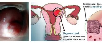
The forming node may be located on the surface of the organ wall. In this case, treatment is faster and the prognosis is favorable. Fibroids with centripetal growth, in which the tumor node grows inside the tissue, are much more difficult to treat. The consequence of the pathological process is a complete breakdown of the functions of the reproductive organ.
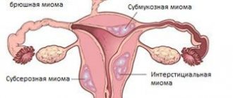
The picture shows how fibroids are located with centripetal growth (interstitial node)
The growth of uterine fibroids with a centripetal node is characterized by a slow pace. However, rapid development of a neoplasm, which is characterized by abnormal development, is also possible. Treatment in most cases is surgical, since the germination of the node creates difficulties for the effect of drugs.