Why is subserous uterine fibroid dangerous? A benign tumor of the muscle cells of the uterus is one of the most common non-inflammatory diseases in gynecology. It causes infertility in many women of reproductive age, and also provokes uterine bleeding, menstrual irregularities and chronic pain syndrome. How does subserous fibroid manifest itself and how can it be eliminated without consequences for health?
General information about uterine fibroids
Uterine fibroids are a tumor consisting of smooth muscle and fibrous cells, having a developed network of blood vessels and a pseudocapsule. It is benign - it does not destroy underlying tissues and does not metastasize. Its growth is due to increased production of ovarian hormones (estrogen and progesterone), as well as metabolic disorders and an unhealthy lifestyle. Under the influence of unfavorable factors, abnormal cells appear, which, quickly dividing and overgrowing with blood vessels, form a tumor node.
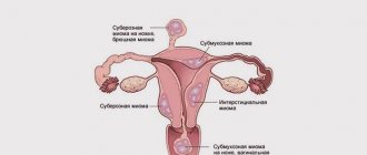
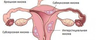
In relation to the layers of the uterus, fibroids are:
- Interstitial - among the myometrial fibers.
- Submucosal - protrudes into the cavity.
- Subserous - located on the surface of the organ.
The nodes most often grow in the body of the uterus; the isthmus and cervix are less often affected. Sometimes tumors are observed growing in the thickness of the uterine ligaments, as well as sprouting other organs (parasitic). Each location is characterized by its own symptoms, which begin to appear as the diameter of the fibroids increases.
What is subserous uterine fibroid?
A subserous myomatous node is a uterine formation, more than 50% of which extends to the surface of the organ. It is located on a dense, wide or thin connective tissue base (pedicle). Sometimes it grows into the uterine ligaments, peritoneum or neighboring organs.
Pedicled fibroids are less common than neoplasms growing inside the muscular layer of the uterus. However, it does not change the organ cavity, and its clinical manifestations are mainly caused by compression of the neurovascular plexuses and nearby organs or disruption of the trophism of its tissues.
Multiple or multinodular fibroids
Among all other types, multiple fibroids are the most common. It is characterized by the development of several nodes concentrated on different parts of the reproductive organ.
As medical practice shows, multinodular neoplasms occur in 12-25% of cases. The peak of the disease occurs during the reproductive and premenopausal age of women. However, recently, cases of this pathology have begun to be diagnosed in women under 33 years of age, which cannot but worry doctors. Among girls under 20 years of age, the risk of encountering this disease ranges from 0.9 to 1.4% of cases.
In this case, the nodes can have different sizes:
- small – less than 20 mm;
- medium - no more than 60 mm;
- large - from 60 mm and larger.
As for pregnancy, due to multiple neoplasms, seminal fluid does not pass well through the fallopian tube, as a result of which fertilization of the egg becomes significantly more difficult, but this only applies to large subserous nodes. Smaller uterine fibroids do not have the same effect. But even if conception is successful, the node begins to actively develop, sometimes coming into contact with the placenta, which can lead to complications.
Causes of subserous fibroids
Any fibroid node begins to form in the muscular layer of the uterus. Subserous myomatous nodes are formed if the focus of atypical cells was closer to the superficial, serous layer of the uterus.
The most common causes of fibroid formation are:
- Physiological hormonal changes: period of menopause, pregnancy.
- Diseases of the endocrine glands.
- Hormonally active ovarian tumors.
- Diseases of the nervous system that disrupt the functioning of the hypothalamus and pituitary gland.
- Obesity.
- Smoking.
- Alcoholism.
- Long-term use of hormonal drugs.
Important! A significant role in the development of tumors in the uterus is played by disruption of the immune system. Stress, hypothermia, chronic somatic and infectious diseases suppress the immune system, preventing it from recognizing and destroying atypical cells in a timely manner.
Prevention
To prevent the development of a pathological process, a woman must closely monitor her health throughout her life. Even if a myomatous node was discovered during examination, it is necessary to adhere to some recommendations that will prevent its transition to a malignant form:
- lead a healthy lifestyle (quit smoking and alcohol, play sports);
- eat only healthy food;
- treat in a timely manner ;
- Be regularly examined by a gynecologist, which will allow you to identify diseases in the early stages of development.
Classification
According to the degree of cell maturity, subserous fibroids are of the following types:
- Conventional - a mature benign formation in which intensive growth and division of structures is not observed.
- Proliferating (growing) – in the tissues of the node, cells are actively dividing.
- Malignant (with signs of presarcoma) - when exposed to unfavorable factors, such fibroids can develop into a cancerous tumor.
Depending on the number of pathological foci, it can be single or multiple. By volume, the subserous node is:
- Small - up to 2 cm in diameter.
- Average – 4-6 cm.
- Large - over 6 cm.
The structure and size of the node are determined by ultrasound or by the results of histological examination after hysteroscopy or laparoscopy.
Multiple uterine leiomyoma
It is worth noting that multiple subserous uterine leiomyoma is a diagnosis in which two or more identical or different sized myomatous nodes are identified.
These formations can grow at different rates and may not be located nearby. The clinical symptoms of the pathology and its severity will also depend on the location of the fibroids and their size. Often, with small tumors of the uterus, a woman does not feel any symptoms, but there are cases when the disease manifests itself as follows:
- Multiple uterine leiomyoma is accompanied by intermenstrual bleeding;
- Menstrual flow may last longer than expected;
- There is a feeling of heaviness in the lower abdomen;
- This also applies to increased pressure in the pelvis and pain in the urinary system;
- A woman may face problems conceiving a baby and carrying it to term.
Multiple uterine fibroids with a subserous node can be detected during a gynecological examination, as well as on an OMT ultrasound, during hysteroscopy or laparoscopy. The choice of treatment method depends on many factors, including age and reproductive plans. Based on a number of indicators, preference is given to drug therapy or surgical intervention.
Symptoms of subserous fibroids
Clinical signs clearly manifest themselves as subserous fibroids develop. Small tumors may not manifest themselves for a long time and not interfere with a woman’s normal life.
The main symptoms of subserous myomatous tumor are:
- Nagging painful sensations in the abdomen and lower back.
- Compression of the bladder due to the growth of a formation along the anterior wall of the uterus: frequent urge to go to the toilet, compression of the ureters, complicated by hydronephrosis and pyelonephritis.
- Compression of the intestines when a node is located on the back wall of the uterus: regular constipation, discomfort during bowel movements.
- Acute pain syndrome due to torsion of the leg of the node, development of symptoms of “acute abdomen”: fever, nausea and vomiting, difficulty urinating and defecating.
Subserous tumors rarely disrupt the menstrual cycle. Myometrial contractility deteriorates, which is manifested by heavy, prolonged discharge during menstruation and uterine bleeding, regardless of the menstrual cycle.
Complications
The most common complication of subserous myoma is torsion of its stalk. In this case, a sharp, acute pain occurs in the lower abdomen. The patient's condition deteriorates sharply: the temperature rises, weakness and dizziness occur. With further development of the process, symptoms of inflammation of the peritoneum (peritonitis) appear: nausea, vomiting, diarrhea or constipation, painful urge to urinate.
Such manifestations are associated with the death of the tissues of the node when the vessels passing through the leg are compressed. Inflammation and necrosis develop, which quickly spreads to the peritoneum and neighboring organs. If the process lasts for a long time, a bacterial infection may occur, which significantly aggravates the woman’s condition.
Important! Large subserous uterine fibroids are characterized by compression of the intestines and bladder. This is manifested not only by a delay in their emptying, but also by the formation of fistulas. This condition requires urgent surgical intervention.
The influence of subserous fibroids on pregnancy is possible with the cervical-isthmus location of the node. Sperm cannot enter the uterine cavity and tubes, which makes conception impossible. Fibroids, which compress the fallopian tubes, also prevent fertilization. Subserous nodes can provoke a complicated course of pregnancy, disrupting the uteroplacental blood flow (the occurrence of hypoxia and delayed fetal development) and compressing the organs and neurovascular plexuses of the pelvis. During pregnancy, superficial fibroids have a high risk of torsion and necrosis.
Does a subserous intramural node affect pregnancy?
Uterine fibroids are a real challenge for expectant mothers. Since there are known cases of spontaneous termination of pregnancy due to the rapid growth of a tumor, women’s fears are not unfounded. However, if the subserous node with fibroids is quite small and there are no prerequisites for its enlargement, there is no need to worry. Many women successfully carry and give birth to completely healthy babies, after which they move on to the issue of treating the pathology.
About pregnancy with fibroids >>
If there are multiple subserous nodes located on both walls of the reproductive organ, this means that the expectant mother is at risk of premature birth or miscarriage. In the second and third trimester, in addition to the risk of miscarriage, heavy uterine bleeding often occurs. They come in varying intensities, but do not reduce the risk of serious complications.
Diagnostics
With a bimanual vaginal examination, the gynecologist can detect dense areas of tumor tissue and approximately determine their diameter and number. During the examination, the doctor takes swabs from the vagina and cervical canal to detect possible infections that worsen the disease.
The main diagnostic method confirming the presence of a subserous tumor on the uterus is examination of the pelvic organs using ultrasound. Ultrasound can examine fibroids of any location, accurately determine its structure and occupied volume, and also, using Doppler ultrasound, establish blood flow in the area of the pathological neoplasm.
This makes it possible to distinguish a benign tumor from a cancerous one: the blood flow rate in myoma is much less than in myosarcoma.
Magnetic resonance and computed tomography can be used to determine the structure and location of myomatous subserous tumors of even minimal size.
In addition to instrumental tests, patients with fibroids are prescribed a number of laboratory tests:
- General and biochemical blood test.
- Serological analysis for certain types of infections (HIV, parenteral hepatitis, syphilis).
- Determination of tumor markers.
Treatment of subserous fibroids
The treatment tactics for myomatous tumors are selected depending on the intensity of its growth, size and condition of the patient.
Conservative treatment
Small subserous nodes that do not grow rapidly are treated with drug therapy. Drugs are used that either reduce the synthesis of ovarian hormones or block receptors for them on the tumor itself.
The main medications for eliminating subserous tumors on the uterus are:
- Progestogens. These are synthetic analogues of progesterone, which, when used for a long time, inhibit the production of the ovaries' own hormones. Can be used in the form of tablets, injections and intrauterine contraceptive systems.
- Combined oral contraceptives. They contain precisely adjusted doses of sex hormones that help reduce ovarian activity.
- Gonadotropin releasing hormone antagonists. They disrupt the hypothalamic-pituitary-ovarian connection, inhibiting the synthesis of sex hormones.
- Progesterone antagonists. They block the hormone receptors of the tumor itself, due to which it begins to quickly decrease in size.
The duration of hormonal treatment is determined by the attending physician. In this case, after a course of drugs, a control ultrasound is prescribed, based on the results of which the effectiveness of the therapy is assessed.
When subserous uterine fibroids and pregnancy are combined, hormonal treatment is not prescribed.
It is possible to relieve symptoms with drugs approved during gestation and regular medical and ultrasound monitoring of the activity of the node. If severe complications develop, it is possible to remove fibroids during pregnancy.
To treat the symptoms inherent in subserous nodes, the following medications are prescribed:
- Non-steroidal anti-inflammatory drugs (Diclofenac, Indomethacin, Nimesulide). They can be prescribed in the form of tablets and rectal suppositories. Eliminate pain and inflammation.
- Hemostatic agents (tranexamic acid). Used for uterine bleeding caused by a tumor, reduces the amount of menstrual flow.
Surgery
Indications for surgical removal of subserous uterine fibroids are:
- Large tumor diameter.
- Intensive growth.
- Node activity during the menopausal period.
- Lack of effect from conservative treatment.
- Trophic disturbance and necrotization of the node due to torsion of the leg.
Currently, laparoscopy is considered the most common method for removing uterine nodes. Endoscopic instruments are inserted through small punctures in the navel area, which are used to cut off the node and clamp the bleeding vessels. And they also examine the pelvic cavity for the presence of other pathologies or possible surgical injuries.
A minimally invasive way to eliminate subserous uterine nodes is uterine artery embolization. The catheter is inserted under X-ray control and passed through the femoral artery to the vessels of the uterus. When the pathological focus is reached, an embolic substance enters the tumor vessels through it, disrupting the blood flow in the node. Myoma undergoes regression.
Why does the tumor grow?
The reasons for the appearance of a tumor in a woman’s body have not been fully established. However, recent studies have found that a major role in maintaining fibroid cell division belongs to the disturbed balance of estrogen and progesterone, with the relative predominance of the former. Estrogens have proliferative activity; they stimulate cell growth and division. But progesterone also plays an important role in this process. It has been proven that after the tumor begins to grow, the number of receptors for this hormone in it increases, which leads to additional stimulation of the cells of which it consists and the maintenance of their division. Progesterone also reduces local immune defense and disrupts the mechanism of apoptosis - programmed cell death. Therefore, cells in fibroids do not die, as should happen normally.
With an imbalance of estrogen and progesterone - hormones that are responsible for a woman’s reproductive health - there is a high risk of tumors.
Numerous studies have found that there are certain factors that can increase or decrease the risk of developing fibroids. Most often, factors predisposing to the appearance of pathology act as a complex.
The following conditions increase the risk of fibroids:
- Early onset of menstruation;
- Absence of childbirth in patients over 30 years of age;
- Late reproductive age (after 35 years);
- Obesity and endocrine pathology;
- Treatment with Tamoxifen and some other drugs that change hormonal levels;
- African American race.
Reduce the risk of developing fibroids:
- A large number of births;
- No miscarriages and no induced abortions;
- Menopause;
- Long-term breastfeeding (more than 6 months).
On a note
There is no exact information about the influence of combined oral contraceptives, hormonal therapy, dietary habits, or region of residence on the development of leiomyoma.
Myoma growth can occur in different directions. Depending on its location relative to the uterine wall, it is divided into several types:
- Submucous - protrudes into the uterine cavity, can be located on a stalk;
- Intramural – grows in the thickness of the muscle layer;
- Subserous - located mainly under the peritoneum or extends beyond the uterus, connecting to it with a thin stalk;
- Interligamentous – located between the layers of the uterine ligament;
- Cervical - grows in the area of the cervix.
Myomatous nodes, depending on their location, are classified into several types, which may differ in symptoms, clinical form, and treatment methods.
Extracorporeal cervical nodes account for only 5% of the total number of myomatous formations. The main location of the tumor is the body of the uterus.
As mentioned above, each form of leiomyoma has its own characteristics of the course of the disease and characteristic symptoms, as well as approaches to treatment. More complete information about the different types of fibroids can be found in a separate article. The most blurred picture of manifestations is in a tumor with a subserous location. In ICD-10 it is assigned code D25.2.