Uterine fibroids occupy a leading place among gynecological diseases in women of the childbearing age group. Without a clear clinical picture in most cases, uterine fibroids progress in many possible types and variants.
Myoma is a hormone-dependent pathology in which the formation and further development of a tumor-like neoplasm occurs in the thickness of the uterine myometrium. Myoma is not an oncologically dangerous disease, however, if it is not promptly treated with medications and folk remedies, surgical intervention may be necessary.
The myometrium is the middle muscular layer of the body of the uterus. It is located between the perimeter and endometrium, ensuring the growth of the uterus during pregnancy.
The development of uterine fibroids occurs in several successive stages:
- the formation of a growth zone, where there is an increase in blood flow and vascular permeability;
- development of a tiny node that is visible only microscopically;
- the tumor reaches a size sufficient to be detected macroscopically.
The formation can be localized both in the uterine body and in the cervical part. Nodes located in the body of the uterus are of several types.
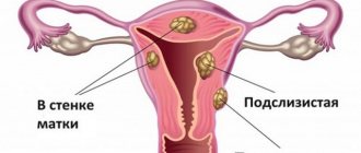
- Intramural or interstitial. Such neoplasms develop inside the myometrium. Intramural uterine fibroids are a common type of benign tumor. An intramural node is diagnosed in approximately 60% of the total number of fibroids.
- Submucosal or submucosal. Tumors of this variety grow under the mucous membrane. Submucosal fibroids have good blood flow and, due to their localization, progress faster than other nodes.
- Intraligamentary. This is a rather rare type of fibroid that can be found between the layers of the broad ligament of the uterine body.
- Retroperitoneal. These neoplasms grow from the lower section, which is represented by the uterine cervix.
- Subserous or subserous. Fibroids, located subserosally, progress under the peritoneum or serosa. Some subserous fibroids combine two types: subserous and intramural (interstitial) nodes.
Interstitial subserous uterine fibroids - what is it? Interstitial subserous uterine fibroids mean a fibroid that is subserosal, and its node is located intramural.
Interstitial-subserous uterine fibroids are characterized by latent progression, which causes its late diagnosis and treatment, when medications and folk remedies are powerless.
Intramural-subserous fibroids are usually multiple in nature. Single interstitial subserous fibroids of the uterine body respond better to treatment with folk remedies and medications. In addition, the interstitial or intramural subserous form can be treated surgically using organ-preserving tactics.
What it is
Fibroids are divided into types depending on the location of the benign formation relative to the uterine wall. Therefore, to better understand how one type of fibroid differs from another type, you should consider the structure of the uterine wall.
It consists of 3 layers:
- external - it is called serous or perimetry;
- middle - muscle layer, myometrium or interstitium;
- internal - endometrium or mucous membrane.
There are 3 main forms of uterine fibroids depending on the direction in which the tumor grows:
- Intramural or interstitial - the tumor develops only in the interstitium.
- Subserous or subserous - the neoplasm is located on the outer surface of the myometrium under the serous membrane and grows towards the abdominal cavity. It is attached to the uterus with a stalk and can extend beyond the organ.
- Submucosal or submucosal - the formation appears under the inner mucous membrane of the uterus, and increases in the direction of the cavity.
A tumor that develops between the layers of the uterine ligament is called intraligamentary or interligamentous, and in the cervix it is called cervical.
An interstitial subserous tumor, having appeared in the thickness of the myometrium, protrudes outward between the muscular and serous membrane of the uterus. It refers to a mixed type of uterine fibroids.
It is united with the subserous variety by the same direction of growth of the node - towards the abdominal cavity. The deep penetration of the base of the interstitial subserous tumor into the myometrium makes it similar to interstitial myoma.
Pregnancy and uterine fibroids
The chances of conception with such a diagnosis, although not great, are still there. If the node is located directly in the body of the uterus, then there are no obstacles to the movement of sperm.
Important!
Interstitial-subserous fibroids during pregnancy can provoke rapid heartbeat and shortness of breath. In addition, the risk of spontaneous abortion or premature birth increases. This is due to the fact that the tumor produces substances due to which the uterus begins to actively contract.
Interstitial-subserous fibroids of the uterine body do not require special treatment before birth, since during the period of bearing a child the consequences of any intervention are dangerous. Doctors systematically monitor the size that the myomatous node acquires. Specialists ensure that the pregnant woman is in a relatively stable condition. If the interstitial-subserous myomatous node acquires a critical size, termination of pregnancy can be performed according to indications.
Despite the risk of complications, the diagnosis may have a favorable prognosis. It is important to be attentive to your health and regularly see a gynecologist in order to begin therapy at the initial stage.
Location of the tumor in the uterus
The subserous-interstitial type of uterine fibroids can be either single or multiple. The second option is more common. In the solitary form of the disease, only one tumor develops. Multiple fibroids are characterized by the presence of 2 or more tumors that have different locations in the uterus, as well as growth rates.
Myomas can be located on a broad base or on a thin stalk with a base in the myometrium.
The second variant of tumor development poses a great danger to the health and life of a woman. It is associated with a high probability of torsion of the leg of the node. With such a complication of the disease, urgent hospitalization of the woman is necessary.
The choice of treatment is the prerogative of professionals
Even taking into account the fact that fibroids are a benign tumor, it is strictly forbidden to ignore them. And if, after the next medical examination, the doctor reveals myomatous nodes, you should immediately begin to restore your health and immediately begin treating the disease. Today there is no panacea that could cure fibroids like the flu in a few days, so it is better to entrust the choice of the most effective method of therapy to an experienced doctor. The specialist not only knows the answer to the question, what is a myomatous node - what is it, but also has enough experience to choose the safest and most effective treatment option for each patient.
Previously, the only way to restore women's health with uterine fibroids was surgery. However, today medicine has advanced greatly, and with small tumors it is possible to solve the problem with medication.
Causes
There are several reasons for the appearance of the interstitial-subserous variety of myomatous nodes. The main cause of the development of the disease is considered to be a hormonal imbalance, in which the level of progesterone is below normal, and estrogen, on the contrary, exceeds normal. It is for this reason that the disease often develops in women after 30 years of age.
The following reasons can provoke the development of a tumor:
- the presence of chronic inflammatory processes in the organs of the reproductive system;
- the intrauterine device was incorrectly installed;
- the woman took hormonal contraceptives for a long time, which she prescribed for herself without checking her hormonal levels and without visiting a gynecologist;
- the woman did not give birth;
- the uterus was injured during difficult childbirth, abortion or surgery;
- the woman suffered a severe nervous shock or was in a stressful state for a long time;
- suffers from diabetes;
- the presence of excess weight and metabolic disorders;
- there is a hereditary predisposition - your mother, sister or grandmother had fibroids.
Irregular sex life or a constant lack of orgasm during sexual intercourse can lead to the development of the disease. Both situations lead to stagnation of blood in the pelvic organs. The risk group includes women who have not had a single pregnancy before the age of 30 and who gave birth for the first time after the age of 35.
Diagnosis and treatment
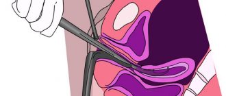
A pathological disorder in the body of the uterus is diagnosed using ultrasound examination.
The specialist uses a vaginal sensor. Additionally, hysteroscopy and examination on a gynecological chair may be indicated. The choice of treatment method takes into account a number of factors:
- woman's age,
- size and type of tumor neoplasm,
- accompanying pathological processes in the body,
- probable risks and threat of complications,
- specific localization of myomatous nodes.
Since subserous-interstitial uterine fibroids are a hormone-dependent tumor, hormonal techniques become the basis of conservative treatment . It allows you to restrain the growth and development of the tumor. If there is no bleeding or severe pain, then treatment can be carried out on an outpatient basis.
Subserosal interstitial fibroids can be treated using the following methods:
- correction of hormone levels in the body,
- taking steroid medications with antitumor properties,
- surgical removal of a tumor that has affected the body of the uterus,
- the uterus may also be removed.
When the tumor is small, for example, only nine millimeters, and the patient does not have pain, bleeding or other unpleasant signs, hormone therapy is usually indicated. It is necessary to artificially reduce the concentration of estrogen. It is important to combine such therapy with a diet recommended by your doctor. This method is especially relevant for women after 50 years of age, because there is a possibility of spontaneous disappearance of myomatous nodes at this age. Girls planning to become pregnant need to take treatment seriously; sometimes a “wait-and-see” approach is inappropriate .
Surgical excision is indicated:
- if there is a risk of complications: torsion of the tumor nodule,
- with dynamic intensification of the symptoms of the pathological process,
- with rapid growth or large size of the tumor even at the diagnostic stage.
Symptoms
The appearance of the interstitial-subserous type of formation is not accompanied by any symptoms. They appear after the fibroid increases in size and begins to put pressure on nearby internal organs: the bladder and intestines in the rectal area.
The following common symptoms of an overgrown tumor can be identified:
- discomfort or pain during sexual intercourse;
- heavy menstrual flow accompanied by painful sensations;
- aching pain in the lower abdomen, which can radiate to the lower back, perineal area or anal area;
- the urge to urinate becomes more frequent, and at the same time, there may be a feeling that the bladder is not emptied completely;
- Pressure on the rectum causes frequent constipation, and sometimes hemorrhoids develop.
Single and multiple fibroids have the same symptoms. The brightness of their manifestation depends on the size of the nodes and their influence on neighboring internal organs.
How does the pathology manifest itself?
Symptoms of an interstitial subserous tumor appear when it reaches a large volume. Due to the fact that such a tumor grows not inside, but outside the organ, it does not necessarily lead to heavy menstrual bleeding. This is due to the fact that due to it the area of the endometrium does not increase.
The following signs are encountered:
- frequent or difficult urination (pressure on the bladder)
- pelvic, sacral pain (pressure on nerve endings)
- constipation (rectum is compressed), swelling of the legs (pressure on the pelvic veins)
- pain from the lower abdomen, radiating to the thigh.
Diagnostic methods
It is most difficult to diagnose interstitial-subserous tumors of small size - up to 2 cm in diameter. A gynecologist cannot always detect them by palpating the uterus during a standard examination due to the peculiarities of localization.
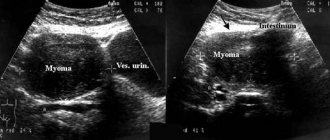
But small myomatous tumors will be detected on an ultrasound of the pelvic organs.
It is very important to monitor your women's health and visit a gynecologist every six months and have an ultrasound of the pelvic organs every year. With this approach, fibroids will be detected at the very beginning of their development, when it is easier to cure.
If the fibroid has grown, during the examination the gynecologist will identify changes in the size and contours of the uterus. The symptoms voiced by the woman, changes in the uterus detected by the doctor and pain during palpation serve as the basis for issuing a referral for an ultrasound of the pelvic organs.
Based on its results, the following studies are prescribed:
- Transvaginal ultrasound is the main study confirming the interstitial subserous appearance of the tumor. The woman sits in a gynecological chair, and an ultrasound machine sensor is inserted into the uterine cavity through her vagina. This study allows you to accurately determine the number of tumors, their size and density.
- MRI of the pelvic organs. Magnetic resonance imaging uses x-rays that are sensitive to soft tissue. The device takes layer-by-layer images of the uterus, and a computer program, by combining frames, allows you to obtain a three-dimensional image. During the study, the nature of all formations, their size, exact location and the relationship between them will be accurately determined.
- Ultrasound Dopplerography of the vessels of the uterus. It is prescribed to assess the intensity of blood flow feeding the fibroid and confirm the benign nature of the formation.
- Hysterosalpingography or metrography. Allows you to determine the degree of deformation of the uterine cavity. X-ray examinations of the uterus are carried out, into which a contrast agent has previously been injected. Interstitial-subserous multiple fibroids lead to a narrowing of the lumen of the uterus, which will not happen with other types of fibroids.
If there is a node on a thin stalk, laparoscopy is prescribed as a diagnostic method. If indicated, fibroids can be removed immediately.
Uterine fibroids - interstitial form: drug treatment
Conservative treatment involves the use of hormonal drugs that block the production of estrogen. There are also medications that reduce the sensitivity of the myometrium to this hormone. This treatment is especially effective for small tumor sizes, in conditions such as regressing interstitial uterine fibroids.
Hormonal treatment will be effective if the intramural fibroid is small. For treatment the following are prescribed:
- gonadotropin-releasing hormone agonists;
- antigonadotropic drugs;
- antiprogestogens;
- progestogens.
The above medications can only be prescribed by a doctor after additional examination. The patient’s treatment regimen is developed taking into account the test results.
What is the danger of the disease
The greatest danger is posed by nodes on the leg.
Due to a sudden movement, serious physical activity, or for another reason, the leg of the fibroid may become twisted, and complications will arise that are dangerous to the life and health of the woman:
- The development of necrosis is the death of tumor tissue caused by disruption of its nutrition due to torsion of the pedicle. Necrosis has a clinical picture of acute abdominal pain: sharp pain, fever, increased heart rate, possible vomiting and loss of consciousness. The patient's prolonged stay in this condition is dangerous for her life.
- Threat of peritonitis. The breakdown of tumor tissue can lead to the appearance of an inflammatory process in the abdominal cavity and the development of peritonitis.
- Heavy bleeding. If the leg is twisted, the vessel feeding the node may rupture, and heavy bleeding will begin, which is quite difficult to stop.
- Risk of death. If hospitalization is not timely, an infection may accompany the bleeding, and the patient’s life cannot always be saved.
In all of the above cases, it is important to immediately call an ambulance. An emergency surgical operation will be scheduled in a hospital setting.
Removal and other methods
Hysterectomy, a radical method of treating tumors of this type, is performed for women in menopause or with a large number of large fibroids. It should be noted that it is the interstitial-subserous neoplasm that can be removed by laparoscopy. Especially if it grows on a stalk. Large tumors are excised using an open method (a full abdominal incision).
If conservative or surgical treatment is not carried out, the tumor is treated with radiation therapy. Principles of the method:
- the node is susceptible to such a dose of radiation that causes infertility,
- as a result, menstruation stops, and the tumor decreases.
Another treatment option: embolization (blocking) of the uterine arteries. Local anesthesia is used. The operation can be described as follows: the arteries feeding the tumor are blocked. The tumor becomes nonviable and dies. The method requires special experience of the surgeon.
The most modern treatment accepted in developed countries is FUS-MRI ablation, when the tumor is heated with ultrasound and it disintegrates due to thermal necrosis. No hospitalization is required, the uterus and fertility are preserved.
What are the differences from other forms of fibroids?
Myomatous node of interstitial-subserous type belongs to the category of mixed tumors. It has some similarities with two main types of fibroids. The base of an interstitial subserous tumor appears deep in the muscular layer of the uterine wall, similar to intramural myoma. This leads to a slight increase in the body of the uterus.
The direction of growth of the interstitial-subserous node is the same as that of the subserous node - towards the abdominal cavity. Both types of fibroids, when growing, compress nearby internal organs, and thereby inhibit their basic functions.
But interstitial subserous tumors can grow to gigantic sizes. In medical practice, fibroids with a diameter of about 25 cm have been encountered, and according to the classification, a node larger than 6 cm is considered large.
Surgical method of treatment
Doctors recommend removing the pathological formation when the size of the diagnosed fibroids reaches 12 or more weeks of pregnancy. Also, indications for surgical intervention may be rapid tumor growth, or a vivid clinical picture of the disease (severe pain, frequent heavy bleeding, etc.). But even if the doctor recommends removal of myomatous nodes, this is not yet a reason for concern. Modern surgical capabilities are quite extensive, so the consequences of the operation will be insignificant.
Treatment
Treatment of uterine fibroids of the subserous-interstitial type is carried out using the following methods: conservative, surgical and UAE. The decision on the method of treatment is made by the doctor, taking into account the number of fibroids, their size, location and the presence of a pedicle.
Drugs
Drug treatment is prescribed to women whose tumors do not exceed 2-3 cm in diameter and who plan to become pregnant in the future. After a thorough examination and blood tests for hormones, the woman is selected for combined hormonal contraceptives.
Taking them allows you to normalize hormonal levels and stop tumor growth. In some cases, during therapy, the tumor gradually decreases in size.
The following drugs have a good effect: Desogestrel, Ethinyl estradiol and Norgestrel. The drug and its dose should be prescribed exclusively by a doctor.
Recipe for folk remedies with herbs
When it was possible to diagnose a small fibroid - up to 2 cm in diameter, then you can use folk remedies.
An alcohol tincture of the root of peony evasive, popularly called marina root, is popular. Despite the name, not alcohol is used for preparation, but 40% vodka. The tincture can be purchased at a pharmacy or prepared yourself. You need to pour 50 grams of marina root into 0.5 liters of vodka and leave in a dark place for 14 days, shaking the infusion daily.
Then strain and store in a dark glass bottle in a dark, dry place. Take 1 teaspoon of infusion, diluted in a small amount of water, 3 times a day, half an hour before meals. The course of therapy is 4 weeks, and then a week break and a new course. According to this scheme, the drug is taken until complete recovery. It is important to monitor the condition of the tumor by performing control ultrasounds.
A good effect is obtained by taking propolis tincture orally, which is sold in pharmacies. The tincture is taken with a small amount of water or milk. 20% tincture is taken 20 drops 3 times a day before meals. The course of therapy is 20 days, and after a 10-day break, a repeat course. 30% tincture is taken once a day: 2 hours before bedtime, dilute 1 tbsp. spoon of tincture in water and drink. The course of therapy is 10 days, then a 5-day break and a new course. In total, you need to conduct 3 treatment 10-day courses.
Before choosing any traditional recipe, consult your doctor.
Surgical intervention
The method of surgery depends on the number of tumors and their location. Hysterectomy or removal of the uterus is extremely rare. It is prescribed for the development of several large nodes, leading to complications that threaten the patient’s health. More often, myomectomy is prescribed - an operation to remove fibroids, in which the uterus is preserved.
It is carried out in 2 ways:
- Laparoscopic - punctures are made in the lower abdomen and a special device - a laparoscope - is inserted through them. Under the control of a video camera, the surgeon, using a laparoscope, excises and removes the tumor.
- Laparotomy is an abdominal operation in which an incision is made in the lower abdomen.
The choice of method for removing fibroids depends on their location, growth in the myometrium and deformation of the uterus. To remove nodes on the leg, the laparoscopic method is chosen. When the operation cannot be performed for some reason, the UAE procedure is prescribed.
EMA
UAE is a uterine artery embolization procedure. During the painless procedure, special small synthetic balls - 300-700 microns - are injected into the uterine arteries through a puncture in the femoral artery. Together with the blood, they enter the terminal vessels that supply the myomatous node and block its blood supply.
As a result of the cessation of the supply of oxygen and nutrients, the reverse development of fibroids begins. After a few weeks, the smooth muscle cells of the tumor are replaced by connective tissue. Externally, the node, reduced in size, has a rounded shape, but in structure it is no longer a fibroid. It does not cause heavy periods or other symptoms, and does not cause problems during pregnancy and childbirth.
Reasons for growth
Uterine fibroids of any type progress due to hormonal disorders. Interstitial fibroids are formed during the embryonic formation of the girl’s genital organs. It has been established that uterine fibroids are associated with increased levels of estrogen hormones, which trigger the processes of proliferation of smooth muscle cells.
However, in modern gynecology, other hypotheses are also being developed to explain the occurrence of neoplasms of the uterine body. In particular, experts consider hyperplasia of muscle cells as the cause.
The etiology and pathogenesis of a disease such as uterine fibroids have not been sufficiently studied. In modern gynecology, three hypotheses are actively being developed:
- congenital structural features of the uterine wall caused by intrauterine developmental disorders;
- a tumor of the muscular wall of the uterus is considered by some authors as hyperplasia rather than a tumor formation;
- acquired pathology that develops as a result of repeated surgical interventions.
Interstitial or intramural uterine fibroids go through several stages of development:
- the appearance of a zone of increased growth in the area of localization of small-caliber spiral arteries of the uterus;
- progression of a microscopic nodule;
- macroscopic appearance of the neoplasm.
The following factors can provoke the growth of interstitial or intramural fibroids along the anterior and posterior wall of the uterus:
- endocrine disorders;
- multiple surgical procedures;
- excess weight;
- lack of physical activity;
- high blood pressure in women under 30 years of age;
- family history aggravated by the disease;
- onset of menstruation before age 11;
- late first birth;
- stress;
- venous stagnation.
Interstitial and intramural myomatous tumors have different rates of progression. The volume of neoplasms is measured in weeks of pregnancy. Large uterine fibroids cause deformation of the uterus.
Before starting treatment for nodules, both large and small, it is necessary to eliminate the provoking factors.
What effect does it have on pregnancy?
If there are small fibroids of the intestinal-subserous type, a woman will be able to become pregnant. Quite often, expectant mothers learn about the presence of fibroids during pregnancy. If the tumor is not prone to rapid growth, then the pregnancy is not in danger, and the child’s development meets all standards.
Large tumors can compress the fallopian tubes, which will lead to disruption of their patency and problems with conception. If a woman does manage to get pregnant, there will be threats of miscarriage. Myomatous cells secrete a special substance that increases the contractility of the muscle tissue of the uterus.
There may also be problems with the development of the placenta due to pressure on the uterus from a large myomatous node and the hormonal imbalance that accompanies the disease. Without receiving enough nutrition, the fetus may develop abnormalities. Torsion of the tumor stalk or its active growth are indications for termination of pregnancy for medical reasons.
During pregnancy, the intertial-subserous type of uterine fibroids is not treated, but only monitored for its development. After all, hormone therapy during pregnancy is contraindicated.
Symptoms
Usually, the symptoms of the uterus do not manifest themselves clearly, or the pathology is generally asymptomatic. The situation is complicated by the fact that subserous-interstitial uterine fibroids are difficult to diagnose and are also difficult to remove surgically.
Characteristic signs for the disease:
- pain in the abdomen, in the lower part, manifesting itself with different irradiations, radiating to the anal area, lumbar area, perineum;
- problems with the outflow of urine or increased excretion - this is due to the fact that the myomatous node, growing, puts pressure on the kidneys or ureter;
- long, heavy menstruation - due to problems with the contractile function of the smooth muscle tissue that covers the uterus.
As the myomatous node develops, the intestines, rectum, and bladder function worse. If at the initial stages the body of the uterus did not experience such pressure due to the small size of the neoplasm, then with growth the painful sensations gradually increase, although many women do not attach importance to them, since they consider them to be a variant of the norm. Serious disruptions in the menstrual cycle also do not occur until the tumor reaches a large size and grows.
What is an interstitial node of uterine fibroids and why is it so scary?
The interstitial node of fibroids of the anterior and posterior wall of the uterus is a benign neoplasm.
Until recently, it was believed that such a formation could degenerate into a cancerous tumor, and accordingly, the only possible method of treatment was considered to be surgery to remove the tumor along with the uterus. Organ preservation was considered only in the case of surgical intervention in young girls planning a future pregnancy. It is for this reason that the diagnosis of uterine fibroids sounded like a death sentence. Attitudes towards this disease began to change only in the early 90s, when the results of scientific research proved that the interstitial node cannot degenerate into oncology. The likelihood of developing cancer with uterine fibroids is equal to the development of a malignant tumor even in its absence from healthy cells of the female body.
Despite this scientific discovery, most domestic doctors continue to treat the tumor using the method of removing it during surgery. On the part of doctors, this method of “treatment” is a crime against their patients. Surgery should be a last resort when other treatments have failed. Surgical intervention in most cases deprives women of the opportunity to have children in the future, and also leads to a number of complications due to the removal of the reproductive organ.
Today, fibroids (interstitial node) can be successfully treated in all clinics around the world using uterine artery embolization. This technique is also successfully used by doctors in our medical institutions. Clinics for the treatment of fibroids, where this method is practiced, are equipped with modern medical equipment and include highly qualified doctors on their staff.
Numerous studies have revealed the true nature of benign uterine tumors. It represents the reaction of the reproductive organ to damage. In this case, such damage is repeated menstruation.
The fact is that nature has laid down the main function of the female body - procreation. Thus, with the onset of reproductive age, pregnancy should follow, then a period of breastfeeding, two or three menstruation and pregnancy again. According to this plan, girls should have had no more than forty menstruation in their entire lives.
Of course, in practice, not a single woman performs her reproductive function with such enthusiasm. On average, by the age of thirty, she becomes a mother once or twice, and the period of breastfeeding on average takes about a year. With this development of events, approximately 400 menstruation is observed throughout life.
It is unlikely that nature intended for the fair sex to experience a whole “bouquet” of negative sensations caused by menstruation every month. These include headaches, attacks of nausea and vomiting, pain in the lower abdomen and lower back, dizziness, frequent mood swings, tearfulness, changes in taste preferences, etc.
Every month, the female body prepares for pregnancy, “tuning” each of its organs. When fertilization does not occur and pregnancy does not occur, the body returns to its “original settings.”
Like any other repeated process, monthly menstruation can create errors, especially in combination with surgical interventions and inflammatory processes. This is how most diseases of the female reproductive system arise, including uterine fibroids (the interstitial node of the anterior and posterior walls of the organ).
The interstitial node along the posterior wall of the uterus, as well as the interstitial node along the anterior wall of the uterus, is initially formed from one individual cell of the uterus in the form of tiny rudiments. Tumors begin to grow against the background of hormonal fluctuations. Such changes in hormones occur during menstruation. Moreover, fibroids can grow in different ways - some faster, others slower, and others can disappear as unexpectedly as they appeared.
The growth of tumor primordia can be accelerated by certain provoking factors, including:
- Abortions, gynecological curettages, operations, difficult childbirth;
- Endometriosis;
- Inflammatory processes.
Separately, juvenile forms of interstitial fibroid nodes are distinguished. It is believed that damage to the cells of the uterus, from which neoplasms subsequently develop, occurs during the prenatal period. With the onset of menstruation, that is, with a sharp surge in hormone levels, the interstitial nodes of the fibroids begin to grow. Juvenile forms of neoplasms are diagnosed in girls under the age of 25.
Women planning to conceive in the future are prescribed the following operations:
- Laparoscopy, in which the myomatous node is removed using a laparoscope. The woman is given only a few punctures in the abdomen, through which the formation is directly removed. The doctor monitors the progress of the operation on the screen;
- Hysteroscopy – a special instrument is inserted through the vagina to remove the tumor. The process is also monitored on a monitor;
- Blocking the blood flow to the tumor is scientifically called embolization of the uterine arteries. Due to the blocking of blood supply, the fibroid simply dries out.
If the formation has grown to an impressive size, then before surgery you will have to take special medications that reduce the size characteristics of the node.
How is subserous-interstitial uterine fibroid treated?
If the interstitial myomatous node is small, hormonal therapy is performed. Since tumors are sensitive to hormones, normalizing the patient’s endocrine background often leads to a reduction in fibroids. Additionally, treatment such as physiotherapy and vitamin therapy is carried out. When large nodes are detected, surgery is indicated, since the tumor stalk can twist.
During surgery, healthy uterine tissue is preserved, and reproductive function is not impaired. Only the neoplasm is excised. If pregnancy occurs with fibroids, surgical treatment is performed only if there are absolute contraindications. With this pathology, many women give birth successfully, but doctors prescribe the following medications:
- tocolytics;
- antiplatelet agents;
- antispasmodics;
- antibiotics (rarely).
How to treat uterine fibroids with folk remedies
Therapy for small myomatous nodes can be done using folk remedies. Excellent in combating grass pathology: marigold, yarrow, nettle. Crushed dry plants, taken in equal proportions, are poured with boiling water and then infused. The broth is cooled, filtered, then drunk 3 times a day for 1-2 months.
Another effective folk remedy that normalizes hormonal levels is tincture of walnuts. To prepare it, you need to grind 30 g of partitions, then pour 1 glass of alcohol into them. The medicine is infused for 10 days in the dark, after which it is wrung out and filtered. You need to drink 30 drops of the tincture before meals until it runs out.
How is interstitial-subserous uterine fibroid diagnosed?
In diagnostic centers, the disease is diagnosed using MRI, ultrasound, and hysteroscopy. With interstitial fibroids, it is easy to determine the deformation of the uterus and an increase in size during a gynecological examination. Ultrasound visualizes even very small myomatous nodes, the direction of growth, the calcification process and the composition of the uterine myometrium. On ultrasound, fibroids look like a different structure in relation to the surrounding tissues. Using hysteroscopy, you can assess the condition of the intramural and submucosal node. Such diagnostics often help to identify the initial stage of necrosis.