It is now difficult to surprise anyone with a diagnosis of uterine fibroids. About 25% of the entire female population often faced a similar diagnosis. Often this disease occurs latently, revealing itself only during a routine examination. We suggest you learn about the features of ultrasound for uterine fibroids, indications, contraindications and interpretation of results from this article.
Myoma and its manifestations
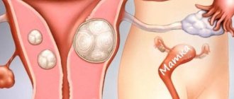
Uterine fibroids are a benign neoplasm of the muscular layer. It usually affects women of reproductive age (30-45 years). Recently, such tumors have become not uncommon in girls 20-25 years old (usually those who have not given birth or are not sexually active). This is a hormone-dependent pathology: it is associated with the quantitative ratio of hormones. Along with the growth of muscle cells, the tumor can grow and form knots of intertwined smooth muscle fibers. Tumor formations can be single or multiple, and their sizes can vary from a few millimeters to several kilograms.
The reasons leading to the appearance of fibroids are still not fully known. They are based on hormonal imbalances, traumatization of the mucous membrane, or exposure to a virus.
The most common “push” factors for the appearance of fibroids are:
- imbalance of steroid hormones;
- late or complicated labor;
- interventions in the uterine cavity (abortion, curettage);
- inflammation in the uterus;
- combination of excess weight and stress;
- uterine bleeding during menstruation;
- associated ailments (obesity, diabetes, thyroid disease, etc.)
Most often, for the appearance of a myomatous node, a combination of several unfavorable factors is necessary: for example, abortion and subsequent accumulation of infection.
Also important are genetic factors that contribute to a chain of disorders in the immune-hormonal system, leading to impaired epithelial growth.
The most common complications of fibroids are:
- anemia;
- infertility;
- accelerated growth;
- cancerous degeneration.
How often to do an ultrasound for fibroids
Usually, when, or more precisely on what day of the cycle, an ultrasound is done for uterine fibroids, greatly affects the accuracy of the study and the chance of detecting a tumor. But in some cases, the examination can be performed even during menstruation using the transabdominal method.
It is better to undergo examination on days 3-5 or 5-7 of the cycle, when ultrasound images of the pelvis will provide the most accurate data about fibroids in the uterus.
What are uterine fibroids?
This is a tumor that is localized in the myometrium. Therefore, for a more reliable diagnosis, you need to know when it is better to do an ultrasound for uterine fibroids.
The neoplasm may consist of only fibers in the muscles. The tumor is then called leiomyoma. Or contain connective tissue (fibroids).
The tumor is considered benign, but can still be dangerous, especially during pregnancy. Because due to the pressure of the neoplasm on the uterus, there is a risk of damage to the fetus.
The appearance of fibroids is due to a number of reasons. Doctors include hormonal imbalance, gynecological operations, heredity, inflammation of the uterus and even poor nutrition as risk factors.
The risk of developing fibroids increases greatly as the patient ages. In women aged 20-35 years, the neoplasm is diagnosed in 15-20% of cases. The risk of a tumor doubles when a woman turns 40, so in adulthood you need to know exactly when and how often it is best to do an ultrasound for uterine fibroids.
Often fibroids develop unnoticed by a woman and can be detected in the early stages during a random examination. Often a tumor is found when they start doing an ultrasound, not in search of fibroids, but in other diseases of the uterus.
As the tumor progresses, it can cause heavy periods and bleeding in the middle of the cycle. Severe pain may occur, including in the lumbar region. When the fibroid reaches a significant size, the patient may experience difficulty urinating.
Which ultrasound is best for studying uterine fibroids?
Because of the way uterine fibroids look on an ultrasound, choosing how to test can be somewhat difficult. While the tumor is still small, a transvaginal examination is recommended. In later stages, transabdominal ultrasound
.
Transvaginal ultrasound
During transvaginal examination
a specialist can assess the dimensions of the uterus and its cervix, detect and localize fibroids, and determine their structure.
Transabdominal ultrasound
Using this method, a specialist can assess the condition of the uterine body and detect fibroid nodes larger than a centimeter in size.
Ultrasound hysterography
This method of examination is rarely used when performing an ultrasound using other methods for uterine fibroids is impossible or ineffective. Before the examination, a contrast agent is injected into the uterine cavity. Thanks to it, uterine fibroids are not always visible, but ultrasound shows changes caused by the tumor.
Preparing for an ultrasound
In most cases, preparation for an ultrasound is not required. If the examination will be performed transabdominally
, the patient is advised to come with a full bladder. This will make the image more informative.
How is the procedure done?
During the examination, the woman takes a horizontal position. When an ultrasound is performed for abdominal uterine fibroids, the sensor is placed on the stomach of the subject.
With the transvaginal method
or ultrasound hysterography, a special sensor is inserted into the vagina. In the second case, before the examination, a contrast agent is injected into the uterine cavity.
Decoding the result
In cases where the tumor is visible on an ultrasound scan, uterine fibroids are a heterogeneous round object with a clear contour. In addition, the presence of a neoplasm is indicated by changes in the boundaries and size of the uterus.
What does uterine fibroid look like on an ultrasound?
The tumor looks like a round formation. Its color on ultrasound will vary, since echogenicity changes depending on the type.
Accuracy of fibroid size by ultrasound
When an ultrasound is performed, only a two-dimensional image of uterine fibroids can be obtained. Therefore, it may be impossible to accurately assess the size of the tumor after a single examination. To establish all the parameters of fibroids, you need to compare the results of several ultrasound scans or resort to additional diagnostic methods.
Ultrasound of fibroids during pregnancy
Mi oma can cause serious harm to the fetus during pregnancy. Therefore, when a patient is just planning to conceive, it is imperative to do an ultrasound in order to detect and treat uterine fibroids in time.
During pregnancy, an ultrasound examination to look for a tumor is carried out at the 12th, 23rd and 30th weeks.
It is now difficult to surprise anyone with a diagnosis of uterine fibroids. About 25% of the entire female population often faced a similar diagnosis. Often this disease occurs latently, revealing itself only during a routine examination. We suggest you learn about the features of ultrasound for uterine fibroids, indications, contraindications and interpretation of results from this article.
Myoma and its manifestations
Uterine fibroids are a benign neoplasm of the muscular layer. It usually affects women of reproductive age (30-45 years). Recently, such tumors have become not uncommon in girls 20-25 years old (usually those who have not given birth or are not sexually active).
This is a hormone-dependent pathology: it is associated with the quantitative ratio of hormones. Along with the growth of muscle cells, the tumor can grow and form knots of intertwined smooth muscle fibers.
Tumor formations can be single or multiple, and their sizes can vary from a few millimeters to several kilograms.
Source: https://venyvarikoz.ru/kak-chasto-delat-uzi-pri-miome/
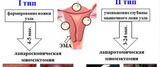
Types of fibroids
There are several classifications of fibroids. This is important for diagnosis, therapeutic or surgical treatment.
The most important types of classifications of fibroids are:
By structure:
nodular (in the focus) or diffuse (scattered throughout the organ).
By location:
- in the body (most often), in the isthmus or cervix;
- interstitial or intramural (within the myometrium);
- subserous (near the serous uterine lining),
- submucosal (close to the uterine mucosa);
- intraligamentous (grows in the broad ligament).
Also, nodes can be multiple or single, on a broad base or on a stalk.
Fibroids can consist of purely smooth muscle or connective tissue in varying proportions (uterine fibroids).
According to their morphology (structure), nodes can be:
- simple;
- proliferating;
- presarcoma.
Uterine fibroids are characterized by:
- women of childbearing age or before menopause (range from 35 to 55 years);
- varied symptoms (often asymptomatic);
- gives complications during childbirth;
- may give a growth spurt;
- reduction in size during menopause (or its disappearance).
Symptoms of fibroids
Usually, a woman finds out that she has fibroids by chance, after another ultrasound. Women do not notice most of the manifestations of this disease or explain them for other reasons.
Most often, uterine fibroids manifest:
- Menorrhagia (heavy periods). This often leads to anemia (anemia). Sometimes bleeding even requires medical attention due to its profuseness. The muscles of the uterus contract poorly due to the presence of nodes in them.
- Metrorrhagia or bleeding outside the cycle. In this case, the uterine mucosa itself “bleeds”.
- Painful manifestations (in the lower abdomen or lower back), sometimes sudden. During menstruation, the pain is nagging, sometimes cramping (when it grows into the mucous membrane).
In the early stages of fibroids, there is most often no pain.
- Constipation, urinary disturbances, feeling of pressure on the bottom.
- Associated anemia with manifestations of weakness, headache, pallor. Heart pain and increased blood pressure are common.
- Secondary infertility (with submucosal fibroids, when the fallopian tube is compressed) or miscarriage.
Ultrasound as a “safeguard” for women’s health with uterine fibroids
It is now difficult to surprise anyone with a diagnosis of uterine fibroids. About 25% of the entire female population often faced a similar diagnosis. Often this disease occurs latently, revealing itself only during a routine examination. We suggest you learn about the features of ultrasound for uterine fibroids, indications, contraindications and interpretation of results from this article.
Types of fibroids
There are several classifications of fibroids. This is important for diagnosis, therapeutic or surgical treatment.
The most important types of classifications of fibroids are:
By structure:
nodular (in the focus) or diffuse (scattered throughout the organ).
By location:
- in the body (most often), in the isthmus or cervix;
- interstitial or intramural (within the myometrium);
- subserous (near the serous uterine lining),
- submucosal (close to the uterine mucosa);
- intraligamentous (grows in the broad ligament).
Also, nodes can be multiple or single, on a broad base or on a stalk.
Fibroids can consist of purely smooth muscle or connective tissue in varying proportions (uterine fibroids).
According to their morphology (structure), nodes can be:
- simple;
- proliferating;
- presarcoma.
Uterine fibroids are characterized by:
- women of childbearing age or before menopause (range from 35 to 55 years);
- varied symptoms (often asymptomatic);
- gives complications during childbirth;
- may give a growth spurt;
- reduction in size during menopause (or its disappearance).
Symptoms of fibroids
Usually, a woman finds out that she has fibroids by chance, after another ultrasound. Women do not notice most of the manifestations of this disease or explain them for other reasons.
Most often, uterine fibroids manifest:
- Menorrhagia (heavy periods). This often leads to anemia (anemia). Sometimes bleeding even requires medical attention due to its profuseness. The muscles of the uterus contract poorly due to the presence of nodes in them.
- Metrorrhagia or bleeding outside the cycle. In this case, the uterine mucosa itself “bleeds”.
- Painful manifestations (in the lower abdomen or lower back), sometimes sudden. During menstruation, the pain is nagging, sometimes cramping (when it grows into the mucous membrane).
In the early stages of fibroids, there is most often no pain.
- Constipation, urinary disturbances, feeling of pressure on the bottom.
- Associated anemia with manifestations of weakness, headache, pallor. Heart pain and increased blood pressure are common.
- Secondary infertility (with submucosal fibroids, when the fallopian tube is compressed) or miscarriage.
Examination to detect fibroids
Often, a preliminary diagnosis of fibroids is possible during a gynecological examination. It is noticed by the unevenness and increase in the uterine surface. It is not uncommon for this disease to have a blood test with low hemoglobin.
However, to confirm the diagnosis of fibroids, the following additional methods are necessary:
- hysterography (x-ray with contrast agent);
- tomography (usually MRI);
- Ultrasound (transabdominal or transvaginal);
- laparoscopy of uterine fibroids.
Ultrasound for uterine fibroids is the most common method. It is based on the properties of ultrasound to be reflected differently from heterogeneous tissues. The ultrasound method has advantages:
- rapidity;
- minimum contraindications;
- painlessness;
- passes without surgical intervention;
- high data reliability.
How is an ultrasound performed for uterine fibroids?
Each type of ultrasound examination for fibroids has its own characteristics.
Transrectal ultrasound
This method is used infrequently, more often for virgins or women during their period.
The study uses a thin sensor. The woman takes off her underwear and lies down on her right side on the couch. A condom is placed on the sensor and lubricated with gel. Then it is inserted through the rectum and the examination begins.
Internal examination with a sterile umbrella
This type of examination is performed only when indicated for certain ailments (fibroids are included in this list).
On what day is an ultrasound done for fibroids?
Often, fibroids are discovered by chance when visiting a gynecologist or during a routine ultrasound examination (on days 3-5 or 12-14 of the cycle).
If the gynecologist suspects fibroids during the examination, then the patient is sent for an ultrasound examination to confirm the diagnosis immediately, without taking into account the day of the cycle.
Standards for ultrasound results when examining the uterus
Every sonologist (ultrasound specialist) is well aware of the indicators that are the norm for examining the uterus. The following ultrasound results are normal:
- The normal parameters of the uterus are considered to be the following dimensions: 4.5 - 6.5 cm in width, 4.5-6.7 cm in length, with a thickness of up to 4 cm. During menopause, the size of the uterus is significantly reduced.
- The thickness of the endometrium may vary depending on the phase of the menstrual cycle.
- A pear-shaped uterus of a homogeneous structure, having uniform echogenicity and normal muscle tone is considered normal.
- The cervical canal should have a diameter of up to 3 mm, with mucous contents.
Interpretation of ultrasound for fibroids
Uterine fibroids are an ailment that is quite easily detected by ultrasound on any day of the cycle.
Most often, fibroids are detected after 25 years, often in women who have given birth.
Myoma can be judged by the following signs on the monitor:
- enlargement of the uterus and its density;
- decreased uterine tone (as in the postpartum period);
- existing hypoechoic formations and stripes of different echogenicity (hyper- or hypogenic);
- difficulties in determining the far contour;
- the presence of calcifications and cysts in myomatous nodes.
Ultrasound examination is the most accessible, safe and painless method that allows timely diagnosis of fibroids in women and monitoring of its dynamics. Ignoring a gynecological ultrasound examination for women of any age is too risky and unacceptable. You can pay for such frivolity with your life.
- Gonal 33%, 38353835 33%3835 – 33% of all
- Clostilbegit 25%, 28862886 25%2886 – 25% of all
- Menopur 16%, 18651865 16%1865 – 16% of all
- Puregon 14%, 16491649 14%1649 – 14% of all
- Rotten 8%, 964 votes964 votes 8%964 votes – 8% of all
- Menogon 3%, 351 votes351 votes 3%351 votes – 3% of all
Source: https://BornInVitro.ru/diagnostika-besplodiya/uzi-pri-miome-matki/
Examination to detect fibroids
Often, a preliminary diagnosis of fibroids is possible during a gynecological examination. It is noticed by the unevenness and increase in the uterine surface. It is not uncommon for this disease to have a blood test with low hemoglobin. However, to confirm the diagnosis of fibroids, the following additional methods are necessary:
- hysterography (x-ray with contrast agent);
- tomography (usually MRI);
- Ultrasound (transabdominal or transvaginal);
- laparoscopy of uterine fibroids.
Ultrasound for uterine fibroids is the most common method. It is based on the properties of ultrasound to be reflected differently from heterogeneous tissues. The ultrasound method has advantages:
- rapidity;
- minimum contraindications;
- painlessness;
- passes without surgical intervention;
- high data reliability.
Preparing for an ultrasound
During ultrasound of the uterus, two types of this study can be used:
- transvaginal;
- transabdominal.
Occasionally, transrectal ultrasound and examination with the introduction of a sterile umbrella into the pelvis are used.
Preparation for a transvaginal ultrasound includes:
- complete emptying of the bladder before the study;
- drugs to remove gases from the intestines (Smecta, Espumisan) in the evening before the ultrasound;
- cleansing enema in the evening.
Preparation for transabdominal ultrasound includes:
- diet for 2-3 days before the study with the exclusion of legumes, fatty meats, and raw vegetables from the diet;
- drink plenty of clean, still water (carbonated and caffeinated drinks are excluded);
- limiting dairy and fermented milk dishes;
- taking medications for the accumulation of gases in the intestines;
- light breakfast before the ultrasound;
- Maximum filling of the bladder (drink a liter of still water and restrain the urge for 2-3 hours).
Preparation for transrectal ultrasound includes:
- cool water enema 6-8 hours before the test;
- an alternative to enema in the form of pharmacy enemas "Norgalax" or "Microlax", a laxative ("Senade"), the introduction of glycerin suppositories to stimulate bowel movements.
How to prepare for an ultrasound examination
Proper preparation is an important stage of the examination. The diagnostic result, and therefore the prescribed treatment, largely depends on it.
Medical training
Typically, ultrasound examination for fibroids is carried out with a transvaginal sensor - through the vagina. No special preparation is required: it is enough to empty your bladder immediately before the examination. The diagnosis is painless for the patient, but it may be a little unpleasant when the sensor is inserted.
Sometimes an ultrasound is performed transabdominally - through the abdomen. The doctor moves the sensor along the anterior abdominal wall, assessing the condition of the pelvic organs. This is not painful at all, but the information content of this method is lower - it is not always possible to see small myomatous nodes through the abdominal wall.
Transabdominal ultrasound requires preparation - you need to drink 1.5 liters of fluid before the procedure and not urinate until it is completed. Three days before the examination, you should not eat gas-forming foods, otherwise the intestinal loops will block the pelvic organs, and the doctor will not see the fibroids.
During the examination, the woman lies on the couch. In a public clinic, you need to take a diaper with you and lay it on the couch. Private medical centers provide disposable diapers. The whole procedure lasts 10-15 minutes. During the examination, you need to lie still, listen and follow the doctor's instructions. After the examination, get dressed and wait for the report printed on paper.
Psychological preparation
When going for an examination, prepare a list of questions for the doctor. It’s better to write them down on paper or on your phone so you don’t forget. Ask questions one at a time after completing the examination. Directly during the procedure, you should not distract the doctor - while talking with you, he may miss changes in the pelvic organs.
Directly during the examination, the doctor will ask you questions - this is necessary to assess your general condition, clarify the symptoms of the disease, and identify concomitant pathologies. Here's what the doctor will ask:
- When was your last menstruation? Look at the calendar in advance and write down the date - it may slip out of your head at the time of the examination.
- How do your periods go: heavy or moderate, how many days do they last, do they come regularly? Tell your doctor everything in detail - this will help make a diagnosis.
- How many pregnancies did you have, including births, abortions and miscarriages? If you are pregnant now or just planning to, tell your doctor about it.
- What gynecological diseases did you have before? List everything that happened during your life - ovarian cysts, cervical erosion, inflammatory processes.
- When was the last time you looked at myoma on an ultrasound? Take the results of the previous examination with you and show it to the doctor - this way the doctor will be able to assess the growth rate of the myomatous node.
- What's worrying you now? If you have any complaints (pain, cycle disruption), tell us about it.
Prepare in advance - keep your answers short but to the point.
After the examination, ask your doctor:
- If you only suspect fibroids: is there a node, how many tumors are there in the uterus, where and how are they located, and are there any concomitant diseases?
- If you do a control ultrasound: has the fibroid grown or decreased, have new nodes appeared, are there any negative changes (torsion of the fibroid stalk, impaired blood flow in the uterus, node degeneration)?
How is an ultrasound performed for uterine fibroids?
Each type of ultrasound examination for fibroids has its own characteristics.
Transabdominal ultrasound
In this case, the patient is naked to the waist or opens her stomach. The woman lies on her back, and a special gel is applied to her stomach for better conductivity of ultrasound waves. With transabdominal ultrasound, you have to view the uterus through the abdominal wall, and the probe remains outside. The doctor studies the ultrasound data, records them and interprets them using special tables.
The transabdominal method is less informative than the transvaginal method due to the non-penetrating method of examination.
Transvaginal ultrasound
During this type of ultrasound, the patient takes off her underwear and lies down on the couch. Then the woman bends her knees, after which the sensor (it is placed in a condom) is inserted into the vagina. The condom is disposable and prevents the spread of infection among patients. With this type of examination, you can clearly see the uterus (thanks to the shortest path to the internal organs). This method is the most preferred for fibroids.
Transrectal ultrasound
This method is used infrequently, more often for virgins or women during their period.
The study uses a thin sensor. The woman takes off her underwear and lies down on her right side on the couch. A condom is placed on the sensor and lubricated with gel. Then it is inserted through the rectum and the examination begins.
Internal examination with a sterile umbrella
This type of examination is performed only when indicated for certain ailments (fibroids are included in this list).
On what day is an ultrasound done for fibroids?
Often, fibroids are discovered by chance when visiting a gynecologist or during a routine ultrasound examination (on days 3-5 or 12-14 of the cycle).
If the gynecologist suspects fibroids during the examination, then the patient is sent for an ultrasound examination to confirm the diagnosis immediately, without taking into account the day of the cycle.
When to do an ultrasound for uterine fibroids?
Ultrasound examination is prescribed in the following situations:
- Suspicion of uterine fibroids. Ultrasound is indicated in the presence of characteristic complaints - menstrual irregularities, pain in the lower abdomen. Upon examination, you can discover the cause of this condition - see myomatous nodes in the muscular layer of the uterus. If the doctor detects signs of a tumor during manual examination (enlarged uterus, palpable formation in the projection of the pelvic organs), he will also refer the patient for an ultrasound.
- Controlling fibroid growth. Not all nodes need to be treated immediately with hormones or removed. Sometimes it is enough to watch them - they grow, move, decrease. Control ultrasound examination is prescribed every 6-12 months. If the doctor notices the growth of fibroids, he will change the patient’s management tactics.
- Monitoring the effectiveness of treatment. Ultrasound examination is indicated before and after the prescription of hormonal drugs and surgery, and the use of restorative mediation methods. It is necessary to evaluate what the tumor was like before the start of therapy and what it became after its completion.
- Pregnancy planning. Before conceiving a child, you need to do an ultrasound, assess the size and location of myomatous nodes, and, if necessary, undergo treatment.
Standards for ultrasound results when examining the uterus
Every sonologist (ultrasound specialist) is well aware of the indicators that are the norm for examining the uterus. The following ultrasound results are normal:
- The normal parameters of the uterus are considered to be the following dimensions: 4.5 - 6.5 cm in width, 4.5-6.7 cm in length, with a thickness of up to 4 cm. During menopause, the size of the uterus is significantly reduced.
- The thickness of the endometrium may vary depending on the phase of the menstrual cycle.
- A pear-shaped uterus of a homogeneous structure, having uniform echogenicity and normal muscle tone is considered normal.
- The cervical canal should have a diameter of up to 3 mm, with mucous contents.
Interpretation of ultrasound for fibroids
Uterine fibroids are an ailment that is quite easily detected by ultrasound on any day of the cycle.
Most often, fibroids are detected after 25 years, often in women who have given birth. Myoma can be judged by the following signs on the monitor:
- enlargement of the uterus and its density;
- decreased uterine tone (as in the postpartum period);
- existing hypoechoic formations and stripes of different echogenicity (hyper- or hypogenic);
- difficulties in determining the far contour;
- the presence of calcifications and cysts in myomatous nodes.
Ultrasound examination is the most accessible, safe and painless method that allows timely diagnosis of fibroids in women and monitoring of its dynamics. Ignoring a gynecological ultrasound examination for women of any age is too risky and unacceptable. You can pay for such frivolity with your life.
How to do an ultrasound correctly for uterine fibroids
Ultrasound for uterine fibroids is prescribed in the first phase of the menstrual cycle - on the 5-7th day from its beginning (from the first day of menstruation). At this time, the mucous membrane of the uterus is thinned, and nothing prevents the doctor from carefully examining the nodes. But if a woman’s menstruation lasts 6-7 days or more, you can wait until it ends and do an ultrasound on the 8-9th day of the cycle. There is no need to do an ultrasound during menstruation - it is unpleasant for the patient and not very informative for the doctor.
Sometimes the doctor prescribes an examination at the end of the menstrual cycle - on the 21-23rd day or immediately before menstruation. This is necessary to clarify the diagnosis. For example, endometrioid nodes in the uterus look like small fibroids, and in the first phase of the cycle they can be confused. But endometriosis is clearly visible after ovulation - the nodes increase in size and are clearly visualized on ultrasound.
With the onset of menopause, ultrasound examination can be done on any convenient day. The endometrium always remains thin (if there is no pathology), and myomatous nodes are clearly visible.
It is important to know:
- Ultrasound examination is a subjective diagnostic method. The result largely depends on the qualifications of the doctor. Where one doctor sees a small fibroid, another will see nothing. Therefore, get examined by a competent specialist - choose based on reviews, recommendations from friends and acquaintances. It’s good if the ultrasound diagnostic doctor has a specialization in gynecology - he knows what to look for.
- Ultrasound results depend not only on the doctor’s qualifications, but also on the quality of the equipment. Modern 3D and 4D technologies make it possible to obtain information about the fibroid and adjacent tissues and determine the spatial location of the node. Be sure to do an ultrasound with Doppler ultrasound - this technology makes it possible to assess the condition of the blood vessels in the uterus, identify signs of node degeneration, and detect a malignant tumor.
- A control ultrasound should be done on the same day of the cycle, with the same doctor and on the same machine. This will eliminate errors associated with technical errors and subjective perception of the ultrasound picture.