brief information
1.1 Definition
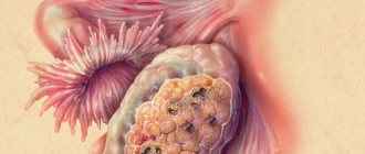
Squamous cell carcinoma of the vagina is a malignant neoplasm of the integumentary squamous epithelium of the vagina.
1.2 Etiology
The etiology and pathogenesis are unclear due to the low incidence.
Commonality with the etiopathogenesis of cancer of the vulva and cervix is assumed, with the exception of clear cell adenocarcinoma of the vagina, caused by exposure to DES, which was used to maintain pregnancy in the 1940-1950s.
Possible risk factors:
- infection with HPV, HSV-2 and HIV with genital warts;
- postmenopausal hypoestrogenism;
- severe chronic senile colpitis;
- involutive, dystrophic post-castration and age-related processes;
- chronic nonspecific vaginitis;
- 10-30 years after combined radiation therapy for cervical cancer
1.3 Epidemiology
In the United States in 2021, there were 4,620 cases of vaginal cancer and 950 deaths.
Primary vaginal cancer accounts for 1% of cancers of the female genital organs.
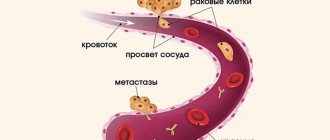
Frequency of metastatic lesions of the vagina:
- For all cancers up to 20%
- endometrial cancer and choriocarcinoma 24 - 55%
- cervical cancer 33%
- bladder and kidney cancer 5%
- rectal cancer 2%
- breast cancer 1%
- ovarian cancer 1%.
Almost 90% are squamous cell carcinoma of the vagina.
1.4 Coding according to ICD - O
C52 – malignant neoplasm of the vagina
1.5 Classification
International morphological classification of vaginal cancer
1. Epithelial tumors 1. Squamous cell carcinoma (epidermoid) (8070/3) 2. Adenocarcinoma (8140/3)
a) columnar cell type;
b) endometriotic adenocarcinoma (8380/3);
c) clear cell (mesonephroid) adenocarcinoma (8310/3).
1. Nonepithelial tumors
III. Mixed tumors
1. Tumors of the melanogenesis system 2. Other tumors 3. Secondary tumors. 4. Unclassified tumors.
International clinical classification by TNM (2010) and FIGO stages (2009)
T0 The primary tumor is not detected
Tis Preinvasive carcinoma
T1 (FIGO I) Limited to the vagina
T2 (II) Involves paravaginal tissues without extension to the pelvic wall
T3 (III) Extends to the pelvic wall
T4 (IVA) Invades the lining of the bladder or rectum and/or extends beyond the pelvis
T4 (IVB) Distant metastases
N - regional lymph nodes
Upper 2/3 of vagina : N1 in pelvic lymph nodes
The lower 2/3 of the vagina - in the inguinal lymph nodes
- N1 on one side
- N2 on both sides
Grouping by stage of vaginal cancer (FIGO and TNM)
Stage I: TisN0M0; T1N0M0
Stage II: T2N0M0
Stage III: T3N0M0; T1N1M0; T2N1M0; T3N1M0
IVA stage: T4NanyM0
IVB stage: AnyNanyM1
Symptoms of vaginal cancer
Early symptoms of vaginal cancer are rarely mistaken for signs of cancer because they are nonspecific. For example, bleeding between periods, spotting, and leucorrhoea can be explained by many other diseases that are more common.
At an early stage, the disease can be completely asymptomatic for a long time. The first signs include ichor discharge, sometimes with the presence of pus, watery discharge, sometimes with blood.
Bleeding may occur spontaneously between periods or occur during sexual intercourse. The discharge may have a strong, unpleasant odor.
Other symptoms are painful intercourse, discomfort, burning, itching or pain in the perineum and pubic area. The pain may radiate to the groin.
Damage to the lymph nodes impedes the drainage of lymph from the legs, which leads to their swelling, blueness, heaviness, and, at a later stage, to elephantiasis (lymphostasis).
Tumor growth in the bladder leads to frequent and/or painful urination. There may be blood in the urine.
Damage to the rectum causes constipation. The spread of cancer cells into bone tissue causes bone pain. These are the late symptoms of vaginal cancer, which require the most radical treatments.
Diagnostics
2.1 Complaints and anamnesis
Identification of factors influencing the choice of treatment tactics.
2.2 Physical examination
General physical examination
Medical gynecological examination:
- examination and palpation of the vulva
- examination of the vagina with Simpson speculum, but not Cusco speculum, which poorly visualizes the anterior and posterior walls
- examination of the cervix in speculums
- bimanual examination
- palpation of peripheral lymph nodes.
Criteria for vaginal cancer:
- the primary focus is located only in the vagina;
- intact epithelium of the cervix and cervical canal, endometrium;
- may be asymptomatic.
2.3 Instrumental diagnostics
(Verification)
- Imprint smears from the tumor for cytological and/or morphological examination.
- Smears from the cervix and cervical canal
- Aspiration biopsy of the endometrium with cytological and morphological studies.
- Tumor biopsy with morphological examination is the main diagnostic criterion.
- Puncture of enlarged lymph nodes with cytological examination.
- Histological examination of a surgically removed tumor specimen
Assessment of the prevalence of the tumor process
(Tumor Imaging)
- Vulvoscopy.
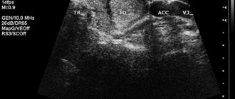
- Vaginoscopy is the simplest method for visualizing a tumor.
- Colposcopy.
zones :
- pelvic organs
- abdominal cavity (adrenal glands - with adenocarcinoma)
- inguinal-femoral and retroperitoneal lymph nodes
- supraclavicular lymph nodes (adenocarcinoma metastases).
For common stages:
- cystoscopy;
- sigmoidoscopy;
- excretory intravenous urography.
If you suspect metastatic cancer:
- separate diagnostic curettage of the uterine mucosa and cervical canal;
- breast examination;
- examination of the ovaries and other organs.
What diseases can it be associated with?
As noted earlier, primary vaginal cancer occurs only in every hundredth diagnosed case. Otherwise, vaginal cancer develops either as a result of metastases of existing tumors (uterine cancer, cervical cancer, ovarian cancer), or against the background of such disorders as:
- vaginal adenosis is a precursor to vaginal adenocarcinoma, manifested by a predominance of glandular epithelium in place of squamous epithelium;
- human papillomavirus;
- endocrine disorders - diabetes mellitus and hypoestrogenism;
- chronic infections localized in the genitourinary system.
Treatment
Radiation therapy is the main method of radical treatment.
Surgical treatment is possible for small primary tumors.
TisN0M0 stage
- VAIN I – monitoring;
- VAIN II and III - depends on the prevalence:
- laser surgery;
- wide local excision with/without skin grafting;
- partial/complete vaginectomy with/without skin grafting;
- HT;
- intracavitary RT.
T1N0M0 (I) stage
Combined RT; Intracavitary RT - for superficial growth <0.5 cm; Interstitial RT (brachytherapy) in its own version - mainly for local tumors; Surgery:
- wide local excision;
- vaginectomy with vaginal reconstruction + neoadjuvant RT (if indicated);
- vaginectomy and lymph node dissection, with/without vaginal reconstruction
Adjuvant RT - for positive resection margins
T2N0M0 (II) stage
- Combined RT - the type of brachytherapy is selected taking into account distribution and localization;
- Combined treatment - vaginectomy and exenteration of the pelvic organs with intracavitary and/or remote RT;
- Chemotherapy – to improve local control.
Stage III (T3N0M0; T1N1M0; T2N1M0; T3N1M0)
- DHT in combination with/without intracavitary RT;
- Surgery with DHT, if indicated, is supplemented with intracavitary RT.
IVA stage (T4, any N, M0)
- Combined RT;
- Surgery with DHT/or intracavitary RT, chemotherapy with platinum drugs is possible.
Stage IVB (any T, any NM1) (distant metastases)
- RT palliative/brachytherapy to relieve symptoms and improve quality of life;
- HT.
Recurrence of squamous cell carcinoma of the vagina
- Individual RT and CT regimens;
- Evisceration of the pelvic organs with local resectable residual tumor.
Volume of DHT for tumor:
- upper 2/3 of the vagina - pelvic irradiation;
- lower third of the vagina - irradiation of the pelvis and groin areas.
Topometric preparation for RT:
lying on your back with immobilization of the upper and lower body (headrest and knee support); it is possible to use radiopaque markers on the skin of the abdomen to verify the lower border of the tumor (GTV border) and introitus; a full bladder will allow you to more accurately determine the location of the body and cervix during irradiation; intravenous contrast for better visualization of pelvic vessels; the supporting role of PET-CT in determining the primary tumor volume of GTV; contouring by analogy with cervical and uterine cancer; CTV-T includes the entire vaginal tube, cervix and paracervical region, paravaginal tissues; in CTV-T for a tumor in the lower third, the introitus is additionally included; CTV-N includes pelvic lymph nodes below the level of the common iliac lymph nodes, taking into account the location of the primary tumor:
- in the upper 2/3 – obturator, external and internal iliac, presacral and pararectal lymph nodes;
- in the lower third – inguinal-femoral and distal external iliac lymph nodes;
- on the posterior wall of the vagina - presacral and pararectal pelvic lymph nodes.
PTV1 = CTV-T + 15 mm PTV2 = CTV-N + 7 mm PTV = PTV1 + PTV2 contouring of risk organs: bladder, rectum, small intestinal loops, anus, bone marrow (sacrum, coccyx, L5 body, acetabulum , proximal part of the femur), head of the femur.
Depending on localization and distribution:
- VaIN and superficial with a depth of invasion up to 5 mm – brachytherapy with vaginal applicators;
- invasion more than 5 mm – interstitial RT (usually after DHT);
- upper 2/3 of the vagina - intracavitary RT, similar to the treatment of cervical cancer.
- the middle and lower thirds of the vagina, a tumor limited by its posterior wall - a cylindrical applicator is possible vaginally and rectally.
Doses-fractionation:
- DHT of the primary tumor and the area of regional lymph nodes RD 2 Gy 5 days a week SD 46 - 50 Gy. In the absence of brachytherapy - local boost.
Brachytherapy:
- Independent to a depth of 5 mm from the mucosa RD 5.5 Gy SD 33 Gy 10 – 19 days;
- Intracavitary RT RD 7 Gy 3-4 fractions once every 5-6 days;
- Interstitial RT 20 - 30 Gy.
3.1 Chemotherapy
2-3 courses of neoadjuvant chemotherapy for advanced squamous cell RVL.
6 courses of adjuvant chemotherapy or until progression.
Minimum volume of chemotherapy 6 courses:
- cisplatin 50 mg/m2 every 21 days;
- cisplatin 50 mg/m2 on day 1 + 5-FU 500 mg/m2 on days 1-3 with an interval of 21 days.
The optimal volume of chemotherapy with an interval of 3 weeks:
- paclitaxel 175 mg/m² + cisplatin 75 mg/m² on 1 day;
- paclitaxel 175 mg/m2 + carboplatin AUC 5-6 per day;
- cisplatin 50 mg/m2 on day 1 + gemcitabine 1000 mg/m2 on days 1.8;
- xeloda 2500 mg/m2 days 1-14.
For advanced cancer - RT against the background of sensitizing chemotherapy, as in squamous cell carcinoma of the cervix:
- cisplatin 50 mg/m2 once a week;
- chemotherapy similar to regimens for cervical cancer.
3.2 Symptomatic treatment
For pain syndrome:
- DHT,
- drug therapy depending on its cause.
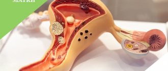
Causes of uterine fibroids
Fibroma that arises in the uterus has a mature, benign nature. Its structure is of a connective type, coming from the walls of the uterus itself. The etiology of the development of such a tumor is currently unclear. It has been noted that women of the Negroid race are more likely to develop fibroids than women of the European race. Researchers have linked fibroids to hereditary factors and increased sensitivity of hormonal levels to estrogen. Although the presence of these reasons does not always lead to the development of fibroids.
There are a number of additional factors that influence the occurrence of a tumor. These include artificial termination of pregnancy, childbirth with complications, chronic gynecological diseases, frequent curettage performed for diagnostic purposes. Indirect reasons include the late onset of the menstrual cycle, lack of sexual activity or its irregularity, and absence of childbirth before the age of thirty. Additional reasons also include obesity, physical inactivity, diabetes, frequent stress, etc.
The reason for the growth of the tumor may lie in the use of tablets containing estrogen to prevent pregnancy or treat menopause. Doctors' observations show a connection between the development of fibroids and the menstrual cycle. Until it is fully established, at a young age, it does not arise. Once menopause occurs, the risk of uterine fibroids is also unlikely due to the natural decline in estrogen levels. But during pregnancy, its level increases and the tumor, if present in the uterus, can begin to actively grow.