Increasingly in recent years, women have experienced problems with conception, pregnancy and childbirth. There are many reasons for this: inflammatory diseases, age, poor health, and so on. In most cases, modern medicine still helps the fairer sex overcome her illness. However, after some treatment methods, scars appear on the uterus. You will learn from the article how they arise and what they threaten. It is worth mentioning separately how a scar on the uterus can be dangerous during pregnancy.
What is a scar?
A scar is tissue damage that has subsequently been repaired. Most often, the surgical method of suturing is used for this. Less commonly, cut areas are glued together using special adhesives and so-called glue. In simple cases, with minor injuries, the rupture heals on its own, forming a scar.
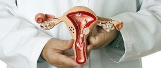
Such a formation can be located anywhere: on the human body or organs. For women, the scar on the uterus is of particular importance. A photo of this formation will be presented to you in the article. Damage can be diagnosed using ultrasound, palpation, and various types of tomography. Moreover, each method has its advantages. So, during an ultrasound, the doctor can assess the position of the scar, its size and thickness. Tomography helps to determine the relief of formation.
General information
According to various studies in the field of gynecology, the frequency of post-traumatic and congenital cicatricial changes in the cervix ranges from 15.3 to 54.9%, while in reproductive age it can reach 70%. The disease is more often detected in women who have given birth to a child for the first time over the age of 30. In patients with cervicitis, the likelihood of post-traumatic replacement of normal epithelium with scar tissue increases. The high importance of prevention, timely diagnosis and treatment of ADSM is due to the significant impact of the disease on increasing the risk of infertility, inflammatory and oncological processes.
Reasons for appearance
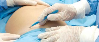
Why do some women develop scars on their uterus? Such damage becomes a consequence of medical interventions. This is usually a caesarean section. In this case, the type of operation plays an important role. It can be planned or emergency. During planned delivery, the uterus is incised in the lower segment of the abdominal cavity. After the fetus is removed, it is sutured layer-by-layer. This scar is called transverse. In an emergency caesarean section, a longitudinal incision is often made. In this case, the scar has the same name.
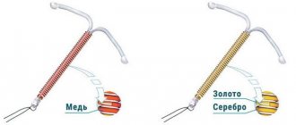
Fused lesions can result from perforation of the uterine wall during gynecological procedures: curettage, hysteroscopy, insertion of an IUD. Also, scars always remain after surgical removal of fibroids. In these cases, the position of the scar does not depend on specialists. It forms where the operation was performed.
Treatment of RDSM
Since the disease is accompanied by anatomical changes, surgical methods are the most effective for its treatment. The choice of a specific technique is determined by the degree of deformation, the woman’s reproductive plans and the presence of complications. The following types of operations are recommended:
- Ablative methods
. To remove scar tissue, ectropion, areas of the endocervix with polyps, dysplasia or leukoplakia, radio wave and argon plasma treatment, laser vaporization, cryodestruction, and diathermocoagulation are used. Ablation is effective for grade I deformity in patients of reproductive age planning pregnancy. - Tracheloplasty
. During reconstructive operations, scar tissue is removed using the method of partial or complete dissection while preserving the muscle layer and mucous membrane, and the cervical canal is restored. The method is indicated for women of childbearing age with II-III degree of scar deformity. - Conization and trachelectomy
. Excision of the affected areas or amputation is performed when the deformity is combined with intraepithelial neoplasia or failure of the pelvic floor muscles. Radical operations are more often performed in patients who have passed reproductive age. - Purse-string sutures
. If signs of isthmic-cervical insufficiency appear during pregnancy, the locking function of the cervix is restored mechanically. An alternative to surgery in this case may be the installation of an obstetric pessary.
Auxiliary drug treatments are aimed at stopping the inflammatory process. After sanitization of the vagina, patients are prescribed medications to restore its normal microflora.
Pregnancy and scar
If you have scars on your uterus, the possibility of having a baby depends on their condition. Before planning, you should definitely consult a gynecologist. The specialist will certainly conduct an ultrasound examination to determine the condition and position of the scar. You will also need to undergo some tests. Before planning begins, it is imperative to cure infections. Subsequently, they can cause problems with pregnancy.
If the scar is in the lower segment and has a transverse position, then problems usually do not arise. The fairer sex is examined and released to plan a pregnancy. In the case where the scar turns out to be insolvent, thinned and consisting mainly of connective tissue, pregnancy may be contraindicated. However, in some cases, the hands of surgeons work wonders. And a woman can still give birth.
Symptoms
This pathology is not accompanied by severe symptoms. This significantly complicates the timely diagnosis of cicatricial deformation of the cervix.
The initial stage is accompanied by an increase in the amount of mucous secretions. Other signs do not appear at this stage.
The average degree of damage may be accompanied by nagging, aching pain in the lower abdomen. Sometimes such pain can radiate to the lumbar region.
Often the pathology is accompanied by infection, and cervical discharge may change. They become cloudy, yellow and gray. The menstrual cycle is disrupted. Painful sensations occur during sexual intercourse.
This disease can cause the inability to conceive a child.
Management of pregnancy and childbirth with a uterine scar
If you have a scar on the reproductive organ, then you need to inform the specialist who will manage your pregnancy about this. At the same time, you need to tell about the existing fact immediately, at the first visit, and not before the birth itself. Pregnancy management in women with a history of uterine injuries occurs somewhat differently. They get more attention. Also, this category of expectant mothers regularly have to visit an ultrasound diagnostic room. Such visits become especially frequent in the third trimester. Before childbirth, an ultrasound of the uterine scar is performed almost every two weeks. It is worth noting that other diagnostic methods are unacceptable during pregnancy. X-rays and tomography are contraindicated. The only exceptions are special, difficult situations when it comes not only to a woman’s health, but also to her life.
Delivery can be carried out by two methods: natural and operative. Most often, women themselves choose the second option. However, if the scar is intact and the expectant mother is in normal health, a natural birth is quite acceptable. To make the right choice, you need to consult with an experienced specialist. Also, during labor and increasing contractions, it is worth conducting periodic ultrasound monitoring of the condition of the scar and the uterus. Doctors also monitor the fetal heartbeat.
Damage to the cervix
As practice shows, some women who give birth on their own have a scar on the cervix. It occurs due to tissue rupture. During the process of childbirth, a woman feels painful contractions. Attempts begin behind them. If the cervix is not fully dilated at the moment, then they can lead to its rupture. This does not threaten anything for the child. However, the woman subsequently has a scar on her cervix. Of course, after delivery, all tissues are sutured. But in the future this may become a problem during the next birth.
Such a scar at the mouth of the cervical canal can also appear after other gynecological manipulations: cauterization of erosion, removal of a polyp, and so on. In all cases, the resulting scar appears to be connective tissue. During subsequent delivery, it simply does not stretch, leaving the area of the cervix undilated. Otherwise, the damage does not pose any danger to the mother and her unborn child. Let's try to find out why scars located on the reproductive organ can be dangerous.
Diagnosis of the disease
A doctor may suspect the presence of this disease by carefully examining a woman's medical history. Characteristic factors are complicated childbirth and invasive procedures. It is especially important to exclude the presence of abortions. Diagnostic methods:
- Examination by a gynecologist. Examination in a chair using mirrors will help assess the condition of the cervix. Allows you to determine the presence of scars and tears. In this technique, the doctor palpates the pharynx.
- Colposcopy. The diagnostic method under a microscope allows you to study the etiology of scars on the cervix and cervical canal.
- Bacterial studies of mucus. To do this, the doctor takes smears to determine characteristic damage.
- Laboratory diagnostics. Tests and bacteriological cultures of the flora will help detect the presence of infection.
Rough scars in this case help to quickly establish a diagnosis.
Attachment of the fertilized egg and its growth
If there are scars on the uterus, then after fertilization a set of cells can attach to them. So, this happens in about two out of ten cases. At the same time, the forecasts turn out to be very dire. On the surface of the scar there is a mass of damaged vessels and capillaries. It is through them that the fertilized egg is nourished. Most often, such a pregnancy is terminated on its own during the first trimester. The consequence can be called not only unpleasant, but also dangerous. After all, a woman needs emergency medical care. Decaying fetal tissue can lead to sepsis.
Incorrect attachment of the placenta
A scar on the uterus after a cesarean section is dangerous because during the next pregnancy it can cause improper attachment of the baby's place. Often women are faced with the fact that the placenta is fixed close to the birth canal. Moreover, as pregnancy progresses, it migrates higher. The scar may prevent such movement.
The presence of a scar after damage to the reproductive organ often leads to placenta accreta. The baby's place is located precisely on the scar area. Doctors distinguish basal, muscular and complete placenta accreta. In the first case, the forecasts may be good. However, natural childbirth is no longer possible. If placenta accreta is complete, the uterus may need to be removed.
Complications
Cicatricial deformity is often complicated by the addition of a secondary infection with the development of chronic cervicitis. The insufficiency of the protective function of the cervical canal leads to the spread of the inflammatory process to the endometrium, fallopian tubes and ovaries. Since the endocervix is constantly exposed to the acidic environment of the vagina, the likelihood of developing erosion, dysplasia, leukoplakia, polyps, and malignant tumors increases sharply. A scarred cervix during childbirth exhibits functional failure - natural childbirth is delayed or becomes impossible. The disease is one of the causes of cervical infertility.
Uterine growth
In a normal non-pregnant state, the thickness of the walls of the reproductive organ is about 3 centimeters. By the end of pregnancy they stretch up to 2 millimeters. At the same time, the scar also becomes thinner. As is known, fused damage is replaced by connective tissue. However, normally a large area of the scar is represented by a muscle layer. In this case, the scar is considered valid. If the damage thins down to 1 millimeter, this is not a very good sign. In most cases, experts prescribe bed rest and supportive medications to the expectant mother. Depending on the length of pregnancy and the thickness of the uterine scar, a decision may be made to deliver prematurely. This condition has dangerous consequences for the baby.
After childbirth…
Scarring on the uterus after childbirth can also be dangerous. Despite the fact that the baby has already been born, consequences may arise for his mother. Scars are damage to the mucous membrane. As you know, after childbirth, every woman experiences bleeding. The process of separation of mucus and remnants of membranes occurs. These secretions are called lochia. In some situations, mucus may linger on the scar area. This leads to an inflammatory process. The woman requires curettage, her body temperature rises, and her health worsens. In the absence of timely treatment, blood poisoning begins.
Classification
When determining the degree of RDS, criteria such as the consistency of the external pharynx, the number and size of scars, the condition of the endo- and exocervix and surrounding tissues are taken into account. There are four degrees of scar deformation changes:
- I degree.
The external pharynx allows the tip or entire finger of the doctor to pass through. The cervical canal has the shape of a cone, the apex of which is the internal uterine os. The depth of single or multiple old tears does not exceed 2 cm. Signs of ectropion of the lower parts of the cervical canal are revealed. - II degree.
The external os cannot be identified. The cervix is “split” into separate anterior and posterior lips, with old tears extending into the fornix. The endocervix is completely everted. - III degree.
Old tears reach the vaginal vaults. The external os is not defined. One of the lips of the neck is hypertrophied. Epithelial dysplasia and signs of an inflammatory process are noted. - IV degree.
It manifests itself as a combination of old ruptures that extend to the vaginal vaults, with insufficiency of the pelvic floor muscles.
Summarize
You learned about what a uterine scar is, in what situations it appears and why it is dangerous. Let us note that if you properly prepare for pregnancy and listen to the advice of an experienced doctor when managing it, then the outcome in most cases is good. The new mother and baby are discharged from the maternity ward in about a week. Don't be too upset if you have a uterine scar. Before you start planning, be sure to consult your doctor, undergo routine examinations, and take all tests. After this you can get pregnant.
Experts do not advise starting to plan a pregnancy earlier than two years after receiving such an injury. Also, don’t delay this. Doctors say that after 4-5 years it will be almost impossible to stretch the scar. This is when problems can begin during pregnancy and childbirth. All the best to you!


