Ideally, in order to start planning for a child, you need to be in excellent health. However, this is rare these days, so giving birth with uterine fibroids is one of the normal options.
Often in recent years, women give birth to their first child at the age of 30 or more. Lost time and female processes not started on time (lactation, growth of the baby in the womb) create an excellent springboard for the development of such a pathology as uterine fibroids. Sometimes after delivery by caesarean section it can be removed surgically. When is this possible to do after childbirth, and what is the feedback for this event?
Preparation for the procedure
Planned hospitalization of a pregnant woman occurs at the 37th week of pregnancy and the expectant mother is thoroughly examined before giving birth.
During an ultrasound, a surgeon must be present - specialists study not only the condition of the fetus, but also determine the size, location, number of myomatous nodes, and also examine the vessels feeding the tumor. Standard diagnostics of the state of health and development of the fetus includes fetometry, cardiotocography and Doppler measurements of the umbilical cord vessels. The results of past ultrasounds and the latest study are necessarily compared - this allows specialists to determine how much the tumor has progressed in growth and how this affected the development of the fetus.
Can it be removed during pregnancy?
There are various tactics for managing pregnant women with uterine fibroids. Indeed, sometimes nodes can lead to impaired growth and development of the fetus, the threat of premature birth, miscarriage, etc.
Removing fibroids during pregnancy is a risky procedure that can cause miscarriage, developmental arrest and other pathologies. The optimal time frame for such operations is 16 - 18 weeks. At this time, the formation of the baby’s basic structures has already occurred, so all methods used will cause the least possible harm to him.
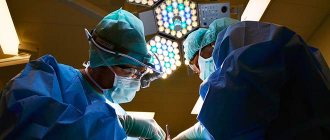
Progress of the operation
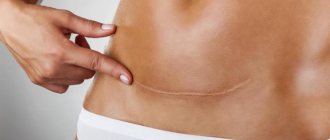
The operation is performed using epidural (spinal) anesthesia or endotracheal anesthesia. Through a transverse incision in the abdomen along the “bikini line,” the newborn baby is removed, and then the placenta.
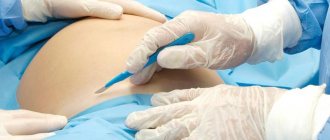
If the myomatous nodes are small, they are resected after delivery.
The following stages of caesarean section are distinguished:
- Abdominal incision using the Joel-Cohen method in thin girls without bladder trauma. The Pfannenstiel incision is performed in large women.
- Section of the uterine tissue in accordance with the localization of myomatous nodes, their size and depth of occurrence in the muscle structure. As a rule, an arc-shaped transverse incision is made.
- Extraction of the fetus.
- Separation of the placenta.
- Removal of myomatous nodes.
- Layer-by-layer suturing of the walls of the uterus and abdominal wall.
Reviews from doctors and patients
Below are reviews about the removal of tumors during a cesarean section: Evgeniya, 28 I am an experienced surgeon, I advise removing fibroids a couple of years after a cesarean section - this way the recovery period for the woman in labor is faster and easier, she successfully begins to breastfeed the baby, which contributes to growth inhibition tumors. In my practice, I have encountered cases where fibroids disappeared without a trace a year after the birth of the baby. Olga, 52 years old I had a tumor on the outside of my uterus. The doctors said that she was not an obstacle to pregnancy and recommended that she get pregnant. It was not possible to conceive a child right away, but after 2 years of trying, I became a happy mother. The doctor removed the fibroid node during a caesarean section. He was prescribed urgently due to weak labor and the absence of strong contractions for 8 hours. Elena, 32 She gave birth to her first child late. When I went to register, I heard a terrible diagnosis: fibroids on the outer wall of the uterus, quite large in size. Doctors said that it was necessary to remove it before conception, but now nothing can be done. I spent my entire pregnancy visiting doctors, taking tests all day long. I spent the second and third trimesters in the hospital - my doctor was afraid for me. He said I could give birth at home before term. But everything went well, my son and I survived the caesarean section safely. The surgeon left the fibroid in place for now; I will remove it when my son turns 1.5-2 years old.
Contraindications to fibroid removal during cesarean section
Removal of a myomatous node during cesarean section is contraindicated if a woman has the following conditions:
- severe manifestations of iron deficiency anemia;
- severe blood loss during fetal extraction;
- divergence of the vascular network feeding the nodes in the lower part of the uterus;
- extragenital pathology;
- premature placental abruption;
- the patient is over forty years old;
- multiple nodes that deform the uterus;
- distant localization of nodes from the incision site.
It is not advisable to carry out the procedure if there is one node up to two centimeters, since after delivery it can resolve on its own.
How to treat fibroids?
The choice of drugs for the treatment of uterine fibroids depends on many factors; in each specific case, the decision is made by the gynecologist observing the woman.
Taken into account:
- stage of the disease (at the initial stage, conservative treatment methods are used);
- the woman’s age and desire to have children (during childbearing age, doctors try to give the woman a chance and preserve the uterus);
- general health;
- presence or absence of fibroid complications
Conservative treatment includes the prescription of hormonal drugs and symptomatic drugs (anti-inflammatory, analgesic, antispasmodic and others).
However, extensive tumors and age after menopause become indications for surgery. In addition, there is a possible outcome in which it is possible to save the uterus by removing only part of the wall with fibroids.
Types of operations
Myomectomy is an abdominal operation that involves excision of fibroid nodes while preserving the uterus. A woman retains the opportunity to become pregnant and give birth to a healthy child naturally. This type of surgery is performed in several ways:
- laparoscopically - performed through small incisions in the abdominal wall. The operation is performed under general anesthesia and is characterized by low morbidity, a short hospital stay and a short rehabilitation period.
- laparotomy - through a long incision in the abdominal cavity (open method), which is performed longitudinally from the navel to the pubic bones or transversely slightly below the abdominal fold in the bikini area. In the second case, the seams can be hidden under the underwear, and they are practically invisible to the prying eye.
- hysteroscopically - through the vagina using a special hysteroscope device.
See also on the blog: What a woman after 50 needs to know about her health
Hysterectomy – removal of fibroids along with the uterus. In this case, the woman loses the opportunity to have children in the future.
- abdominal surgery under epidural or general anesthesia;
- laparoscopic intervention
Indications for myomectomy are:
- severe pain in the lower abdomen, cramps, pressure that does not go away after taking medications;
- the development of anemia that threatens a woman’s health and cannot be cured with medications;
- formation of fibroids with subsequent deformation of the uterine wall.
Indications for hysterectomy after cesarean section
Indications for removal of the uterus affected by fibroids immediately after extraction of the fetus are:
- presence of atypical tumor cells;
- necrosis of the node and development of septic processes in the uterus;
- tumor recurrence after a previous myomectomy with aggressive development and damage to several areas of the uterus;
- concentration of the node in the plexus of large arteries and vessels;
- detection of uterine cancer during diagnosis in preparation for caesarean section.
Stages of tumor removal
Myomectomy is performed in stages as follows:
- Separation of the myometrium from the node, on the one hand.
- Figure-of-eight sutures - the crossing of threads occurs inside the tissues.
- Separation of the myometrium from the node, on the other hand.
- Re-suturing in a figure-eight pattern uses self-absorbable suture material.
- Layer-by-layer sutures on the muscular and muscular-serous layer in the area where the node is located.
- Application of an additional U-shaped suture around the internal sutures when removing large fibroids.
- Layer-by-layer suturing of the wound.
This type of suturing prevents the formation bed from deepening inward, the development of major blood loss and the formation of hematomas. If a subserous node on a thin stalk is removed, an incision is made above the formation several centimeters. An electric knife is used to reduce blood loss. The duration of the procedure is about one and a half hours.
Recovery after fibroid removal during cesarean section
At the end of the operation, the woman in labor is moved to the intensive care unit. She spends one day there, after which she is transferred to the postpartum ward.
The mother's condition is most often normal, and the condition of the born child is characterized as satisfactory, since signs of mild hypoxia are observed. Oxygen starvation is eliminated quickly without consequences. The course of the postoperative period does not differ from rehabilitation after a regular cesarean section. As a rule, no complications are observed.
The woman in labor must be prescribed a weekly course of antibacterial therapy, as well as painkillers and drugs to contract the myometrium for two to three days. If there are signs of anemia, additional iron supplements are administered through an IV. It is worth noting that the doctor prescribes medications that do not affect lactation and the properties of breast milk.
There is a drainage tube in the abdominal cavity for about two days. A bandage is applied to the wound. It should not be wetted, touched, much less removed. A woman should monitor the hygiene of the skin around the suture every day. On the third day, a control ultrasound is performed.
The diet of a nursing mother should be enriched with nutrients, but at the same time not cause gas formation. Constipation and excessive straining during bowel movements should be avoided.
Uterine fibroids (or fibroids, fibroids, leiomyomas) are a benign tumor of the muscular (connective) layer of the uterus (myometrium). It occurs as a result of spontaneous cell division, and the reasons causing this process are not fully understood. However, it has been established for certain that the “blame” for everything is the increased production of the hormone estrogen. It is this hormone that causes the growth of fibroids, while progesterone causes the opposite effect. However, even if there is a normal balance of estrogen and progesterone in the blood, one cannot safely say that the woman does not have fibroids.
Uterine fibroids and childbirth are completely compatible concepts. It is only recommended to undergo an additional ultrasound before childbirth - this will clarify the location and size of the tumor nodes.
How do pregnancy and childbirth affect fibroids?
During pregnancy, certain changes occur in a woman's body:
- hormonal changes,
in which the level of estrogen and, to an even greater extent, progesterone increases, and this affects the condition of fibroids; - mechanical restructuring of the uterus
- its enlargement and stretching.
To supply the enlarged organ with blood, new vessels grow to the muscles. All these changes can affect an existing fibroid, but the degree of its changes will depend on where and how exactly the tumor is located, and how much it “invades” the uterus.
During pregnancy, uterine fibroids practically do not grow. Its slight growth can be observed in the 1st and 2nd trimesters, but in the 3rd trimester the fibroids become smaller. In general, the growth of fibroids has virtually no effect on the development of pregnancy.
During the postpartum period, fibroids may undergo changes, but they are unpredictable. For example, tumors that caused trouble during pregnancy may not show a single symptom after childbirth. However, as a result of the reverse development of the uterus in the first months after childbirth, fibroids often change their location.
How does fibroid behave?
It is extremely difficult to predict what will happen to the node during pregnancy and after childbirth. Often fibroids either decrease or increase in different trimesters. This depends on many factors, namely:
- From where the node is located - on the surface of the uterus or in the thickness of the myometrium, at the fundus, isthmus, on the side wall, between ligaments, etc. This determines the blood supply, which affects growth.
- What is the initial size of fibroids? Small nodes are less susceptible to change than large ones. The latter can increase two or more times.
- From the woman's hormonal background.
- Was the pregnancy natural or IVF? In the latter case, serious hormonal support occurs, which does not go unnoticed for the nodes.
During pregnancy
Under the influence of all factors, in 2/3 of cases, the size of fibromatous nodes decreases by approximately 20 - 30% during pregnancy. In the remaining third of women, fibroids increase in size, often doubling or even more.
But this is the general picture that can be obtained if you measure the nodes at the beginning and then at the end of pregnancy. In fact, there are fluctuations in the diameter of formations by trimester.
The general trends are as follows:
- In the first trimester, fibroids grow due to the action of estrogen.
- In the second, the pace decreases and even regression of formations occurs.
- In the third, the estrogen content increases again, and the fibroid grows.
Watch the video about uterine fibroids and the reasons for their development:
In the early postpartum period
In the early postpartum period, a slight increase in the size of fibroids can be observed. This may be a consequence of swelling of the nodes during myometrial contraction. Sometimes these processes are so pronounced that necrosis of the fibroid is observed - a condition requiring immediate surgical treatment.
As the uterus contracts, the nodes also become smaller, but more often they remain somewhat larger than before pregnancy.
Uterine fibroids in the early postpartum period can cause another dangerous condition - bleeding. It occurs when myometrial contraction is impaired. Sometimes you even have to resort to removing the uterus in order to save a woman’s life.
In the late postpartum period
The size of fibroids in the late postpartum period largely depends on whether the woman is breastfeeding. The fact is that during lactation the production of LH and FSH is inhibited, which directly affects the nodes.
As a result, women who have fibroids of any size are advised to continue breastfeeding as long as possible. This is beneficial for both her and the baby.
Delivery with uterine fibroids
Pregnancy that occurs against the background of uterine fibroids can be accompanied by a number of complications, and at the same time, the risk of its interruption remains throughout the entire period. However, if this happens, then a miscarriage occurs due to a malnutrition of the endometrium in the early stages. Sometimes the cause of a miscarriage is an inconvenient place of attachment of the embryo (for example, the so-called cervical - in the area of the cervix, which makes it impossible to bear the fetus). With fibroids, the risk of tubal pregnancy increases.
When the tumor is localized in the cervical area, it opens painlessly even before the onset of labor, and in the early stages this can provoke a miscarriage, and after 22 weeks there is a threat of premature birth.
With large size nodes and endometrial pathology, increased uterine tone remains throughout the entire period, which often leads to premature birth. This is explained by the fact that the large size of the tumor prevents the baby from taking the correct position in the uterus, and most often it is located either obliquely or transversely, which is an indication for a cesarean section. Moreover, the tumor located in the muscular layer of the uterus interferes with the normal functioning of the placenta: the supply of nutrients and oxygen to the fetus is disrupted, hypoxia (oxygen deficiency) develops, which leads to delayed development of the fetus (it lags behind in height and weight). In the future - after birth - this will affect the baby’s health, his physical and mental development.
Another danger that arises against the background of fibroid growth is changes in the endometrium and tight attachment of the placenta. This makes it difficult for the placenta to come out on its own after childbirth and provokes heavy bleeding. In this case, the doctor performs a manual examination of the uterus and removes the placenta under general anesthesia.
Why do uterine fibroids appear after childbirth?
Uterine fibroids are a multifactorial disease. And hormonal disorders play a significant role in the formation of nodes. It is reliably known that, once fibroids appear, they begin to respond inadequately to changes in the concentration of sex hormones. This causes changes in the size of the nodes, including during pregnancy and after childbirth. Thus, the concentration of the following hormones in a woman’s blood is important:
- Estrogens. During pregnancy, their number increases, especially in the first and third trimesters. Estrogens are responsible for hyperplasia of muscle fibers and connective tissue, which leads to the growth of fibromatous nodes. During lactation their levels drop.
- Concentrations of LH and FSH (luteinizing and follicle-stimulating hormones). During pregnancy and after childbirth, their formation decreases. Oral contraceptives have a similar effect. This leads to some reduction in nodes.
- The balance between estrogens and gestagens is important, the violation of which also leads to the growth of nodes.
- Gestagens. Their high concentrations during pregnancy (and this is necessary for pregnancy) contribute to the degeneration of nodes (reduction in size).
During pregnancy, and then during lactation, constant changes in the concentration of certain hormones occur, which is necessary for the normal growth and development of the baby, and changes in the size of the uterus itself. This cannot but affect the growth of fibromatous nodes.
We recommend reading the article about fibroids during early pregnancy. From it you will learn about the causes and symptoms of the pathology, treatment methods and risks for the woman and the fetus.
And here is more information about the causes of painful periods after childbirth.
Uterine fibroids can be diagnosed for the first time in a woman during pregnancy and childbirth for the following reasons:
- If a woman has never undergone an ultrasound examination of the pelvic organs before. Perhaps she had knots for a long time. And during pregnancy and after childbirth, they grow, and when examining the baby, a tumor is detected.
- Sometimes a woman learns that she has fibroids only during a cesarean section and a visual examination of the uterine body. As a rule, small (up to 2 - 3 cm) nodes with subserous growth are detected.
Types of uterine fibroids
Can fibroids affect natural childbirth?
Often, in pregnant women with uterine fibroids, labor occurs on time and occurs without any complications, but the expectant mother is hospitalized at 37-39 weeks.
If the condition of the fetus is satisfactory and the size of the fibroids is small, spontaneous childbirth is allowed. In some cases, delivery in the presence of a tumor has some features:
1. Premature rupture of water.
2. There is a possibility of premature birth (before 37 weeks).
3. Approximately half of pregnant women with fibroids experience prolonged labor, and if there are large tumors or numerous nodes in the tumor, a caesarean section is often necessary.
This is mainly due to the presentation of the fetus - transverse, pelvic, facial, in which natural childbirth is not possible. Moreover, if the incision area during surgery is on a fibroid, the doctor can immediately remove the tumor.
- the pregnant woman had previously undergone surgery to remove fibroids, and scars formed on the uterus;
- previous pregnancy ended in caesarean section;
- myoma necrosis occurs;
- fibroids degenerate into a malignant tumor;
- besides fibroids, there are other complications of pregnancy;
- a serious condition of the fetus is diagnosed.
5. Another feature that occurs when the tumor is located at the placenta attachment site is its detachment.
Is it possible to remove fibroids during a caesarean section?
Most experts are against removing fibroids during cesarean section, as this carries a high risk of dangerous bleeding. However, in exceptional cases the tumor can be removed, for example if:
- pedunculated fibroids (subserous);
- the tumor prevents suturing the uterus,
- if the sectional incision is along the fibroid.
Myoma in the postpartum period
The presence of fibroids often in the early postpartum period, due to decreased tone of the uterus, provokes delayed passage of the placenta, caused by its tight attachment or accretion, and postpartum hemorrhage. However, these conditions are successfully treated.
In the late postpartum period, incomplete involution of the uterus (when it cannot return to its original size) and infection of its cavity may occur.
Quite often, after childbirth, fibroids significantly decrease in size.
Catad_tema Pathology of pregnancy - articles
The article is devoted to obstetric tactics in the management of pregnant women with uterine fibroids. 153 pregnant women with uterine tumors were examined. At 16-18 weeks of gestation, 25 pregnant women underwent myomectomy. After the operation, the pregnancy in 15 women was prolonged to full term and a caesarean section was performed. In 48 pregnant women, abdominal delivery was performed when uterine fibroids were combined with obstetric or extragenital pathology. 80 patients were delivered through the vaginal canal, also in the presence of a uterine tumor. Outcomes of both operative and spontaneous births were favorable for both mothers and their newborns. L.S.
Logutova, S.N. Buyanova, I.I. Levashova, T.N. Senchakova, S.V. Novikova, T.N. Gorbunova, K.N. Akhvlediani Moscow Regional Research Institute of Obstetrics and Gynecology of the Ministry of Health of Russia (director of the institute - corresponding member of the Russian Academy of Medical Sciences, Prof. V.I. Krasnopolsky).
In recent years, obstetricians have increasingly had to decide on the possibility of prolonging pregnancy when it is combined with uterine fibroids. This is due to the fact that the number of women of fertile age suffering from uterine tumors is increasing from year to year. The course of pregnancy, obstetric tactics, as well as methods of delivery have their own characteristics. Features of the course of pregnancy when combined with uterine fibroids include the threat of miscarriage at various stages of gestation, fetoplacental insufficiency (FPI) and fetal growth restriction syndrome (FGR), rapid tumor growth, malnutrition and necrosis of the myomatous node, placental abruption, especially in those cases when it is partially located in the area of the myomatous node, incorrect position and presentation of the fetus. Childbirth in pregnant women with uterine fibroids also occurs with complications (untimely rupture of water, abnormalities of uterine contractility, fetal distress, tight attachment of the placenta, hypotonic bleeding, subinvolution of the uterus in the postpartum period, etc.).
The complicated course of pregnancy and childbirth determines the high frequency of surgical interventions and obstetric care in pregnant women with uterine tumors. Caesarean section in the presence of uterine fibroids, as a rule, ends with an expansion of the scope of surgical intervention (myomectomy, removal of the uterus). The complicated course of pregnancy and childbirth requires a strictly differentiated approach to the management of pregnant women with uterine fibroids and determines individual obstetric tactics in each specific case. First of all, this concerns resolving questions about the need, possibility and conditions of myomectomy during gestation. Indications for this operation may arise in situations where prolongation of pregnancy is practically impossible (cervical-isthmus or intraligamentary location of the myomatous node, centripetal growth of interstitial fibroids, large sizes of subserous-interstitial tumors). Pregnancy in these women, as a rule, proceeds with a pronounced threat of miscarriage, but when a miscarriage begins, curettage of the walls of the uterine cavity is sometimes technically impossible (cervical-isthmus location of the node). Gynecologists have to resort to radical operations (removal of the uterus along with the fertilized egg), which is a great tragedy for women who do not have children. At the same time, in many women, with a small tumor size and no signs of malnutrition of the nodes, pregnancy proceeds favorably and, as a rule, ends in spontaneous birth.
We observed 153 pregnant women with uterine fibroids. In 80 women, pregnancy ended with spontaneous birth, 63 had a cesarean section, 10 women continue to be monitored for pregnancy (they underwent myomectomy at 15-18 weeks of pregnancy). Another 15 patients underwent surgical treatment during gestation; their pregnancies had already ended with surgical birth. Thus, 25 women underwent myomectomy during pregnancy.
All pregnant women at various stages of gestation were observed in the scientific advisory department and the department of pathology of pregnant women of MONIIAG, 143 pregnant women gave birth at the institute. There were 33 (23.1%) women aged from 20 to 29 years, 89 (62.2%) from 30 to 39 years old, and 21 (14.7%) pregnant women were over 40 years old. Thus, the age of 76.9% of women exceeded 30 years, 80 (55.9%) pregnant women were about to give birth for the first time. In 128 patients, uterine fibroids were detected before pregnancy and only in 25 in early gestation. In addition to uterine fibroids, 15 (10.4%) patients suffered from adenomyosis, 23 (16.0%) had infertility, and 19 (13.3%) had ovarian dysfunction. Of the extragenital diseases, 13 (9.1%) pregnant women had myopia, 17 (11.9%) had hypertension, 11 (7.7%) had an enlarged thyroid gland, and two had mitral valve prolapse.
When examining pregnant women with uterine fibroids, attention was paid to the following features: localization of myomatous nodes, their structure, location of the placenta, tone and excitability of the myometrium. In 6 pregnant women, at the first examination, isthmus uterine fibroids were discovered, but the size of the tumor was small and did not interfere with the development of pregnancy. In 12 women, the nodes were subserous-interstitial (from 8 to 15 cm in diameter), located in the fundus or in the body of the uterus, nutritional disturbances in the nodes were not noted, and the pregnancy was also prolonged to full term. In 106 patients, uterine fibroids were multiple, myomatous nodes were small in size, predominantly subserous-intrastial. In 4 pregnant women, centripetal growth of fibroids was detected, but the fertilized egg was implanted on the opposite wall of the uterus, and the pregnancy was also able to be prolonged until the period at which the fetus became viable.
And finally, in 25 patients at 7-14 weeks of gestation, giant tumors were found, located intraligamentously, preventing the development of pregnancy, with symptoms of compression of the pelvic organs. These pregnant women underwent conservative myomectomy at 16-18 weeks. 3-5 days before the operation, “conservation therapy” was carried out, including tocolytic drugs, which were prescribed to all pregnant women with symptoms of threatened miscarriage and for prophylactic purposes. Tocolytics - partusisten, bricanil, ginipral - were used either per os, 1/2 tablet 4-6 times a day together with verapamil, or intravenously at a dose of 0.5 mg of a tocolytic drug with 40 mg of verapamil in 400 ml of isotonic sodium chloride solution. The most favorable results were obtained by alternating intravenous administration of partusisten with a solution of magnesium sulfate (30.0 g of magnesium sulfate diluted in 200 ml of isotonic sodium chloride solution). At the end of infusion therapy, drugs such as baralgin or spazgan were used in a dose of 5 ml intravenously. They are anti-prostaglandin agents and normalize the tone of the uterus. In addition, the complex of therapy aimed at prolonging pregnancy included drugs such as Magne-B6; vitamin E, spazgan 1 tablet per day.
Considering the adverse effect of uterine fibroids on the state of fetoplacental blood flow, especially when the placenta is localized in the area of the myomatous node, therapy was carried out aimed at its improvement (chirantil 25 mg or trental 300 mg 3 times a day), as well as the prevention of intrauterine fetal hypoxia (sigetin, cocarboxylase , ascorbic acid).
We considered the optimal time for conservative myomectomy to be 16-19 weeks of pregnancy, when the concentration of progesterone produced by the placenta increases approximately 2 times. The latter is considered a “protector” of pregnancy. Under the influence of progesterone, the contractile activity of the uterus decreases, the tone and excitability of the myometrium decreases, the extensibility of muscle structures increases, and the obturator function of the internal pharynx increases. The last date for possible surgery during pregnancy is 22 weeks, since in the event of premature labor, a very premature newborn is born.
The surgical tactics of conservative myomectomy during pregnancy differ significantly from those performed outside of pregnancy. This is due to the need to perform an operation in compliance with the following conditions: 1) minimal trauma to the fetus and blood loss; 2) selection of a rational incision on the uterus, taking into account subsequent abdominal delivery: 3) suture material with sufficient strength, minimal allergenicity, and capable of forming a full-fledged scar on the uterus. Features of surgical interventions during pregnancy were as follows.
1. The operation was performed under endotracheal anesthesia or epidural anesthesia. This type of anesthesia, from our point of view, is the most preferable, as it allows for maximum relaxation and minimal impact on the fetus.
2. To create the most gentle conditions for the pregnant uterus and fetus, as well as optimal access to atypically located fibroid nodes, lower median laparotomy was used. In this case, the body of the uterus with the fetus located in it was not fixed, but was freely located in the abdominal cavity. Given the pronounced vascular network with well-developed collaterals, in order to avoid additional blood loss, fibroid nodes were captured with gauze swabs moistened with warm isotonic sodium chloride solution, without the use of clamps such as Museau and “corkscrew”.
3. If the myomatous node is located cervically on the anterior wall of the uterus, the peritoneum was opened in the transverse direction between the round ligaments, and the bladder was bluntly relegated to the womb. Then, the capsule of the node was dissected with a longitudinal incision along the midline. The myomatous node was isolated using a sharp and blunt method with simultaneous ligation of all vessels located in the myometrium. Careful hemostasis was carried out, taking into account the severity of the blood supply to the nodes during pregnancy.
4. If the node is located intraligamentously, the round ligament of the uterus was transected above the node. In some cases, with a large tumor size and its intraligamentary location, it became necessary to intersect the ligament of the ovary and the tube, or the vascular bundle (in those cases when the listed formations are located on top of the node). The node was peeled out using a partially blunt and partially sharp method. The bed of the latter was sutured with interrupted vicryl sutures in two rows. Careful hemostasis and peritonization of the parametrium were performed.
5. If the node is located subserosally-interstitially, the incision was made longitudinally, bypassing the vessels dilated during pregnancy, reducing trauma to the uterus.
6. An important point in surgical tactics during pregnancy, to which we would like to pay special attention, is the advisability of removing only large nodes (from 5 cm in diameter or more) that prevent the bearing of a real pregnancy. Removal of all nodes (smaller ones) creates unfavorable conditions for the blood supply to the myometrium, wound healing on the uterus and fetal development.
7. We assigned an important place in the outcome of the operation and pregnancy to the suture material and the technique of suturing the uterus. The main suture material used for surgical interventions during pregnancy was vicryl N 0 and 1. Sutures were applied to the uterus in one or two rows. Only interrupted sutures were applied, since in this case the closure of the wounds was considered more reliable. The distance of the sutures from each other was 1-1.5 cm. Thus, the tissues were kept in a state of reposition, and ischemia of the sutured and adjacent areas did not occur.
Postoperative management of pregnant women who underwent conservative myomectomy had its own specific features, due to the need to create favorable conditions for tissue repair, prevention of purulent-septic complications, and adequate intestinal functioning. At the same time, a complex of therapeutic measures aimed at developing pregnancy and improving uteroplacental blood flow was continued. After surgery, intensive infusion therapy was carried out for 2-3 days, including protein, crystalloid drugs and agents that improve microcirculation and tissue regeneration (reopolyglucin in combination with trental and chimes, native plasma, 5-20% glucose solutions, actovegin or solcoseryl ). The question of the duration of infusion therapy was decided individually in each specific case and depended on the volume of surgery and blood loss. In order to prevent purulent-septic complications, a course of antibiotic prophylaxis was prescribed (preferably synthetic penicillins or cephalosporins). Bowel stimulants (cerucal, oral magnesium sulfate) were used with caution.
Depending on the severity of clinical signs of threatened miscarriage, therapy aimed at preserving pregnancy (tocolytics, antispasmodics, magnesium sulfate according to generally accepted regimens) was continued from the first hours after surgery. Oral medication was prescribed until 36 weeks of gestation with a gradual dose reduction. Taking into account hyperestrogenism in pregnant women with uterine fibroids, progestin drugs (turinal) were used together with minimal doses of glucocorticoids or duphaston until 24-25 weeks of pregnancy. On days 12-14 after surgery, pregnant women with progressive pregnancy were discharged for outpatient treatment.
At 36-37 weeks of gestation, 15 pregnant women were hospitalized at the institute for delivery. In case of full-term pregnancy, a caesarean section was performed. Newborns with a high score on the Algar scale (8 and 9 points) weighing 2800-3750 g were removed. The incision of the anterior abdominal wall was inferomedian with excision of the skin scar. When opening the abdominal cavity, only three women had a slight adhesive process in the abdominal cavity. Scars on the uterus after myomectomy were practically not visualized. The duration of cesarean section was 65-90 minutes; blood loss during surgery is 650-900 ml. Pregnancies combined with uterine fibroids in another 48 patients were completed by Caesarean section. The localization of the tumor was different: in the body of the uterus or the lower segment there were small subserous-interstitial nodes (less than 10 cm in diameter): large subserous-interstitial nodes were located mainly in the fundus of the uterus, as well as in its body, but at a considerable distance from lower segment. In neither case did the presence of a tumor prevent the prolongation of pregnancy and there was no need for surgical treatment before the due date. The gestational age before delivery was 37-39 weeks. In only one case, in an elderly primigravida with a history of long-term infertility, with FPN due to the localization of the placenta in the area of a large interstitial myomatous node (15 cm in diameter), a cesarean section was performed at 34-35 weeks of pregnancy. A newborn weighing 1750 g was extracted with an Algar score of 5 and 7 points at 1 and 5 minutes, respectively.
In 32 (66.7%) pregnant women, cesarean section was planned. Indications for surgery in 6 women were the isthmus location of the myomatous node, which prevented the advancement of the fetal head along the birth canal; in 2 - rapid tumor growth at the end of pregnancy with signs of malnutrition; In 24 pregnant women, the indications for cesarean section were combined: breech presentation of the fetus, advanced age of the primipara, a history of long-term infertility, unpreparedness of the body for childbirth, FPN, high myopia, etc. In 16 (33.3%) women in labor, cesarean section was performed during labor, mainly due to labor anomalies (13 women) and fetal hypoxia (3 women in labor). In 30 women giving birth, the scope of the operation was expanded: 24 women underwent myomectomy, 5 had supravaginal amputation, and one had hysterectomy. 34 (70.8%) children were extracted in satisfactory condition (state assessment on the Algar scale - 8 and 9 points at the 1st and 5th minutes, respectively), 13 (27.1%) - in a state of mild hypoxia and only one child with moderate hypoxia. The weight of the newborns was 2670-4090 g. The course of the postoperative period in 45 women was uncomplicated; in two with myomectomy during cesarean section, subinvolution of the uterus was noted and in one there was a wound infection.
Pregnancy in combination with uterine fibroids in 80 women ended in spontaneous birth. Myomatous nodes, as a rule, were small in size and located in the body of the uterus, without interfering with the spontaneous birth of the fetus. In this group, 28 (35%) pregnant women were elderly primiparas: 13 suffered from hypertension, 10 had an enlarged thyroid gland, and 9 had myopia. In all pregnant women, at 37-38 weeks of gestation, preparation for childbirth began with antispasmodic and sedative drugs; 6 women were prepared with intravenous drip administration of enza-prost. Childbirth in 34 (42.5%) women was complicated by premature rupture of water, and in 4 (5%) by bleeding in the placenta and early postpartum periods. The average duration of labor was 10,425 minutes +/- 1 hour 7 minutes, the anhydrous interval was 15 hours 12 minutes +/- 1 hour 34 minutes. 56 (70%) children were born in satisfactory condition, 22 (27.5%) were born in a state of mild hypoxia, and two newborns were born with moderate hypoxia. The weight of newborns ranged from 2050 to 4040 g. In four, the weight exceeded 4000 g. In all postpartum women, the course of the postpartum period was uncomplicated. 78 (97.5%) newborns were discharged home on days 5-7 in satisfactory condition, two children were transferred to staged nursing and then also discharged.
Thus, the increasing incidence of uterine fibroids in women of fertile age increasingly raises the question of obstetricians and gynecologists about the possibility of prolonging pregnancy with this pathology. Conservative myomectomy, especially in women with the last and often only opportunity to have a child, is a method that makes it possible to realize this opportunity.
LITERATURE
1, Ivanova N.V., Bugerenko A.E., Aziev O.V., Shtyrov S.V. // Vestn. Ross. accots, obstetrics-gin. 1996. N 4. P. 58-59. 2. Smitsky GA. // News. Ross. assoc. obstetrics-gin. 1997. N3. pp. 84-86.
Recently, cases of diagnosis of uterine fibroids during pregnancy have become more frequent. This is often associated with an increase in the average age of women in labor, because fibroids appear just after the age of thirty. In addition, the level of diagnostics has increased significantly these days.
The reasons for the development of fibroids, according to experts, are very diverse. These include disgusting ecology, various inflammatory diseases of the genital area, problems of the endocrine system and much more. Myoma itself is relatively safe, but during pregnancy it can cause some trouble.
Difficulties associated with fibroids during pregnancy
First of all, regardless of the period, there is a threat of miscarriage. In addition, the presence of myomatous nodes is dangerous due to difficult and protracted labor. Therefore, from the moment this neoplasm is detected, the pregnant woman should be under the supervision of doctors, and, most likely, she will have to give birth by cesarean section. And here many people ask the question: is it possible to remove uterine fibroids during a cesarean section?
It should be noted that doctors have a very negative attitude towards this possibility, for several reasons:
- It is not without reason that surgeons believe that removal of fibroids by caesarean section is fraught with significant blood loss.
- In addition, fibroids cause a completely different reaction from the body to blood loss than in the absence of it.
- The mere presence of fibroids already provokes heavy bleeding during surgery. Therefore, any increase in its volume is dangerous for both the woman in labor and her baby.
- Removal of fibroids during cesarean section will almost certainly result in the formation of adhesions. In turn, they can provoke obstruction of the tubes, that is, infertility.
In addition, even if the nodes are removed, there is a high probability that a recurrence of the disease will occur in the future, and, therefore, within 1-2 years, the issue of treating fibroids will still have to be resolved again. Of course, if the fibroid nodes are in the path of the cesarean section incision, then the surgeon will certainly remove them. Otherwise, most likely, he will not do this. Doctors instead recommend uterine artery embolization some time after birth. This is a much more gentle procedure that allows you to get rid of fibroids and makes it possible to get pregnant and give birth in the future without problems.
Treatment of fibroids after childbirth
The only way to get rid of uterine fibroids forever is to remove the nodes, which can be done after childbirth, but not earlier than six months later or according to indications. All other methods, including surgical ones, lead only to a temporary reduction of nodes or their elimination. But new foci of myomatosis appear even after surgical treatment, since the tumor itself is removed, but the cause of its formation is not.
Conservative treatment
Used in the following cases:
- to prevent the appearance of new nodes after surgical treatment;
- to prepare a woman for surgery;
- for symptomatic fibroids.
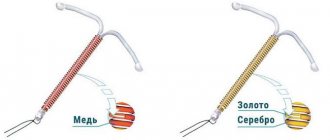
Hormonal drugs are used to prevent the growth of new foci of myomatosis. As a rule, oral contraceptives take into account the woman’s constitution. The most commonly prescribed drugs are Jess, Yarina, Dimia, Midiana, Regulon; the Mirena intrauterine system can be used - this is a hormonal-acting device.
If a woman does not pursue a contraceptive effect, only gestagens can be prescribed - Duphaston, Utrozhestan and others.
Hormonal treatment is especially indicated in the following cases:
- if fibroids are accompanied by endometrial hyperplasia or polyposis;
- if additional contraception is needed;
- if a woman complains of heavy menstruation.
Duration of treatment is from three months. When using oral contraceptives - according to the standard regimen, when using gestagens, the doctor prescribes them individually depending on the clinical situation.
As preoperative preparation, other drugs are used, more serious ones - they cause a state of artificial menopause in the woman’s body, which leads to a slight decrease in the nodes. This makes surgical intervention technically less difficult. Buserelin, Zoladex and others are used according to individual schemes lasting from a month to three and less often - up to six months.
To reduce the severity of fibroid symptoms, hirudotherapy and physiotherapy (magnets, electrophoresis) can be used . They improve well-being and can slow down the growth of nodes.
In other cases, symptomatic treatment is carried out. For example, if uterine fibroids are accompanied by heavy menstruation, which leads to blood loss, iron supplements are prescribed to maintain hemoglobin levels. If there is a malnutrition of the nodes without clinical symptoms of an acute abdomen - antispasmodics, antibiotics, vitamins. Lack of treatment (both with anemia and malnutrition of the nodes) is a direct indication for surgical treatment.
Surgical removal
Indications for surgery are the following conditions:
- individual nodes are more than 5-6 cm in diameter;
- malnutrition of nodes and their necrosis;
- fibroids, accompanied by constant heavy blood loss and anemia;
- when planning pregnancy (including IVF) and multiple fibroids;
- for subserous nodes from 3 cm.
After childbirth, if there are no emergency indications, myomatous nodes are removed no earlier than six months later and when the woman’s condition is stable.
The following types of operations are possible:
- Laparoscopic . In this case, special modern technology is used. Several small incisions are made in the skin of the abdomen through which manipulators are inserted. With their help, an operation is performed, and the doctor observes the “picture” on the monitor screen. This is a somewhat technically complex, more complex operation that requires special equipment and trained personnel.
However, the degree of blood loss and the severity of pain in the postoperative period are several times less. As is the general recovery period.
- Laparotomy. This is a classic version of the operation. In this case, a longitudinal or transverse incision is made in all layers of the anterior abdominal wall, and the doctor directly carries out all stages of the operation with his hands and instruments. In this case, the intervention may be accompanied by heavy blood loss, a long period of rehabilitation and the need to administer serious drugs for pain relief.
However, this is the method of choice if a woman is planning a pregnancy, since the quality of the scars on the uterus and the sutures are higher.
- Hysteroscopy . Used only when nodes are located inside the uterine cavity. However, they should not be very large in size, otherwise the manipulation will be technically impossible.
Hysteroscopic myomectomy
Removal of fibroids can be in several options:
- total hysterectomy - removal of the uterus and cervix with all nodes;
- subtotal hysterectomy - removal of the uterine body with nodes, leaving the cervix;
- removal of only the nodes while preserving the rest of the myometrium;
- removal of submucosal nodes - inside the uterine cavity.
Removal of nodes or the uterus can be performed as part of a cesarean section immediately after the baby is removed. This makes it possible to avoid another operation.
Watch this video about methods of treating uterine fibroids: