Health
Woman who thought she could never have children again becomes pregnant with twins
.
Australian doctors and scientists have been able to help a woman become pregnant for the first time after she had her ovaries removed during cancer treatment seven years ago
.
This was a real breakthrough, which gives hope to many women who have survived cancer to conceive children.
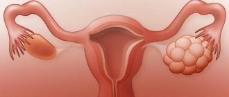
Vali, a 24-year-old woman from Brisbane in Australia, before treatment, asked doctors to save part of the ovarian tissue, which was then transplanted into her abdominal wall
.
What is ovarian resection?
The process involves partial removal of the ovary as a tumor is diagnosed. Previously, patients were offered laparotomy. However, with the development of medicine, laparoscopy became available.
When a tumor is detected, and after drug treatment it does not resolve on its own, then resection is appropriate. As for the indications, they are as follows:
- carcinoma;
- dermoid cyst;
- endometrioma;
- cystadenoma.
It is important to check both generative organs so that the tumor does not develop further. With PCOS, incisions are made, and if a rupture occurs and suppuration appears, then it is necessary to proceed to radical measures.
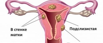
Doctors excise the affected area. This is done when:
- numerous cysts;
- PCOS;
- benign tumors;
- traumatization;
- apoplexy.
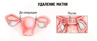
Resection methods
The operation can be performed using two methods. The main one is laparoscopy. This technique is the safest. Doctors make a small incision in the abdomen. It is customary to insert special devices into the holes for excision and translation of internal mechanisms on the screen. The scar will be small from an aesthetic point of view.
The second type is laparotomy. This intervention is abdominal; with its help, doctors make a longitudinal incision, the size of which reaches 10 cm. Through it, the affected part of the ovary is removed. This type is more dangerous and traumatic; the scar remains very large.
Indications can be identified after a long examination to determine whether pregnancy has occurred. In particular, ladies often complain about an irregular menstrual cycle, painful periods, untimely release of eggs or lack of ovulatory process.
Obstacles to conception
As a rule, the presence of only one tube indicates the presence of atopic pregnancy in the past. Ectopic pregnancy is a pathological condition that is characterized by the fixation of a fertilized egg outside the uterine cavity. Most often this occurs in the uterine appendage, but possibly also in the ovaries, abdominal cavity, and cervical canal. The only treatment for tubal pregnancy is laparoscopic removal of the fallopian tube along with the attached fertilized egg.
It will take twice as long to conceive a baby with one appendage than for healthy couples. In addition to patience, such partners should monitor ovulation. If surgical treatment of an ectopic pregnancy was performed, then in the postoperative period you should undergo full treatment, the purpose of which is to restore fertility and prevent adhesions.
For this, both medication and physical therapy are used. You should plan your next pregnancy only with a doctor, carefully following his recommendations.
Your ovulation calendar
Menstruation
Ovulation
Fertile day
Preparation
Initially, the attending physician will send the patient for a thorough examination. He will need the results of a general clinical blood chemistry test to determine whether antibodies to viruses are present. The latter often reduce clotting.
Everything is carried out exclusively under anesthesia, before which you need to relax the muscles located in the field between the esophagus and stomach. Experts advise stopping eating before 8 pm on the eve of the procedure, and drinking water at 10 pm.
Intestinal cleansing will also be required, because its peristalsis will be temporarily inhibited. This is done using an enema with clean water.
How it is done
The girl is under anesthesia and doesn’t feel anything. The doctor makes one main and several small incisions. The next sequence of actions is as follows:
- the organ is freed from neoplasm and small adhesions;
- clamps are applied;
- an incision is made into the ovarian tissue;
- cauterization and suturing of blood vessels is carried out;
- the remaining glands are sewn;
- drainage is installed in the pelvic cavity;
- stitching damaged tissues.
The woman is warned in advance, especially if cancer is suspected, that doctors may proceed to laparotomy. Then human life and health are given priority.
Recovery period
The most important days are the first. The more correctly doctors provide medical care and prescribe treatment, the more successful the result will be.
You are allowed to get out of bed on the second day. A special place here is occupied by the hygiene of postoperative wounds. Nurses are required to change gauze bandages every day and treat stitches with a special solution.
It is believed that the woman will fully recover in a month. Sexual rest must be observed for two weeks; you can play sports after 30 days. It is also recommended not to take a bath for 10 days.
How to get pregnant if one ovary is not working
Questions about the possibility of pregnancy with one gonad concern women not only after oophorectomy. In a number of situations, the organ ceases to function, remaining in its place. This happens with a unilateral inflammatory process, the formation of neoplasms, and hormonal pathologies. Also, the ovary may stop working after surgical treatment of a cyst, when only a small part of the organ remains.
Diagnosing pathology is not difficult. Impaired activity of the reproductive gland is accompanied by the absence of dominant follicles for several cycles and a decrease in size. The ovulatory reserve also decreases, as shown by laboratory blood tests.
If one ovary does not work, and the second functions correctly, then pregnancy is possible. It is necessary to check the fallopian tubes for patency and monitor favorable days. The presence of a nonfunctioning reproductive organ pair does not reduce the chances of success. In the absence of other barriers to conception, doctors recommend an active sex life before and during ovulation.
When does ovulation appear after resection?
Surgical intervention does not affect the release of the egg. Ovarian resection and ovulation are interconnected, since after the procedure reproductive function should be restored. The patient's hormonal levels normalize, so her own follicles begin to mature. High levels of androgens can prevent this. Reducing them will restore function.
With uterine obstruction, there are also chances, but not for long. It is recommended to try to conceive in the first 6-12 months as new adhesions may form. Even in the presence of full-fledged NCs, they will interfere with fertilization.
Removal of appendages and pregnancy: risks and health consequences
Removal of appendages and simultaneous pregnancy are accompanied by a high risk of complications arising on the operating table. Although the percentage of successful removals exceeds the number of complicated interventions, patients should be aware of the possible consequences:
- Damage to the intestines, ureters and bladder;
- Thromboembolism;
- Torsion of the tumor stalk;
- Rupture of the tumor capsule;
- Abortion.
Removal of the appendages leads to changes in the hormonal balance in the body, which can complicate future pregnancies. Most often this manifests itself as psycho-emotional instability, insomnia, irritability or depression.
As estrogen synthesis decreases, this leads to the following complications:
- Lipid metabolism disorders;
- Gaining fat mass;
- The appearance of atherosclerotic plaques in blood vessels;
- IHD;
- Increased calcium levels in the blood;
- Osteoporosis.
In addition, there is a possibility of headaches, heart rhythm disturbances and arterial hypertension.
Onset of pregnancy
If pregnancy is planned after resection, then the woman should be aware that a number of difficulties cannot be excluded. Over the entire period, from 400 to 600 nuclear centers are produced during the reproductive period. When part of it is removed, this amount decreases. If it was carried out before the age of 30, then the chances increase, since the reserve of the nuclear center is sufficient. Doctors often resort to stimulation afterwards to restore egg production. For this purpose, hormonal drugs are prescribed in combination with folk remedies, such as rose, hogweed, sage, and plantain.
Your period comes quite quickly, and in the next menstrual cycle the follicle begins to mature.
Pregnancy often does not occur due to hormonal imbalance or adhesions. They appear as damaged tissue tries to quickly repair itself. They are first tried to be treated with medication, but then gynecologists decide to take more radical measures.
pregnancy after tubal removal
Authors : Lelyukh Natalya Nikolaevna
Today we will think about the topic of fallopian tubes and their patency. The very name “pipe” already suggests a picture in the form of a sausage, a tunnel through which sperm scurry back and forth, eggs swim majestically, so that it is here, in the feather-grass expanses of the epithelium, that the miracle of their meeting and the first division of the resulting embryo occurs. So, in order. Familiar scheme:
So, in truth , as shown on the left - the fallopian tube does not cover the ovary, but lies next to it, conventionally attached to it by a long ovarian fimbria (the fallopian tube strongly resembles a hydra with octopus-shaped legs at the end, these legs are called fimbriae or fimbriae) . Real picture:
The fallopian tube is NEAR (I said, next to!) the ovary of the same name, turned towards it by fimbriae (fimbriae), awaiting ovulation and the egg floating on a wave of follicular fluid. Ovulation (amazing footage found on the internet):
This little yellow sun is an egg. It leaves the ovary into the abdominal cavity and, in favorable conditions, is carried into the vestibule of the fallopian tube by a wave of follicular fluid. And there the fimbriae (aka fimbriae) are already waiting, like this:
There, the egg proudly, but benevolently, allows itself to be hugged, wrapped in fringes and carried away inside the tunnel. And then - interesting things. A healthy fallopian tube from the inside resembles a feather grass steppe, or an eared wheat field, or an untrimmed lawn. Choose your own comparison and metaphor, but with magnifying optics it looks like this:
These villi are special ciliated epithelium . Each cell of which has a long outgrowth, histologists affectionately call it an eyelash. The main task of which is to flicker and create oscillatory movements so that the “sun egg” moves towards the uterus. The sun (read eggs) has no legs, well, what kind of legs can the sun have? Therefore, delicate eyelashes work tirelessly... uh... eyelashes! And when fertilization occurs, the resulting embryo is also carefully carried by its cilia towards the future home. So, conclusions from “cute physiology”: 1. The fallopian tube is not a hollow, smooth substance, reminiscent of an attraction in a water park; inside it has a very delicate and thin structure. 2. The fallopian tube does not enclose the ovary and is not attached to it. 3. The fallopian tube has fimbriae (fimbriae), which are able to attract the egg inside the tube. Conclusions from the conclusions: 1. All, without exception, inflammatory processes in the tubes cause damage to the finely organized cilia, and even the death of some of them. Because of this, bald areas form in the pipe, which leads to...? Right! This leads to the development of an ectopic pregnancy. 2. Inflammation, especially caused by aggressive pathogenic flora (gonorrhea, chlamydia and tuberculosis) causes a pronounced adhesive process around the pipes, and the pipe can be tied several times with adhesions from the outside, which leads to its obstruction. And to the same ectopic pregnancy. 3. Genital endometriosis, and we have already talked about it more than once, also causes inflammation with the formation of adhesions, with the subsequent development of obstruction and everything, as in the points above. 4. Separately about chlamydia. Chlamydia really likes to live in fimbriae (aka fimbriae) and with chronic chlamydia, the fimbriae stick together tightly. And no one waits for the emerging sun and no longer lures him into the abyss of passion. The sun will swim, swim around, don’t take it! And he dies. Because this sun has a lifespan. And now my personal conclusions from the above. The only, most logical and even therapeutic way to check the patency of the fallopian tubes can only be laparoscopy. Because we see a problem, we try to eliminate it, and only then we introduce a contrast agent to check the internal patency of the pipes. To make it completely clear. All “contrast” methods of checking the patency of the fallopian tubes, and in our geographical coordinates this is an x-ray technique and an ultrasound technique, look like this:
This, my friends and girlfriends, not only hurts, but also has the following risks: 1. If the pipe is obstructed due to an external factor, and we also put liquid in there, which due to the obstruction does not pour out. It looks like this:
The doctor now has only one option: to quickly call this patient for an operation to remove such a fallopian tube. And if you were to do it the smart way, then go in, cut the adhesions, lay out the fimbriae and pump in the liquid for your health! 2. The second argument is not in favor of these two methods - if liquid gets into the pipe and does not immediately pour out, or the patient does not want to run after the doctor to the operating room to immediately remove the pipe, then a water hammer occurs on the epithelium of the pipe, and there, you remember - delicate eyelashes. So, they, dear and trembling, perish in such situations. And, if, nevertheless, it was possible to release the liquid from the pipe, then it is now a little bald inside, and already vaguely resembles an attraction in a water park, along which there are screams of “hey”!!! sperm rush, collide with the egg and... no one goes anywhere anymore, because you can’t go back up the pipe in the opposite direction... 3. My third personal argument. Remember we spent a long time discussing the topic of endometriosis and you wrote to me saying how could this be? How can the mucous membrane from the uterus suddenly get into the abdominal cavity? So, it can get there not only during sex during menstruation, but also during hydrotubation. A syringe is used to inject liquid into the uterine cavity under pressure, and this liquid can carry individual fragments of the endometrium into the abdominal cavity. And, in less than half a year, the passable pipes will become not entirely passable, or completely impassable. And at all subsequent consultations about infertility, the patient will show evidence of an X-ray or ultrasound image that, they say, they are passable! Yes, I perfectly understand and realize that laparoscopic testing of the patency of the fallopian tubes is an operation with anesthesia, it is one day in the hospital and five days on sick leave, but! this is an answer to questions and an attempt to fix the problem. Therefore, I am against echohydrotubation and X-ray testing of fallopian tube patency. It's up to you to choose. Again to you. Your doctor Natasha.