Sclerocystic ovary is one of the most common gynecological diseases that occurs as a result of disruption of the endocrine system. It is a compaction of the uterine appendages, the formation of small cysts in their cavity and the absence of ovulation. Based on medical statistics, sclerocystic disease affects 13% of women of childbearing age and more than 70% of women who have experienced menopause. Very often (in 70% of all cases) female representatives who cannot conceive a child for a long time suffer from this particular pathology.
Treatment measures should be started immediately in order to increase the likelihood of a complete cure and the possibility of becoming pregnant in the future. In order to detect the presence of a disease in time, we will try to understand in detail what it is, how the disease manifests itself, is diagnosed and why it appears.
How healthy ovaries work
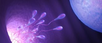
To better understand how ovarian sclerocystosis and pregnancy are connected, you need to know how these organs are structured and how they work if there is no pathology in them. The ovaries are the female paired reproductive organs. They can be imagined as peculiar sacs filled with brain matter. The walls of the ovaries are lined with a layer of dense connective tissue, on which there is a layer of cortical substance. It has a complex structure and is important. It is in this layer that follicles are formed - specific structural elements in which eggs develop. Follicles, called primary follicles, numbering approximately one to two million, are formed in the body of each girl at the fetal stage. Throughout life, from the period of puberty to the period of menopause, they are gradually consumed, and new ones are no longer formed. Therefore, the hour comes when their supply runs out.
This almost never occurs in women of childbearing age, so infertility cannot be caused by a lack of follicles. Another thing is that in their stage-by-stage maturation sometimes failures occur. So they are the culprits that the desired pregnancy does not occur. Moreover, improper development of follicles in one hundred percent of cases leads to gynecological diseases, without treatment of which in women the risk of thrombosis, thrombophlebitis, diabetes mellitus, heart attack, and malignant formations in the mammary glands increases.
Signs of sclerocystic ovaries in women and methods of treating pathology
One of the most common pathological processes that develops due to endocrine disorders is ovarian sclerocystosis. The disease affects 12% of women of reproductive age. According to statistics, most problems with conception in women are caused by ovarian sclerocystosis.
Definition of pathology
Sclerocystic disease is a pathological process that affects both ovaries. As a result of the changes, compaction of the outer protein membrane and the formation of cysts on the surface of the organ are observed. These cystic formations are classified as follicular.
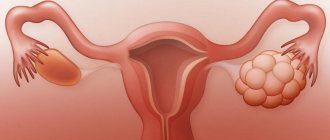
With scleropolycystic ovaries, a large number of follicular cysts filled with light fluid are formed. At the same time, the number of mature follicles decreases. This leads to the proliferation of stromal tissues and an increase in the size of the organ. Such metamorphoses make ovulation impossible.
In addition, against the background of such changes, a woman may develop hyperplasia of the uterine body.
The main factor in the development of pathology is disruption of the functionality of the endocrine system.
Increased levels of male sex hormones (hyperandrogenism) and decreased levels of female estrogens are the main factors influencing the occurrence of the pathological process.
That is why it is very important to undergo a preventive examination once a year, as well as take all the necessary tests in order to notice in time the onset of degenerative changes in a woman’s body.
According to one theory, sclerocystosis develops against the background of insulin resistance (a pathology in which there is no sensitivity to insulin).
Against the background of this disease, there is a disturbance in the functionality of the endocrine gland.
Experts note that diabetes mellitus is one of the predisposing factors in the development of ovarian sclerocystosis. Therefore, it is important to monitor your blood sugar levels.
It is not recommended to ignore the symptoms of the disease. Sclerocystic disease is not a passing disease, but a chronic illness that causes persistent and, in some cases, irreversible changes.
Advanced sclerocystosis of the appendages provokes disturbances in the metabolic system, and also combines not only endocrine disorders, but also somatic pathological conditions.
Unfortunately, there is no complete cure for sclerocystic disease, but in medical practice various medications are widely used to correct and compensate for the symptoms that the patient already has.
A positive result of treatment is the woman’s ability to become pregnant after therapy. Stein-Leventhal syndrome, which is the name given to ovarian sclerosis, was first mentioned in 1935 by American gynecologists.
Causes and types of sclerocystosis
There are two types of sclerocystic ovary syndrome: acquired and hereditary. This pathology usually occurs in girls during puberty and in young women who have not yet become mothers.
The disease can develop with multiple cysts, as well as with enlarged or shriveled ovaries. The surface of the paired organs in both cases is covered with a specific dense membrane, under which cystic follicular neoplasms are visible.
Modern gynecology and reproductive medicine do not name factors that have an absolute influence on the occurrence of pathology.
The causes of ovarian sclerocystosis can be:
- Heredity factor. In this case, the dominant place is given to enzyme deficiency with additional disruption of the functionality of specific hydrogenases and dehydrogenases. These substances are actively involved in the production of steroid hormones. As a result of such disturbances and abnormalities in work, the transition of male androgens to female hormones estrogens is significantly reduced. Such internal changes in hormonal metabolism entail disruptions in the functioning of insulin receptors, leading to a significant decrease in the sensitivity of insulin-dependent cells.
- Chronic infections. Quite often, the cause of the development of ovarian sclerocystosis is not inflammation in the appendages, but neuroendocrine disorders with a change in the functionality of the ovaries. Some medical sources have drawn a connection between the development of sclerocystic disease in women and chronic inflammation of the tonsils.
- Complicated childbirth, abortion, oophoritis, salpingitis, endometritis.
- Excess weight occurs not only as a consequence of disturbances in the hormonal system, but can also be a factor predisposing to the appearance of sclerocystic disease.
- Malfunctions of the hypothalamus and pituitary gland lead to disturbances at the ovarian level. The main causes of failures are hypothalamic and diencephalic syndromes. This type of change occurs quite rarely in patients and is not dangerous.
- Pathological changes in the adrenal cortex. There is an assumption that under the influence of specific hormonal substances produced by the pituitary gland, not the ovaries, but the adrenal glands begin to be stimulated. According to the hypothesis, this occurs during pubertal development.
Source: //TvoiYaichniki.ru/kista/vidy/sklerokistoz
How does an ovarian cyst appear and what does it have to do with pregnancy?
When girls become sexually mature, their body begins to work on the process of maturation of primary follicles, which until now seem to be sleeping. This process always takes place cyclically. In each cycle, up to about 15 follicles “wake up”. Under the influence of the FSH hormone produced by the pituitary gland, they begin to grow, increasing in diameter from 50 to 500 microns. During this period, follicular fluid forms in them, and a cavity appears in the largest of them. This follicle becomes dominant, grows up to 20 millimeters, and protrudes. An egg cell develops rapidly inside it. The remaining follicles from the group of “awakened” ones die and dissolve one by one. If everything goes according to the rules, the endocrine system in the female body comes into play. As a result, the hormones estrogen, progestins and androgens are produced, affecting the further maturation of the dominant follicle. Under the influence of the luteinizing hormone (luteotropin, lutropin, abbreviated as LH), it ruptures, the egg leaves it into the fallopian tube, and it itself turns into the corpus luteum and gradually dissolves.
If a rupture does not occur, the unreleased egg is degenerated, and an ovarian cyst the size of a cherry appears in place of the follicle. Those of the “awakened” follicles that did not have time to die also turn into cysts, only smaller in size. A cyst formed from a follicle sometimes grows to a significant size (40-60 millimeters), but may not manifest itself in any way. Only in some cases do patients complain of pain in the ovarian area. After a woman’s hormone production normalizes, it gradually resolves. If a woman’s ovulation is restored, the follicular cyst present in the ovary at that time does not interfere with pregnancy, but if this cyst has grown to a size of 90 millimeters, it must be removed surgically.
Sclerocystic ovaries and pregnancy
A woman suffering from pathology needs to normalize the cycle and recreate the normal thickness of the endometrium. For this purpose, ovulation inducers are used. If during therapy the growth of the dominant follicle is observed, then an ovulatory dose of hCG-based medication is administered. After 2 days from the moment of injection, the egg matures.
Scleropolycystic ovaries and desired pregnancy are compatible. The operation is performed in case of ineffectiveness of hormonal stimulation, a thick ovarian capsule and in patients over 30 years of age. Pregnancy usually occurs within 3-5 cycles. Further, the excised membrane of the appendage is restored, and conception becomes difficult.
If pregnancy does not occur, then IVF is used. Indications for it also include the presence of tubal infertility and the inability to conceive a child due to reproductive abnormalities in the health of the woman and her partner.
Sclerocystic ovaries are a process of their degeneration, accompanied by the formation of small cystic formations up to 1 cm in size.
With sclerocystosis, the ovaries enlarge, and compacted membranes form on their surface.
This syndrome is sometimes called Stein-Leventhal syndrome. Typically, sclerocystic disease forms as a result of polycystic ovary syndrome as the disorders progress.
The occurrence is 3-5% of all gynecological diseases, in approximately 30% of them ovarian sclerocystosis causes persistent infertility.
Causes of the disease
Scientists know in detail how ovarian sclerocystosis is formed. The reasons for this phenomenon have not yet been precisely established; there are only assumptions. Since hormones play a vital role in the normal development of the follicle and the release of the egg from it, hormonal disorders, and in particular a failure in the mechanism of estrogen synthesis, are considered to be the main cause of ovarian sclerocystosis. The following causes of hormonal disorders are called:
- heredity;
- abnormalities in gene structure;
- disorders in the pituitary-ovarian system;
- mental trauma;
- complications after abortion;
- infectious and gynecological diseases;
- complications after childbirth;
- changes in the functions of the adrenal cortex.
Causes of sclerocystic ovaries
Views on the etiology of the disease vary.
Previously, the leading opinion was that it was caused by disturbances in the circhorial rhythm of luliberin production. Not long ago, another approach was proposed, based on the theory of insulin resistance. It is believed that insufficient processing of glucose leads to an increase in insulin concentration, as a result of which LH rises and the ovaries increase in size. Some scientists believe that the cause of sclerocystic disease is excessive production of FSH by the anterior pituitary gland. This hormone is responsible for the growth of the dominant follicle in the appendages, from which an egg is released at the time of ovulation. However, an increased amount of FSH leads to the appearance of many immature follicles.
The hereditary nature of the disease was established, which prompted the search for genetic defects responsible for a complex hormonal disorder. Modern science considers ovarian sclerocystosis as a multifactorial pathology, in the development of which the leading role is given to a genetic anomaly, leading to the launch of cytochrome P-450 and steroidogenesis in the appendages.
Other causes of pathology are:
- chronic infectious diseases accompanied by neuroendocrine disorders;
- complex childbirth, multiple abortions, chronic pathologies in the field of gynecology;
- obesity;
- diseases of the adrenal glands of a primary nature.
Clinical symptoms
Unfortunately, it is only possible to detect ovarian sclerocystosis in a girl at the beginning of puberty. Symptoms at this stage are vague and mainly consist of menstrual irregularities. But this phenomenon can have many other reasons not related to ovarian disease, including poor nutrition and nervous disorders. By the age of twenty, or at most by the age of twenty-five, girls develop more definite symptoms of ovarian sclerocystosis. The main problem is still a violation of the cyclicity and nature of menstruation (in 96 percent of patients). More often there are long delays in menstruation (about six months or more) or too small amounts of discharge (hypomenstrual syndrome). Much less often, patients complain about the duration and abundance of menstruation.
Other symptoms that may suggest ovarian sclerocystosis are as follows:
- hirsutism (about 90 percent of patients experience hair growth in the area around the nipples, back, abdomen, chin and above the lip);
- overweight (70 percent of patients);
- baldness and acne on the face (occurs in no more than 40 percent of cases);
- some changes in body proportions;
- disturbances in the functioning of the nervous system;
- asthenic syndrome;
- enlarged ovaries (detected by a gynecologist during examination).
In addition, some women may experience symptoms common to many diseases: pain in the lower abdomen, malaise, and unexplained fatigue.
Complications and consequences
Although sclerocystic ovary syndrome is not fraught with transformation into an oncological disease, however, the presence of such a pathology, as well as disruption of hormonal and reproductive functions, increases the likelihood of developing malignant neoplasms.
Due to the fact that the disease adversely affects hormone levels, the following pathological conditions can develop in the body:
- Arterial hypertension;
- Atherosclerosis;
- Disturbances in the functioning of the cardiovascular system;
- Incorrect perception of insulin, which leads to the development of diabetes;
- Endometriosis (pathological thickening and proliferation of the protective layer of the uterus);
- Other disorders in the pelvic organs.
In addition to the conditions described above, the disease, if treated inappropriately, can develop to such a stage when it is no longer possible to save the ovaries. They will have to be excised along with cavitary tumors during surgery. Such manipulations lead to incurable infertility.
Laboratory research
Based on external signs, ovarian sclerocystosis is only suspected, and the final diagnosis is made after additional examinations. These are:
- blood test for testosterone (total should be within 1.3 ng/ml, free in women under 41 years old - within 3.18 ng/ml, and up to 59 years old - no more than 2.6 ng/ml);
- analysis for glucose sensitivity, blood sugar and triglycerides;
- colpocytogram (material is taken from the vagina, the analysis data shows whether there is ovulation or not, as well as the correspondence of the colpocytogram indices to the patient’s age and the phase of her menstrual cycle);
- scraping of the endometrium (allows us to judge dysfunctions in the ovaries);
- monitoring changes in basal temperature;
- tests for some hormones of the thyroid gland, pituitary gland, ovaries (LH, FSH, PSSH, prolactin, cortisol, 17-hydroxyprogesterone);
- determination of the amount of estrogen excretion.
Now patients can independently conduct a simple test to suspect they have cystic ovarian formations. To do this, you will need a microscope (can be purchased at pharmacies). In the morning, just waking up and not having eaten or drunk anything yet, you need to place a drop of your saliva on a laboratory glass and let it dry. During ovulation, estrogen levels always increase, which, in turn, changes the composition of saliva. If there is ovulation, the saliva sample in the microscope will be in the form of fern leaves, and if there is no ovulation, it will be in the form of dots.
Diagnostics
First of all, after visiting the hospital, the doctor conducts a gynecological examination, interviewing the patient and palpating the affected organs.
Further, to confirm or refute the presence of a gynecological disease, the doctor can carry out the following diagnostic measures:
- Blood test for hormones;
- Testing for insulin resistance;
- Ultrasound examination of the pelvic organs;
- X-ray;
- Computed and magnetic resonance imaging;
- Diagnostic laparoscopy.
In order to determine the condition and performance of the endometrial layer, doctors may prescribe a diagnostic curettage procedure, hysteroscopy or biopsy.
Based on the examination results, the doctor prescribes the most appropriate method of therapy.
Hardware diagnostics
As a rule, for an accurate and final diagnosis, patients are prescribed a complex examination using medical equipment.
The most gentle and absolutely painless method is ultrasound diagnostics of sclerocystic ovaries. The procedure can be transabdominal (through the abdomen), transvaginal (the most highly informative method), transrectal (performed only on young girls and elderly women).
Using ultrasound, the size of the ovaries, their shape, structure, the number of follicles in them, the diameter of which is up to 8 mm, the presence or absence of a dominant follicle, the presence or absence of ovulation, the presence of cysts in the ovary are determined.
Another type of examination is a gas pelveogram, which shows deviations from the norm in the size of the ovaries and uterus.
One of the most difficult types of diagnostics is laparoscopy. It is performed in a hospital under general anesthesia. The algorithm is as follows: the surgeon punctures the peritoneal wall of the patient and inserts a device inside that pumps carbon dioxide in order to create volume in the peritoneum and better examine the organs. Next, a laparoscope is inserted into the patient’s body, which shows the condition of the ovaries on the screen. Laparoscopy is the most accurate diagnostic method, but after it the woman requires a rehabilitation period.
Causes
The main role in the development of sclerocystic ovaries is attributed to disruption of the processes of synthesis and release of sex hormones. In addition, disturbances in the functioning of all endocrine organs, one way or another involved in the regulation of the menstrual cycle, play a role.
Some doctors are of the opinion that sclerocystic disease occurs due to excessive production of follicle-stimulating hormone (FSH). This leads to the suppression of the normal functioning of the ovary and the formation of small immature cysts covered with a dense membrane.
In the development of sclerocystosis, a role is also played by disruption of the synthesis of luteinizing hormone (LH).
Another theory identifies the main cause as excessive activity of the adrenal cortex, as well as defects in the formation of steroid hormones and estrogen deficiency. For this reason, the maturation of follicles is disrupted, the level of male sex hormones increases and infertility occurs.
Sclerocystosis can be hereditary or acquired; it usually occurs in girls after puberty and in nulliparous young women.
Sclerocystic disease can occur with enlarged or polycystic ovaries, or with reduced or shrunken ovaries. In both cases, their surface is covered with a dense membrane, under which cystic degenerating follicles can be contoured.
Conservative methods of treatment of sclerocystic ovaries
After a final diagnosis is made, in most cases a woman is first prescribed drug therapy. Its goal is to restore the normal menstrual cycle and resume ovulation. How to treat ovarian sclerocystosis is decided by the gynecologist together with the endocrinologist.
If a patient is obese, the first stage of treatment is weight loss. The woman is prescribed a diet and feasible exercise.
The second stage is increasing insulin perception. Metformin is prescribed, which must be taken for 3-6 months.
The third stage is stimulation of ovulation. Begin therapy with the simplest medicine - Clomiphene. The initial course consists of taking the drug at a dose of 50 mg at night, starting from the 5th day of the cycle for 5 days in a row. If there is no result (menstruation), Clomiphene is taken for a month. If the effect is still not obtained, the dose is increased to 150 mg per day.
The next stage (in the absence of positive dynamics) is the prescription of the drug “Menogon”. It is administered intramuscularly, and at the end of the course, Choragon injections are given. "Menogon" can be replaced by "Menodin" or "Menopur".
Having completed the entire course, blood biochemistry is done, and based on the results of the analysis (if there is not enough LH hormone), Utrozhestan or Duphaston is prescribed.
At the same time, doctors are trying to remove excess hair from the woman, and therefore she is prescribed Ovosiston and Metronidazole.
A mandatory addition to the course is vitamin therapy.
Sclerocystic ovaries: what is it, causes, treatment, effect on pregnancy
Ovarian sclerocystosis is a pathology that is diagnosed in more than half of women suffering from endocrine infertility and problems with conception. The exact cause of the disease remains unclear. The disease can occur in various forms, each of which requires an individual approach to therapy.
What is ovarian sclerocystosis
PCOS (sclerocystic disease, polycystic disease or polycystic ovary syndrome) is a change in the structure and normal functioning of the ovaries, in which there is no maturation and release of the dominant follicle (ovulation).
With the disease, the number of polycystic ovaries increases and the dominant follicle is absent. The altered ovaries increase in volume due to the proliferation of the stroma, their tunica albuginea acquires a pearly hue. In cross-section, they look like honeycombs with cavities of different diameters.
According to the results of various clinical studies, sclerocystic ovaries are primary (Stein-Leventhal disease) and secondary, which develop with neuroendocrine disorders.
In gynecology, sclerocystic ovarian syndrome is clinically divided into three forms:
- Primary form (typical modified ovaries).
- Mixed (combination of pathology of the ovaries and adrenal glands).
- Central form (impaired functioning of the central sections and, as a consequence, secondary sclerocystosis).
Causes
There is no clear, single confirmed cause for the development of this condition. The primary form of sclerocystosis is associated with a genetic lack of enzymes. These enzymes block the conversion of androgens to estrogens. In this case, disruption of the enzyme system leads to hormonal imbalance, which causes characteristic changes.
The cause of the mixed form may be a change in the function of the adrenal cortex, where the synthesis of sex hormones is disrupted.
In scleropolycystic ovarian disease of central origin, brain structures play a role. The cause of disruption of their work can be depression, infectious factors, psychotrauma, abortions.
Symptoms
Clinical symptom complexes of sclerocystic disease can be varied. The key symptom in any form is menstrual disorders, for example, anovulation.
Anovulation is a disorder in which the normal maturation of the egg and its release from the follicle are disrupted, which is a necessary condition for the intended conception.
In the typical form, changes can be observed starting from the first menstruation; in other types of sclerocystic disease, they appear later.
Other signs of the disease may include:
- infertility;
- instability of the menstrual cycle;
- excessive hair growth;
- obesity of varying degrees;
- brittle nails, hair loss, stretch marks on the skin.
Each manifestation depends on the form of the disease and is individual for different patients. In rare cases, active signs of virilization (male-type changes) are observed, such as a change in the timbre of the voice, an increase in the size of the clitoris, and a change in figure like a male.
Diagnostics
The specialist begins the examination for sclerocystosis with a thorough collection of anamnestic data and examination. This allows you to determine the time of onset of the disease. Manifestations of primary polycystic ovaries can be traced starting from the first menstruation in girls, which distinguishes them from the secondary process.
Clinically, sclerocystic disease can be suspected when excess hair growth or other signs of virilization appear in a girl, which can be different. When examined in a gynecological chair, a specialist may note a change in the size of the ovaries (decrease or increase).
The absence of changes in basal temperature, a negative ovulation test, and long-term problems with conception allow us to suspect a lack of ovulation.
Hardware diagnostics
One of the key methods for diagnosing sclerocystosis is ultrasound of the pelvic organs. This study is carried out in several ways using different sensors. Transabdominally (through the abdomen) one can detect a bilateral increase in the size of the ovaries, often with an underdeveloped uterus.
Transvaginal ultrasound shows an increase in the size of the ovaries over 9-10 cm3. An overgrown stroma and underdeveloped follicles under a thickened capsule are determined.
In addition to ultrasound, a specialist may prescribe a pelveogram. It helps detect abnormalities in the size of the uterus and ovaries.
In some situations, diagnostic laparoscopy is performed. This is an intervention that is performed through a puncture in the anterior abdominal wall. With the disease, thickening and smoothing of the ovarian capsule and an increase in their size are observed. With this procedure, it is possible to perform a biopsy followed by histological revision.
Laboratory research
Among laboratory tests, the following are considered mandatory for sclerocystosis:
- Blood test for hormones. The level of testosterone, FSH, LH and other gonadotropic hormones is determined. It was previously believed that certain levels of hormone changes could be considered a diagnostic criterion for polycystic ovary syndrome. But recently, cases have been diagnosed where hormonal parameters are normal, but the clinical picture and signs allow a diagnosis of sclerocystic disease to be made.
- Diagnosis of metabolic disorders: blood test for sugar, triglycerides.
To identify the cause, additional endometrial scrapings and a colpocytogram may be prescribed. Basal temperature measurements and ovulation tests are carried out to confirm its absence.
The final diagnosis is made after a comprehensive comprehensive examination, which takes place with the involvement of specialists from different fields (endocrinologist, neurologist, gynecologist).
Treatment of sclerocystic ovaries
The principles of treatment depend on the symptoms, clinic and age of the patient. The main factor for women of reproductive age is infertility.
In this case, the goal of treatment is to restore normal menstrual function. At the same time, they correct the prevailing symptoms (eliminate obesity, get rid of excess hair).
Drug, non-drug and surgical treatment methods are used.
Therapeutic
At this stage, obesity is corrected using diet therapy and dosed physical activity. At the same time, the use of physical procedures such as massage, baths, and reflexology gives a good effect.
A full-time psychologist should work with such patients to eliminate the psychosomatic component of the disease.
Surgical
Surgical treatment methods are used after or against the background of conservative therapy. Endoscopic approaches are chosen so as not to further injure the pelvic organs and not cause adhesions. For sclerocystosis use:
- Resection of hormone-secreting tissues of polycystic ovaries.
- Decortication (removal of the dense protein layer of the ovaries).
- Removal of individual cysts with laser (laser vaporization).
- Making incisions on the follicles to facilitate the release of the egg from the follicle.
The scope of surgical intervention is determined by the attending physician, after a thorough diagnosis individually for each case. Conservative
Drug treatment is aimed at normalizing hormonal levels. For this purpose, hormonal contraceptives, as well as drugs with pronounced antiandrogenic properties, can be selected.
Source: //ginekola.ru/ginekologiya/yaichniki/sindrom-sklerokistoznyh-yaichnikov.html
Sclerocystic ovaries: treatment with surgical methods
If ovulation is not observed within three months after drug therapy, the woman is prescribed surgery. It is performed using several methods. Which one to use depends on the indications of the condition of the ovaries.
At the present stage, there are the following types of operations:
- cauterization of cysts using a laser;
- Demedulation (removal of the middle part of the ovary);
- wedge resection (removal of a wedge-shaped portion of the affected part from the ovary);
- decortication (the doctor removes the transformed white layer of the ovary, pierces the follicles with a needle and sutures their edges);
- electrocautery (spot destruction in the ovary of the area in which too many hormones are produced).
- notches (the surgeon makes them up to 1 cm deep in places where the follicles are visible, so that when they mature, they can release an egg).
Diagnostic scheme
When making a diagnosis, the criteria established at the Rotterdam consensus in 2004 are followed. According to these data, a woman should have three signs:
- Oligomenorrhea and/or anovulation.
- Hyperandrogenism.
- Ultrasound signs of sclerocystic ovaries.
The following methods are used to detect pathology:
- Gynecological examination. Palpation reveals a normal sized uterus and enlarged ovaries. In advanced situations and severe sclerosis, the gonads become smaller. During the examination, signs of hirsutism and excess body weight attract attention.
- Laboratory research. A blood test shows an increase in luteinizing hormone and a change in the LH/FSH ratio by more than 2.5 times. There is an increase in testosterone, a representative of male sex hormones. A quarter of women experience an increase in prolactin.
- Ultrasonography. Ultrasound reveals enlargement of the ovaries, the appearance of cystic inclusions - small follicles up to 1 cm in size, no less than 10 in number.
- Doppler. When assessing blood flow, a pronounced vascular network is noted in the stroma of the organ.
- Endometrial aspiration biopsy is performed only for uterine bleeding. Sclerocystic ovaries often go along with endometrial hyperplasia, a condition in which the lining of the uterus grows. The longer the duration of the disease, the higher the likelihood of developing such a pathology.
Forecasts
Women who agree to any methods proposed by doctors are interested in the only question: is it possible to get pregnant with sclerocystic ovaries? Statistics show that without treatment, infertility is diagnosed in 90% of cases. Drug therapy with Clomiphene improves ovarian function in 90% of patients, but pregnancy occurs in only 28% of them. However, according to some data, positive results can reach 80%.
The disadvantage of the drug "Clomiphene" is that it is effective only at the very beginning of the disease or after surgery as an adjuvant.
Treatment with stronger drugs, for example Gonadotropin, according to statistics, leads to ovulation in at least 28% of patients, and at most in 97%. In this case, from 7 to 65% of women become pregnant.
If ovarian sclerocystosis is treated surgically, positive results are observed with approximately the same frequency as with conservative therapy. According to statistics, after ovarian surgery, 70-80% of women have a chance to get pregnant.
Symptoms of sclerocystic ovaries
Signs of the disease are often detected during adolescence. The main manifestation of scleropolycystic disease is a cycle disorder such as oligomenorrhea (when the intervals between menstruation are over 40 days) or amenorrhea (in the complete absence of menstruation).
In 15% of women, dysfunctional uterine bleeding is observed, that is, not caused by anatomical changes in the internal organs. In this case, spontaneous pregnancy is possible, but the likelihood of this happening is low. In addition, there is a risk of miscarriage.
Other signs of sclerocystic ovaries are:
- manifestations of increased production of male sex hormones:
- secondary masculinization;
- hirsutism;
- acne;
- thinning hair on the head;
- sometimes – changes in physique, breast hypoplasia;
- impaired glucose tolerance;
- obesity;
Sclerocystosis of the appendages. In an ultrasound picture, the ovaries are almost always enlarged
Reviews
For many women, it becomes a great misfortune to be diagnosed with sclerocystic ovaries. Patient reviews of the treatment are very different. For some, pills helped, for others, surgery helped, and for others, pregnancy did not occur, despite any methods taken.
There is also a small proportion of patients who report becoming pregnant without any treatment at all, although the diagnosis of ovarian sclerocystosis has not been removed. Such opposite results are possible due to the individual characteristics of each person and should not be taken as the norm.
But most women write in their reviews about improved health after treatment. Only some patients report that their periods normalized for a short time, after which they again needed to take hormonal medications.
And finally, there are some reviews in which women note the appearance of long-term painful sensations in the ovaries and peritoneum after treatment with surgery.
Symptoms of sclerocystic disease
Sclerocystosis is characterized by the following symptoms:
- sharp and bilateral enlargement of the ovaries,
- menstrual irregularities,
- infertility,
- increased body hair, often with masculine features,
- hypoplasia of the uterus, genitals and breasts,
- body weight disorder
- problems with hormonal metabolism.
Very often, sclerocystic disease is accompanied by excess weight and obesity, although this is not necessary.
There may be disturbances in general well-being due to an imbalance of androgens and adrenal hormones: general lethargy and weakness, headaches, apathy, neurasthenia, insomnia, decreased sexuality.
One of the most common manifestations of sclerocystosis is the absence of menstruation or its irregularities: first, menstruation lengthens, becomes very abundant or, on the contrary, scanty, then completely disappears.
Sclerocystic disease manifests itself as infertility, which usually occurs in 90% of patients. At the same time, the phenomena of hirsutism - excessive hair growth - gradually increase; hair grows above the lip, on the cheeks and on the chest, there is a lot of it on the arms and legs, and on the stomach. In this case, hypoplasia (underdevelopment) of the uterus or its atrophy is observed, and the mammary glands may be underdeveloped.
Symptoms
Clinical symptom complexes of sclerocystic disease can be varied. The key symptom in any form is menstrual disorders, for example, anovulation.
Anovulation is a disorder in which the normal maturation of the egg and its release from the follicle are disrupted, which is a necessary condition for the intended conception.
In the typical form, changes can be observed starting from the first menstruation; in other types of sclerocystic disease, they appear later.
Other signs of the disease may include:
- infertility;
- instability of the menstrual cycle;
- excessive hair growth;
- obesity of varying degrees;
- brittle nails, hair loss, stretch marks on the skin.
Each manifestation depends on the form of the disease and is individual for different patients. In rare cases, active signs of virilization (male-type changes) are observed, such as a change in the timbre of the voice, an increase in the size of the clitoris, and a change in figure like a male.
Dangerous consequences of pathology for women's health
Polycystic ovary syndrome is not only irregular, scanty menstruation, but also chronic anovulation. Normally, in the body of a healthy woman, an egg should mature monthly and be released into the abdominal cavity. 1-2 anovulatory cycles per year are allowed up to the age of 35 years. In the late reproductive period, the number of cycles without ovulation increases, and this is a natural process of aging of the body.
With sclerocystic ovaries, ovulation occurs extremely rarely, and it is difficult to predict exactly when the egg will mature. Without ovulation, pregnancy does not occur, and many women try to conceive a child for years. And if at the age of 18-25 there are still chances for spontaneous conception, then with age the probability of fertilization decreases. Persistent endocrine infertility develops - the main symptom of scleropolycystic ovarian disease.
It's not just infertility that threatens women. With PCOS, metabolic processes are disrupted, and the risk of developing other conditions increases:
- Hyperplastic process of the endometrium. The growth of the uterine mucosa occurs almost simultaneously with the dysfunction of the ovaries. Accompanied by uterine bleeding, intermenstrual spotting;
- Diseases of the mammary glands. Changes in hormonal levels during scleropolycystic ovaries lead to the proliferation of mammary gland tissue and the development of benign pathology - mastopathy;
Polycystic ovary syndrome can provoke the development of mastopathy.
- Diabetes. Insulin resistance that accompanies PCOS leads to disruption of the pancreas and increased blood glucose levels;
- Pathology of the heart and blood vessels. With PCOS, the risk of developing myocardial infarction, stroke, angina pectoris and other similar diseases increases.
The risk of developing concomitant pathology increases with age and with a long course of the disease. Carrying out specific therapy can not only eliminate infertility, but also reduce the likelihood of developing other complications.
Sclerocystic ovary disease is not associated with the development of gonadal cancer. There is no evidence that the pathology leads to the formation of ovarian carcinoma. However, the medical literature provides evidence that PCOS increases the risk of developing endometrial cancer. Practicing gynecologists associate this with the high frequency of detection of precancerous pathology - atypical endometrial hyperplasia.
Reasons for the occurrence of education
The etiology and pathogenesis of the disease are not fully understood. There are several theories of its origin.
Reason No. 1: disruption of the production of hypothalamic hormones
This theory is based on a disruption in the rhythm of gondotropin-releasing hormone (GnRH). This can be either a genetically programmed disorder or the influence of various factors:
- Past infectious diseases (including neuroinfections);
- Abortions and spontaneous miscarriages leading to hormonal imbalance;
- Complicated course of pregnancy and childbirth;
- Prolonged stress and severe emotional shocks;
- Inadequate physical activity;
- Inflammatory diseases of the pelvic organs;
- Endocrine disorders (pathology of the thyroid gland and adrenal glands).
A common cause of polycystic ovary syndrome is a malfunction of the thyroid gland.
As a result of the failure, the level of luteinizing hormone (LH) in the pituitary gland increases and the concentration of follicle-stimulating hormone (FSH) decreases. An imbalance of FSH and LH leads to increased synthesis of androgens in the ovaries
There is a deficiency of estradiol, an important female hormone necessary for the normal functioning of the reproductive system. By a feedback mechanism, a decrease in estradiol leads to an even greater increase in LH
A vicious circle is formed, from which the woman cannot get out on her own.
Another scheme for the development of pathology is also being considered. Some scientists believe that the primary malfunction is the ovaries, not the hypothalamus. In the gonads, there is increased production of androgens and a decrease in estradiol levels. Against this background, LH increases, which leads to certain changes in the reproductive organs. Atresia of the follicles occurs, the tunica albuginea of the ovaries thickens. Typical symptoms characteristic of PCOS develop.
Reason #2: Insulin resistance
Decreased tissue sensitivity to the hormone insulin occurs in 60% of women with polycystic ovary syndrome. The occurrence of pathology is associated with increased production of steroid hormones against the background of excess insulin in the blood. The LH level rises, the concentration of androgens increases, and estradiol falls. A vicious circle is started, as a result of which changes are formed in the ovarian tissue, and anovulation occurs.
Insulin resistance develops most often in obese women.
Reason #3: ovarian disorders
Recent studies show that excess androgen production is associated with impaired synthesis of cytochrome P450c17. This is a key enzyme that regulates the formation of steroid hormones in the adrenal glands and ovaries. It is assumed that genetic disorders are the basis of this failure. The influence of endogenous factors is also considered.
Some scientists believe that different mechanisms lie behind the development of PCOS with obesity and with normal body weight. In the first case, great importance is given to endocrine pathology and other external and internal factors affecting the rhythm of hormone production of the pituitary gland and hypothalamus. With polycystic disease against the background of normal weight, they talk about genetic disorders.
Women who devote themselves exclusively to work and refuse to bear children are at risk of developing polycystic disease.