Ligamentous apparatus of the uterus
The position of the uterus and appendages depends on the ligamentous apparatus of the uterus, which is represented by supporting, fixing and suspending apparatus.
Thanks to the interaction between the ligaments, fascia and muscles of the pelvic floor, the mobility of the uterus and its normal location in relation to other pelvic organs are maintained.
The hanging and securing apparatuses are represented by ligaments. The supporting apparatus consists of the muscles and fascia of the pelvic floor.
Types of ligaments
The ligaments of the uterus are divided into:
The suspensory apparatus of the uterus is represented by ligaments.
- Round.
- Wide.
- Sacrouterine.
- Proprietary ligaments of the ovary.
- Funnel-pelvic.
The fixing apparatus of the uterus consists of cardinal ligaments and connective tissue cords with an admixture of smooth muscle fibers.
Wide
The broad ligament of the uterus is a duplication of the peritoneum that runs from the ribs of the uterus to the lateral pelvic walls. In the upper section there are fallopian tubes, on the posterior layers there are ovaries, between the leaves there are nerves and blood vessels, and there is fiber.
Round
The round ligaments of the uterus include connective tissue and smooth muscle. The appearance is similar to a cord with a length of 10 to 12 cm.
The ligaments originate from the angle of the uterus, pass under the anterior layer of the broad uterine ligament and approach the internal opening of the inguinal canal.
After the round ligament passes through the inguinal canal, it branches in the form of a fan in the tissue of the pubis and labia majora.
Due to the round ligaments, the uterus tilts anteriorly.
Cardinal
The cardinal ligaments start from the cervix and go to the inner pelvic walls. Posteriorly they go as part of the connective tissue framework of the uterosacral ligaments.
Sacro-uterine
The uterosacral ligaments are folds of the peritoneum that are formed from connective tissue and smooth muscle. The beginning of the ligaments is from the posterior surface of the uterus in the isthmus region. Further, they go posteriorly, cover the rectum and are attached to the anterior surface of the sacrum.
Funnel-pelvic
The infundibulopelvic ligaments are considered a continuation of the broad ligaments. They originate from the fallopian tube and go to the walls of the pelvis.
Clinical picture
In most cases (87%), the Nucca canal cyst is located on the right labia majora.
Diagnostics
Rice. 2 - Diagnostics
Complaints
Patients mainly complain of swelling in the labia majora and pain on palpation.
Inspection and palpation
In girls, the groin area is palpated, especially the inguinal area. It is difficult to recognize a hernia of the uterine appendages when the latter descend into the area of the labia majora. A rectal examination with simultaneous palpation of the hernial protrusion area with the other hand helps clarify the diagnosis. A hernia is characterized by a cord going into the canal. Always examine the contralateral groin area. Positive results of the study, along with updated medical history, make the diagnosis of an inguinal hernia undoubted.
Rice. 3 - Photograph of a Nucca cyst during examination
Differential diagnosis
Basically, Nucca canal cysts are differentiated from:
Rice. 4 — Differential diagnosis of Nucca cyst
The contents of the hernial sac in girls in the Nukka canal, as a rule, are the uterine appendages, which descend into the area of the labia majora and are prone to rotation and rapid necrosis. Differential diagnosis is carried out mainly with Nucca canal cysts, femoral hernias and inguinal lymphadenitis.
Ultrasonography
- Nucca canal cyst
is a subcutaneous hypoechoic retention formation with fluid contents located along the length of the round ligament (Fig. 5).
Rice. 5 - Sonographic picture of a Nucca cyst
- Malignant lymphoma - similar to a cyst, but with pronounced vascularization.
- Hernias contain fluid, there is a hyperechoic component protruding from the hernial opening into the sac, which is the omentum or intestine. When straining, signs of hernia excursion are noted. Sliding inguinal hernias of the female genital organs can be either congenital or acquired. The congenital nature of hernias is evidenced by the fact that they are often observed in childhood, combined with such developmental defects as: non-closure of the Nucca canal, shortening of the round ligament of the uterus, elongation of the ovarian ligament, high position of the genital organs (ovaries, tubes, uterus), long vagina, vaginal atresia, underdevelopment or bicornuity of the uterus. The most common site in inguinal hernias is the ovary, less commonly the fallopian tubes, and even more rarely the uterus. Combinations of these organs can also be observed, for example: slippage of the ovaries and tubes or the uterus and appendages.
Rice. 6 - Hernia of the ovary and fallopian tube Fig. 7 - Sonographic picture of an ovarian hernia
- Abscesses - there is a capsule with hypoechoic walls and a liquid component containing gas.
Endometroid cysts of the Nucca canal - a multi-chamber formation with homogeneous hypoechoic content and diffuse internal echo structures is detected in the inguinal canal and in the labia area.
CT, MRI
Rice. 8 — Tomographic slice
Laparoscopy
Nucca canal cyst on the left, extending through the internal opening of the inguinal canal retroperitoneally.
Laparoscopic diagnosis and treatment of a hydrocele of the canal of Nuck extending in the retroperitoneal space: A case report.
Matsumoto T, Hara T, Hirashita T, Kubo N, Hiroshiqe S, Orita H.
A similar patient in the Children's City Hospital at the age of 37 years with a Nucca canal cyst on the right and an unreducible inguinal hernia. The cyst went deep into the inguinal canal and further retroperitoneal.
Rice. 9 — Surgical removal of a Nucca cyst with capsule
Treatment of Nucca canal cyst is surgical.
The purpose of the operation is to remove the cyst and its capsule.
Stages of surgical intervention for Nucca canal cyst:
Rice. 10 — Surgical removal of a Nucca cyst with capsule
- A skin incision above the Pupart ligament is of sufficient length with a transition to the anterior surface of the labia to completely expose the cyst wall.
- Opening the inguinal canal with subsequent isolation of the round ligament connected to the wall of the cyst.
- Complete enucleation of the cyst with a capsule from the surrounding tissues with partial resection of the round ligament.
- Restoring the continuity of the round ligament followed by plastic surgery of the inguinal canal
Formation of a round ligament cyst
The round ligament of the uterus is paired. It is based on fibrous tissue and smooth muscle. As it exits the inguinal canal, it is surrounded by fatty lobules.
Normally, in women, the peritoneal-inguinal process fusion occurs. When it is incompletely closed from the inner to the outer inguinal ring, the Nucca canal appears. Subsequently, in the presence of risk factors, cysts may form.
Often, at the site of the inguinal canal, a portion of the peritoneum enters the ligament, which is called the nuccal adiverticulum. With this type of pathology, Nucca cysts, or round ligament cysts, can form.
When a cyst forms, the capsule cavity is filled with serous-type fluid, the diameter of which varies widely.
When a cyst forms, a paired or unpaired blind protrusion of the peritoneum is formed towards the labia majora.
Additionally, the following may occur in this location and remain asymptomatic for a long time:
Background:
The Dutch anatomist Anton Nuck (A. Nuck) in 1682 first described a cystic tumor in the inguinal canal, growing along the round ligament and penetrating into the labia majora.
about the author
Nuk Antonius
(Nuck Anton, 1650–1692) - Dutch physician and anatomist. Born in Harderwick. Professor of anatomy and medicine in Leiden. Antonius Nukk's scientific research focuses on the normal anatomy of the reproductive system. In 1691 he published a work on the structure of the uterus. Developed a method for injecting lymphatic vessels with mercury.
Causes
The main risk factors for the occurrence of pathology are:
- hormonal imbalance in a woman’s body;
- inflammatory diseases of the female genital organs;
- mechanical injuries.
Among the reasons that can lead to the appearance of a pathological formation are:
- mechanical damage to the endometrium after curettage of the uterine cavity, abortion;
- abortion in late pregnancy;
- intrauterine contraception;
- traumatic injury to the genital tract;
- inflammatory processes of the genitals: colpitis, cervicitis, endometritis of various etiologies;
- cervical erosion;
- polyps in the uterine cavity;
- adenomyosis.
Women who have given birth are more often susceptible to the pathological process. However, pathology can be detected at any age, regardless of reproductive function.
Symptoms
Most often, the formation is asymptomatic and is a finding during a gynecological examination.
With a prolonged course of the pathological process, characteristic symptoms may appear.
- Pain in the lower abdomen. They have a menstrual-like character. Often occur with a sudden change in body position. During the period of complete rest, the woman has no pain.
- NMC, or menstrual irregularity. The nature of menstruation, its duration and the regularity of the cycle change.
- Changes in the nature of menstrual flow. Menstruation becomes more abundant or scanty.
- Change in body weight without additional changes in diet, physical activity and concomitant diseases.
- Increased body temperature. Rarely seen.
- Infertility. It occurs as a result of hormonal imbalance and NMC.
- Bulging in the groin area. It is necessary to carry out differential diagnosis with inguinal hernia.
Symptoms
In most cases, uterine cysts are asymptomatic and do not pose a danger to the female body. Formations are diagnosed by chance.
Many gynecologists consider an uncomplicated retention cyst as an acceptable physiological norm.
The clinical picture accompanying the disease largely depends on the cause of its formation. In particular, if it develops against the background of an ongoing inflammatory process, then the woman develops symptoms typical of colpitis or endocervicitis:
- leucorrhoea of a serous-purulent nature, which is accompanied by an unpleasant odor. In some cases, blood impurities are visible in their composition;
- unpleasant sensations in the vagina - burning, itching. Painful urination is possible;
- slight pain accompanying sexual intercourse.
Nabothian cysts do not have a typical clinical picture. Only in some cases, women may notice slight spotting after sex.
Endometrioid cysts of the uterus are also asymptomatic in most cases. The tumor enlarges before the onset of monthly bleeding, and then it returns to its previous size. Its typical difference from nabotova is cyclic emptying, i.e. There is a periodic release of a small volume of bloody secretion. This fact explains the slight spotting before and after menstruation, accompanied by moderate pain.
Diagnostics
Diagnostics is complex and includes basic methods.
- Anamnesis collection. Allows you to identify the patient’s complaint, risk factors and possible causes of the development of pathology.
- General inspection. Allows you to identify a protrusion in the groin area.
- Gynecological examination. The cervix is examined in speculum and a two-manual vaginal-abdominal wall examination is performed. Allows you to detect pathological formations.
- Taking smears from the vagina and cervix for flora and oncocytology. Necessary for identifying pathological microflora in the presence of colpitis or cervicitis.
- Screening for STIs.
- Colposcopy is simple and advanced. Done to detect inflammation.
- Ultrasound of the pelvic organs. They allow you to identify the presence of a pathological formation, its size and location. In addition, the fluid contents of the cyst are determined.
- Performing a puncture biopsy. Differential diagnosis with malignant formations is carried out.
- Diagnostic laparoscopy. It is carried out when differential diagnosis is difficult. Allows for biopsy of pathological formation.
Removal methods
The round ligament cyst of the uterus undergoes surgical removal, which is associated with the elimination of risk factors for the development of complications of gynecological pathology.
There are several removal methods.
- Radio wave surgery. Allows you to reduce postoperative risks. Refers to minimally invasive surgical intervention. It is carried out using a radio wave knife, which makes an incision on the formation. Its contents are sucked out by vacuum. After the operation, the walls collapse and stick together.
- Cryodestruction. The exposure is carried out with liquid nitrogen at low temperature. It is a minimally invasive intervention. The cyst is destroyed. It is carried out on an outpatient basis.
- Laser destruction. Produced using a laser knife. It is a minimally invasive and anemic operation. As a result of the intervention, the bleeding vessels are sealed.
- Laparoscopic surgery. It is carried out through 3 punctures on the anterior abdominal wall. Has a short rehabilitation period.
- Laparotomy intervention. It is performed through an incision on the anterior abdominal wall. Gives a complete overview of the pelvic organs.
The choice of treatment method is individual for each patient.
Indications and contraindications for surgery
Indications for surgery include:
- the presence of a round ligament cyst of the uterus;
- cyst rupture;
- torsion of the cyst pedicle;
- suspicion of malignant formation;
- infertility.
Contraindications include:
- the presence of concomitant diseases in the stage of decompensation;
- acute inflammatory processes;
- chronic diseases in the acute stage.
Carrying out
The operation depends on the type of intervention.
During laparoscopy or laparotomy, the woman is hospitalized. The operation is performed under general anesthesia and allows an overview of the pelvic organs.
When accessing the pelvic cavity, the cyst is removed along with the capsule, which reduces the risk of relapse of gynecological pathology.
Minimally invasive interventions can reduce the rehabilitation period and the risks of developing postoperative complications.
Rehabilitation
The rehabilitation period after cyst removal consists of:
- limiting physical activity until the period of complete recovery;
- prevention and treatment of concomitant diseases;
- correction of hormonal levels;
- treatment of inflammatory processes.
Causes of cervical cysts in women
A cervical cyst is not a death sentence. This review contains for you all the information about the disease and treatment methods, based on my medical experience.
Conclusion
- A cervical cyst is a benign formation that does not pose a serious threat to women’s health if treated in a timely manner.
- Nodules can appear outside or inside the canal; they are usually filled with secretory fluid.
- The causes of the pathology are various factors, including infections, hormone levels in the blood, and damage to the cervix.
- The disease most often does not manifest itself at the initial stage. As it progresses, bleeding and pain in the lower abdomen are observed.
- When diagnosing, the main thing is to exclude a malignant process.
- Drug treatment of cysts is ineffective. If there are indications for surgery, Surgitron and laser vaporization are considered the most effective methods.
Why are cervical cysts dangerous?
A cyst is a benign formation in the form of dilated glands filled with secretory fluid. This disease often occurs in both older women and girls.
While the formation is small, it is not dangerous. Cysts in the uterus do not develop into cancerous tumors, do not disrupt the menstrual cycle, and do not cause miscarriage.
But microbes can get into the node, and then inflammation occurs. Frequent inflammatory processes cause chronic diseases of the reproductive system and, as a result, problems with conceiving and bearing a child.
Especially large nodes can burst, however, such cases are rare.
Classification
There are two types of education:
- Retention (Nabothian) cervical cyst is a nodule with mucous secretion inside the canal. Can be single or multiple.
- Endometriotic - a node in the cavity filled with blood.
Based on location they are divided into:
- endocervical - inside the cervix;
- paracervical - outside the cervix and on the vaginal vaults.
Causes of uterine cyst formation
Cysts can appear as a result of one or more factors:
- tissue damage during surgery, childbirth or abortion;
- changes in hormone levels, menopause;
- infections and inflammations;
- intrauterine device;
- endocrine diseases;
- pseudo-erosion.
Diagnosis
When diagnosing, it is important not to confuse the cyst with other formations, especially malignant ones. To establish a diagnosis, the following is carried out:
- Examination of the cervix using a speculum.
- Colposcopy.
- Ultrasound.
- Blood test for tumor markers.
- Histological examination of the smear.
- Biopsy.
- Detection of sexually transmitted infections.
- X-ray of the cervix and fallopian tubes.
Diathermocoagulation
This is cauterization with electric current. The operation lasts several minutes under local anesthesia. Healing occurs within 8 weeks.
Disadvantages of the method:
- risk of postoperative bleeding;
- the appearance of scars and adhesions in the cervical canal;
- exacerbation of diseases of the genitourinary system;
- likelihood of relapse.
Today, diathermocoagulation is rarely used.
Chemical cauterization
This surgical method is painless and does not require anesthesia. Use a Solkovagin solution based on vinegar and nitric acid. Indicated for nulliparous women and for very small cysts.
Like diathermocoagulation, it is considered an outdated method. Its disadvantages:
- When cauterization is carried out, healthy tissue can be damaged with the appearance of scars;
- requires frequent repetition.
Laser vaporization
A highly effective method for removing cervical cysts. The operation lasts 7 minutes and is performed without anesthesia.
Benefits of the intervention:
- the procedure avoids affecting healthy tissue;
- greater accuracy of impact on education;
- contactless and bloodless procedure;
- absence of adhesions, scar formation and postoperative complications;
- possible in diabetes mellitus due to the low likelihood of bleeding.
Cryodestruction - destruction by cold
During this operation, the cystic cavity is treated with liquid nitrogen, frozen and removed.
The procedure does not cause pain, is not accompanied by blood loss, and eliminates the occurrence of adhesions and infections.
Radio wave technique – Surgitron
This is a radio wave technique for influencing pathological tissue using a special Surgitron device. A beam of radio waves pierces the node and removes the contents, stops the bleeding and disinfects the cavity. With this method:
- healthy areas are not damaged;
- rapid tissue restoration occurs;
- inflammation is excluded;
- no scars remain.
Postoperative period
After removal of a cervical cyst, the following symptoms may occur, which are considered normal:
- nagging pain of low intensity for up to 4 days;
- yellowish and brownish discharge for up to 7-10 days.
10 days after surgery, patients are prescribed anti-inflammatory suppositories. But it is also recommended to avoid complications within 20-45 days after surgery:
- exclude physical activity;
- refuse sexual intercourse;
- do not take hot baths;
- do not swim in ponds and pools;
- maintain hygiene.
Possible consequences and complications
A round ligament cyst of the uterus can lead to various complications if left untreated. The consequences that a cyst without treatment can lead to are different.
- Cyst rupture. It can occur when performing heavy physical activity or a sudden change in body position. Accompanied by acute pain in the lower abdomen and deterioration of the woman’s condition. To eliminate the complication, urgent surgery to remove the cyst is necessary.
- Torsion of the cyst stalk. Triggered by physical activity. Accompanied by sharp pain and rapid deterioration in general condition. Nausea, vomiting, and increased body temperature may occur. Hospitalization and surgery are required.
- Infertility. Caused by hormonal imbalance and menstrual irregularities.
- Malignization. The complication is very rare. A cyst of the round ligament of the uterus is a benign formation, however, a long course without treatment can lead to the appearance of atypical cells.
Any complication can lead to death if the woman does not receive medical care.
Round ligament cyst of the uterus
Good afternoon everyone. I don’t know how to do this so that all the interlocutors can see... Well, in general... I removed my round ligament cyst! I had been preparing for this moment for a long time, because I was on guard, but at one point, when it increased and somehow moved to the pubis, causing unbearable pain, I stopped the guard one day and a week later, with all the tests, I ended up on the operating table. Epidural anesthesia (in the back), into the vein of the sibazonchik (sedative) and, one might say, controlled the entire process. The operation lasted 35 minutes. A cosmetic scar 5 cm long remained.
Olga, hello, please tell us how long it took you to return to normal life, to sit and walk for long periods of time? Otherwise, there will also be an operation, and there’s no way to leave work! and where is the final location of the scar?
I don’t know about Olga, but after the operation I left the hospital on my own, driving a few hours later. It was painful to drive, but my one-year-old daughter was waiting at home. The day after the operation, I already lifted her over the bathtub to wash her. I just had to twist my leg in a special way to clamp the seam - I was afraid that it would come apart. And I could sit right away. And walk. The pain goes away in a few days - I was on breastfeeding and did without painkillers. And then, during breastfeeding, my daughter hit me in the groin with her heel - that’s painful :) I have a seam in the groin, more shifted towards the pubis. It is not noticeable and does not interfere with bikini swimsuits. Get well!
Lada, thank you very much.
Girls, hello. I'm on your "chosen" team. A round ligament cyst of the uterus was diagnosed at 16 weeks of the 2nd pregnancy. This is in July 2017. In June 2021 I had an ultrasound and consultation with a surgeon. The cyst has shrunk a little, but it will still have to be removed. The surgeon said to breastfeed quietly, and then we’ll get on with it. And he said that he would try to pump out all the liquid through a puncture. It's kind of scary.
Rimma, tell me, have you already done something with yours? Thank you
Hi :) I also had the fluid pumped out through a puncture. But the cyst filled up again, so I had to cut it. Don't be afraid, it doesn't hurt at all - you only feel a prick (like a regular prick:), and the pumping itself is completely painless
My oncologist said that ideally it is better to remove any cyst, because any benign cysts and tumors tend to degenerate into malignant ones. Those. It won't necessarily happen that way, but the chances are increased. So I would delete it anyway. But at our appointment they always take a puncture, and the fluid is sent for oncological analysis. And since you still need to give this injection, you can pump out the entire liquid. By the way, the cut off cyst is also sent for cancer research.
Definition
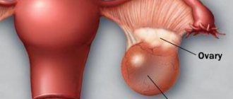
A cystic formation on the round ligament of the genital organ is a benign pathology. This ligament begins in the area of the fallopian tubes, extends along the sides of the pelvis through the inguinal canal, and ends in the area of the labia.
Cysts are round in shape, usually filled with fluid and in some cases reach the size of dropsy.
The secretion that accumulates inside the benign formation is the secretion of the nabothian glands. That is, a cystic tumor causes blockage of the ducts. Because of this, the glands significantly increase in size, and squamous epithelium accumulates in them.
How is a cyst different from a fibroid or tumor? The term tumor usually describes a tissue change. It may be benign or malignant. Malignant tissue changes are called cancer. This diagnosis causes concern for both patients and doctors.
Round ligament cyst or fibroids are officially considered benign tumors. They can be successfully treated and do not threaten a woman with death if such formations are detected in a timely manner.
Characteristics of the disease
Any cystic formation, regardless of location, is a pathological cavity in a capsule format filled with liquid contents. Initially, the cyst is always a benign formation, but when certain factors are formed, it is prone to malignancy.
A cervical cyst appears as a result of the formation of an isolated cavity resulting from blockage and subsequent pathological expansion of the gland. Later, an accumulation of produced secretion occurs in it.
To understand the mechanism of pathology formation, you need to have a minimal understanding of the structure of the uterus. The cervix is a passage cavity that has a cylindrical shape. Essentially, this is a transition chamber from the vagina to the body of the organ. The surface of the cervical canal and the outer part of the cervix is lined with single-layer epithelium. The difference is the presence of special glands: they are present inside the canal, but they are absent on the surface of the vaginal segment of the cervix. The area of contact between these two types of epithelial cells is called the transformation region. This is where the cystic capsule most often forms.
The mechanism of formation of a cervical cyst is quite simple:
- Initially, a woman develops inflammation of the cervical canal, which, in turn, can cause changes in the transformation zone.
- Subsequently, the channels through which mucus is excreted become blocked. The secretion remains inside the duct, which is accompanied by its unnatural expansion.
A non-inflammatory cyst is practically asymptomatic and is detected completely by accident. Characteristic symptoms most often appear with the development of local inflammation.
Symptoms
In most cases, this disease does not cause any complaints and therefore goes unnoticed. Often they are discovered during a woman's examination for another reason. However, if the cysts are numerous or very large, or if they are palpable from the outside, they can cause very painful symptoms.
Symptoms of a round ligament cyst of the uterus:
- Menstrual disorders. Not only the duration of critical days changes, but also the regularity of the cycle. Menstruation may be longer or shorter, and may occur several times a month.
- Pain in the lower abdomen is a common companion to cystic formations of the uterus, and a tumor of the round ligament is no exception. This symptom appears when there is a sudden change in body position. When a woman is resting there are practically no signs of pain.
- Due to cystic formation, hormonal imbalance begins in the body, resulting in weight gain. If there are no changes in the diet, the food is the same as before and physical activity has not decreased, you should think about your health. Perhaps this is the body’s way of signaling violations.
- Cystic formations also lead to diarrhea or constipation.
- An increase in body temperature is rare.
A cyst of the round ligament of the uterus is often confused with an inguinal hernia. Therefore, differential diagnosis cannot be avoided, and these require additional tests and instrumental examination methods.
Types of cervical cysts and is this condition dangerous?
There are different classifications of cervical cysts. The most common types are listed below:
Depending on the location of the pathology, these neoplasms are divided into the following types:
- paracervical or Nabotov. This species often reaches large sizes and has a connection with the cervical canal;
- endometriotic can contain both watery and bloody contents;
- A cyst of the round ligament of the uterus forms in the groin and is often confused with an inguinal hernia.
Many women are interested in the question of how dangerous this condition is.
Gynecologists say that the cyst itself does not pose a danger to the female body. It is not a precancerous condition, since it does not affect hormonal levels or the development of pregnancy.
Also, the cyst does not disrupt the menstrual cycle and does not provoke the development of cancerous tumors in other organs.
The danger for women is the bacteria that accumulate in the cyst, which provoke recurrent inflammatory diseases, such as:
- colpitis;
- endometritis;
- oophoritis;
- endocervicitis;
- salpingitis.
These inflammatory processes can lead to infertility and ectopic pregnancy. Large cysts, from 1 to 2 cm, compress the cervical canal. When pathology is detected during pregnancy, treatment is reserved for the period after childbirth.
Causes and symptoms of the disease
Until now, medicine does not know the exact causes of the tumor. However, there are certain reasons that influence the formation of this pathology. The most common ones are listed below:
Typically, a cyst does not have obvious symptoms and is discovered only during a preventive visit to the gynecologist. The following are symptoms that lead to discomfort in women with this pathology:
- bleeding is often observed, disturbing the woman 2 days before the onset of menstruation;
- menstrual flow is more pronounced;
- periodic pain occurring in the lower abdomen;
- vaginal dryness;
- pain during or after sexual intercourse;
- bleeding after or during sex;
- mucous discharge.
During pregnancy
In most women, cysts are found on the ovaries. Since round ligament cysts are small and do not cause any symptoms, there is no danger to the baby or the expectant mother.
The exception is very large formations, which can lead to complications. If the cyst lies in front of the birth canal or is limiting the baby's growth, a planned cesarean section may be required.
If fever, severe pain, and bleeding occur, doctors perform surgery to remove the benign tumor during pregnancy to avoid other complications.
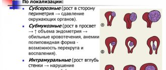
Varieties
There are different types of uterine cyst. This depends on the area of localization of the formation and some other factors.
So-called Nabothian cysts are quite common. They are named after the doctor who discovered them. This pathology is characterized by the appearance of multiple small yellow-white formations.
Nabothian cysts on the cervix do not cause any discomfort in a woman and are perceived as harmless. Sometimes this opinion is a misconception: formations also require therapy adequate to the condition.
Doctors do not know the true reasons for the formation of a nabothian cyst in the cervical canal of the uterus. But presumably they can develop as a result of:
- changes in current hormonal levels;
- chronic disease of the female reproductive system;
- cervical erosion.
For small Nabothian cysts, surgical treatment is not performed. The woman is simply seen by a gynecologist. But if they actively increase in volume, the patient may be prescribed an operation to remove the tumors - puncturing.
Quite often, a retention cyst is a congenital pathology, and therefore can appear at any time. In most cases, the provoking factor is a disruption in the functioning of the endocrine system. It is sometimes difficult to identify a cystic formation of this format, since it practically does not manifest itself at all.
A round ligament cyst of the uterus and an endometrioid cyst of the cervical canal can also be diagnosed, but they are less common.
Diagnostics
First of all, anamnesis is collected. The doctor should clarify the nature of menstrual flow, how often it occurs and what symptoms are present.
Then the genital organs are palpated, during which the doctor can detect a cystic formation, even a small one. This is a round tumor that protrudes into the groin area.
During an in-chair gynecological examination, swabs are taken from the woman to look for other infections and cysts. A mandatory examination of the cervix in a speculum is carried out. Colposcopy may be performed. This procedure involves examining the woman's cervix. Using a colposcope, you can examine not only the vagina and cervix, but also the adjacent muscles.
To find out whether the disease is a consequence of a viral infection, it is necessary to conduct an extensive examination. In this case, acetic acid or Lugol is used.
In cases of abdominal pain, the gynecologist performs an ultrasound examination (sonography). Cysts in the uterus or on the round ligament may be clearly visible. This manipulation with a 92% probability allows you to make the correct diagnosis, as well as distinguish a benign tumor from an inguinal hernia.
Bacteriological analysis is also carried out. This is a mandatory diagnostic measure, which is required for formations on the round ligament and symptoms such as itching, pain and unpleasant discharge.
Diagnosis and treatment
Diagnosing a uterine cyst is not difficult. Many neoplasms are clearly visible during a gynecological examination.
- Nabothian cysts are numerous and small in size. Visually they resemble semicircular formations located in the capsule. A neoplasm is considered large if the diameter of the capsule reaches or exceeds 1 cm.
- The endometrioid formation format is a capsule filled with contents in the form of the endometrium. Size changes occur in accordance with the current phase of the menstrual cycle. Visually, it is a red-brown spot of irregular shape that bleeds slightly during menstruation.
When diagnosing a cystic formation, the following methods are used:
- colposcopy;
- laboratory examination of a smear for microflora and sexually transmitted infections;
- transvaginal ultrasound examination.
In some cases, cystic formations are located directly in the cervical canal, which causes its pathological expansion and deformation.
Due to the absence of a characteristic clinical picture, treatment of the pathology in the early stages is very rare. In addition, conservative therapy does not bring the necessary results.
If the disease is detected, the woman is prescribed surgical treatment. Depending on the type of cyst, the following can be used:
- laparoscopic technique for tumor removal; Diathermoconization of the cervix - contact and non-contact;
- puncture of cysts located outside the cervical region;
- laser excision;
- radio wave surgery.
Early diagnosis of the disease allows you to begin timely treatment of the pathology and avoid the development of complications. That is why it is necessary to visit the gynecologist’s office at least once a year.
(Q50.5) is a congenital malformation of the female genital organs, characterized by the presence of a cyst in the area of the broad ligament of the uterus between the fallopian tube and the ovary.
Provoking factors may be adverse effects on the mother's body during pregnancy (harmful radiation, infections, intoxications, endocrine disorders).
Treatment
Surgical removal of the cyst is the most effective treatment. After which drug therapy is carried out. Modern surgical procedures make it possible to remove benign formations without abdominal intervention.
There are several removal methods. These include radio wave therapy, cryodestruction or laser destruction. The latter procedure is recommended for patients who have problems with blood clotting or diabetes.
Complications
There is a possibility that the cyst will burst. Since it is filled only with tissue fluid, there is no health hazard. However, if a blood vessel on the surface of the cyst is affected, internal bleeding may occur. Such pathologies require emergency surgery.
A cyst is a benign disease, but in some cases it can develop into a malignant tumor. Rupture of a malignant cyst can lead to the spread of carcinoma.
So-called pedicle torsion is another possible complication. Here the cyst turns once around its own axis and narrows the blood vessels. Symptoms may include fever, necrosis, vomiting, and nausea. In this case, immediate surgery is also necessary. When removing a benign tumor, doctors also remove the affected tissue.