Cervical cancer is a dangerous and uncommon disease, more typical for women of reproductive age. It has low responsiveness to treatment and a not very favorable prognosis, but can be cured in the early stages. For timely diagnosis, it is necessary to undergo periodic examinations to identify the prerequisites for it, which can be different. For example, one of these prerequisites is koilocytosis of the cervix, which will be discussed in this material. Is this disease dangerous, and what consequences can it lead to?
Koilocytosis is the main symptom of HPV infection
Human papillomavirus infection is the leading cause of cervical cancer.
For this discovery, the Nobel Prize in Physiology or Medicine was awarded to the German researcher Harald zur Hausen in 2008. The virus infects stratified squamous epithelial cells, which leads to damage to their genome and subsequent tumor transformation. In most cases, elimination of the virus occurs on its own within several years, and the infection is transient (temporary) in nature. Only a small proportion of women (about 10%) develop a persistent infection, characterized by a long, indolent course. This option is unfavorable, since the immune system is not able to eliminate the virus, which over years and decades of persistence leads to irreversible damage to the genome.
There is no effective treatment for HPV, so the only option is to monitor cellular changes and intervene only if there is tumor potential. An annual cytological examination (smear for oncocytology) allows you to suspect cellular atypia in the early stages and prevent the development of cancer.
One of the most important morphological signs of viral damage is koilocytosis (koilocytic atypia).
Koilocytes are squamous epithelial cells affected by the human papilloma virus.
These are quite large cells with enlarged dark nuclei with a folded outline and a perinuclear halo (a light zone around the nucleus). Binucleated or multinucleated cells are very common, which is quite specific for HPV. Koilocytes are usually located in the superficial layers of stratified squamous epithelium and can be widespread or local focal in nature. These changes are usually referred to as mild dysplasia, or mild intraepithelial lesion (LSIL).
Koilocytes are a consequence of the cytopathic effect of the virus and appear only during the active phase of its reproduction. If the HPV test is positive, but the cells are cytologically unchanged, this may indicate that the virus is in the latent phase. It is able to remain in this state for quite a long time and, with a decrease in protective mechanisms, again enter the phase of active replication.
The photo shows the mucous membrane of the cervix, lined with stratified squamous epithelium. On the right is normal epithelium with cells of the same size, the cytoplasm of which is rich in glycogen, and the nuclei in the form of small dark dots. On the left is squamous epithelium with pronounced koilocytosis. As can be seen, koilocytes are randomly located in the epithelium, their nuclei are sharply enlarged, irregular in shape, and split. An immunohistochemical study detects papilloma virus antigens in such cells.
Koilocytes are a consequence of the cytopathic effect of the virus and appear only during the active phase of its reproduction. If the HPV test is positive, but the cells are cytologically unchanged, this may indicate that the virus is in the latent phase. It is able to remain in this state for quite a long time and, with a decrease in protective mechanisms, again enter the phase of active replication.
In various dystrophic processes or chronic cervicitis, cells similar to koilocytes with reactive changes may appear in the epithelium. This should not be a cause for concern, nor should the finding of single koilocytes. The described changes are nonspecific and are characteristic of many benign reactive processes, which do not require specific treatment.
As mentioned above, in most women, koilocytosis goes away on its own within several years. Active treatment is only necessary in the presence of grade 2 and 3 dysplasia (HSIL), which indicates precancerous lesions of the cervix. Until then, all that remains is observation and cytological examination.
comments 42
- Comments 17
- Pingbacks 0
Hello, Doctor. I really need advice. I am 23 years old, I have not given birth, there have been no abortions, the method of birth control is barrier, I have had 3 partners in my entire life. As a result of an annual examination (I undergo it on my own initiative at a paid medical center), the doctor sent me for an HPV test for no apparent reason, and they were Types 16 and 66 were detected, STI test - Gardenella was detected. Ectopic cervix was detected. Cytology is normal. A colposcopy was prescribed and completed (attached photo), CT - 2, an iodine-negative area was discovered, this area was taken for a biopsy. Here is the result: Macrodescription: A fragment of gray lamellar tissue measuring 0.7x0.5x0.3 cm Microdescription: The material is represented by a fragment of the cervix with a transformation zone, with a focal moderate inflammatory infiltrate of lymphocytes, plasma cells, macrophages, fibroblasts and focal accumulations of neutrophils; in one of the fragments in the transformation zone in the endocervical epithelium, an area of erosion with necrosis of the surface epithelium, hemorrhages, fibrin, infiltration with polymorphonuclear leukocytes is determined; in one of the edges of the eroded area, metaplastic epithelium is partially determined with cell proliferation with a mild violation of the nuclear-cytoplasmic ratio, mitoses, parakeratosis . Multilayered squamous epithelium in some areas with a small number of koilocytes and signs of parakeratosis in the superficial layers and focally with single atypical epithelial cells. Conclusion: Morphological picture of chronic moderately severe moderately active cervicitis, focal simple leukoplakia of metaplastic stratified squamous epithelium with a small number of koilocytes and focal mild dysplasia (CINI, LSIL). The doctor who performed the biopsy says he completely removed the damaged area and prescribed epigen spray and isoprinosine. Could you express your opinion: 1. What else can (and should) be done to prevent further development of the disease - cauterization, laser, radio wave treatment, something else? 2. Do I need to consult an oncologist? 3. The biopsy was on August 25, today is September 5, and despite the small size of the area that was taken for the biopsy, I still have light brown discharge every day, there is no pain, is this normal after the biopsy? 4. If I consult another doctor (examination with colposcopy), how long after the biopsy can this be done? I understand that my questions will seem stupid to you, but I am simply terrified and panicked.
Hello! Thanks for the detailed description! 1. At this stage you do not need anything. HPV is difficult to treat; the main role in getting rid of the virus is your own immune system. We can only observe and take more serious measures as the process progresses. Observation means preventive cytological examination (cytology smear) 2 times a year. Many gynecologists practice ablation (cauterization), but this is not highly effective in combating the virus. 2. You don’t need an oncologist yet. If there is CIN 2-3 in the smear or biopsy, you can also contact an oncologist. 3. Such allocations are acceptable. 4. In a month or two you can be examined again. The mucous membrane must recover in order to adequately conduct an examination. There's no need to panic. This is a common situation. Observe for now, there is no need to take active measures.
Conclusion
As is clear from what is written above, koilocytosis is a symptom of a serious disease, therefore, if such cells are detected, treatment should begin immediately. Otherwise, serious complications may develop, including an oncological process, which is difficult to treat and does not have a very favorable prognosis.
Human papillomavirus infection is the leading cause of cervical cancer. For this discovery, the Nobel Prize in Physiology or Medicine was awarded to the German researcher Harald zur Hausen in 2008.
The virus infects stratified squamous epithelial cells, which leads to damage to their genome and subsequent tumor transformation. In most cases, elimination of the virus occurs on its own within several years, and the infection is transient (temporary) in nature. Only a small proportion of women (about 10%) develop a persistent infection, characterized by a long, indolent course. This option is unfavorable, since the immune system is not able to eliminate the virus, which over years and decades of persistence leads to irreversible damage to the genome.
There is no effective treatment for HPV, so the only option is to monitor cellular changes and intervene only if there is tumor potential. An annual cytological examination (smear for oncocytology) allows you to suspect cellular atypia in the early stages and prevent the development of cancer.
One of the most important morphological signs of viral damage is koilocytosis (koilocytic atypia).
Koilocytes are squamous epithelial cells affected by the human papilloma virus.
These are quite large cells with enlarged dark nuclei with a folded outline and a perinuclear halo (a light zone around the nucleus). Binucleated or multinucleated cells are very common, which is quite specific for HPV. Koilocytes are usually located in the superficial layers of stratified squamous epithelium and can be widespread or local focal in nature. These changes are usually referred to as mild dysplasia, or mild intraepithelial lesion (LSIL).
Cervical koilocytosis: what is it and how to treat it?
Cervical cancer is a dangerous and uncommon disease, more typical for women of reproductive age. It has low responsiveness to treatment and a not very favorable prognosis, but can be cured in the early stages. For timely diagnosis, it is necessary to undergo periodic examinations to identify the prerequisites for it, which can be different. For example, one of these prerequisites is koilocytosis of the cervix, which will be discussed in this material. Is this disease dangerous, and what consequences can it lead to?
Koilocytosis of the cervix - what is it, cancer or precancer
What does cervical koilocytosis mean? How to treat and diagnose it. Why is this pathology dangerous?
Every year, approximately 500 thousand cases of cervical cancer (CC) are recorded in the world, of which 27 thousand are fatal due to too late contact with a gynecologist. Every 15 minutes, 1 woman in Europe dies from this cancer. The detection of malignant tumors in young women aged 35 to 40 years has increased by 35-40%. CC is the main reason for the early death of women in full force. As it becomes known from official sources, malignant tumors are to blame.
Of course, medicine works wonders! Today, cervical cancer is successfully treated surgically and even prevented. If you seek qualified help from a specialist in a timely manner.
Identifying the symptoms of a precancerous process, dysplasia, helps prevent the process from becoming malignant. This became possible thanks to the fact that scientists have proven and identified the cause of this disease. Human papillomavirus infection (PVI) is to blame for the suffering of women.
There is no cure for PVI. It's a virus. The main thing is that its negative impact on the cervix does not go unnoticed. Koilocytes are squamous epithelial cells with a modified nucleus. Diagnosed by PAP test (Panicolau smear, cytological examination). Along with altered squamous epithelial cells, cytologists detect pathological cells of multilayered epithelium - dyskeratocytes. These processes on the cervix are called koilocytosis and dyskeratosis, or parakeratosis (when talking about leukoplakia). Koilocytes and dyskeratocytes are signs of PVI and dysplasia (usually mild, CINI). In the picture on the left you see normal cells, and on the right - with enlarged nuclei. These are koilocytes as a cytologist sees them.
Cytological examination of smears from the cervix should be carried out, in case of detection of papillomavirus infection (also known as HPV - human papillomavirus) of oncogenic types, regularly - once every 6 months. In this way, it is possible to identify dysplasia of varying severity and a malignant tumor at the initial stage of its development, before it metastasizes.
If there are signs of HPV and dysplasia, a biopsy is taken from the cervix (intravital sampling of cells or tissues from the body) to clarify the diagnosis and treatment tactics. The changed cervical tissue is removed. In mild cases, a procedure for destruction of the cervix is possible (removal of the dysplastic area using radio waves, the drug Solkovagin, electric current, etc.). If there is chronic cervicitis, it is treated with medication. The drug is selected depending on the type of causative agent of the inflammatory process. If it is E. coli, it is removed with antimicrobial agents, antibiotics used in the form of vaginal suppositories. If fungi of the genus Candida (thrush) - antifungal drugs.
In severe dysplasia, treatment of koilocytosis is surgical - conization or even amputation of the cervix.
Sources used: viskablivanie.ru
SEE ALSO: How to avoid getting infected with HPV through everyday contact Can the type of HPV change HPV fungus HPV is a smear or blood test HPV is detected but there are no warts HPV DNA titer
Definition
Human papillomavirus (HPV) is one of the main causes of cancer of the reproductive system in women. It is papillomas that most often degenerate into malignant neoplasms, although this is not typical for any papillomas, but only for those that were caused by certain strains of viruses. Therefore, in the presence of papillomas, it is necessary to immediately carry out a diagnosis in order to determine which strain caused the disease and draw a conclusion about how dangerous the process is.
Koilocytosis is a process of changes in epithelial cells that is caused by the human papillomavirus. In this condition, atypical koilocytes begin to be detected in the epithelium. When examined under a microscope, they are easily identified by the following features:
- Excessively enlarged nuclei;
- Significantly darker nuclei than in other epithelial cells;
- Folded kernels;
- A peculiar glow (halo) around the core;
- Multinuclear formations;
- Cellular formations of irregular shape.
These and some other changes are caused by the papilloma virus. Such atypical cells can be located both focally and diffusely, that is, more or less evenly. It is worth noting that such cells appear only in the active stage of the virus. That is, when tests reveal HPV, but there are no koilocytes, we can say that the virus is in the latent stage.
Causes
As mentioned above, koilocytosis does not develop as an independent disease. Strictly speaking, this is a condition that is a symptom of the presence of the human papillomavirus in the active stage of reproduction, that is, when its cells are dividing. Moreover, not any form of this virus, but only certain strains of it. Thus, the main cause of koilocytosis is HPV.
For what reasons does HPV appear?
- This is a viral disease, therefore it can only be infected from the outside, the most common way is unprotected sexual intercourse;
- In addition, in much rarer cases, infection can occur as a result of the use of unsterile instruments during examination or surgical intervention in gynecology (but such causes, as well as infection through household contact, are a rare exception, since in most cases this virus dies on contact with air);
- Under normal conditions, the body’s immune system is capable of suppressing viruses, but with reduced immunity (as a result of infectious, inflammatory diseases, surgical interventions), it is unable to do this;
- The same thing happens with reduced local tissue immunity and disturbed microflora, since strong microflora is also capable of suppressing the division of viral agents.
If the immune system cannot cope with the load, then after contact with the mucous membranes, the virus, which cannot be suppressed by anything, begins to multiply. At the same time, it spreads both deep into the tissues and over their area. Its activity causes changes that result in the formation of koilocytes.
Symptoms
In most cases, cervical koilocytes do not manifest themselves in any way. They do not cause severe specific symptoms. This is also typical for the human papillomavirus, which does not manifest itself in any way, either subjectively or externally, in most cases. However, sometimes HPV can still manifest itself nonspecifically. In this case, it gives the following symptoms:
- The presence of neoplasms – papillomas and warts, in single or multiple quantities;
- Discomfort during sexual intercourse in some cases;
- Discomfort in the lower abdomen;
- Increased symptoms of premenstrual syndrome.
But most often the condition is discovered by chance, during a routine examination or when visiting a doctor for another issue. If papillomas are detected, epithelial tissue is also examined, during which the presence of koilocytes can be detected.
Diagnostics
How to diagnose this condition? The following methods are used:
- General and biochemical blood tests, general clinical urine analysis, coagulogram, Wasserman reaction and other nonspecific studies to establish the general condition of the body;
- Blood tests for sexually transmitted diseases - HIV, syphilis, hepatitis C and so on;
- Testing for sexually transmitted infections: bacteriological culture of genital tract discharge, PCR testing of smears for all types of infections, including all strains and types of HPV;
- Cytological examination of cervical tissue;
- Visual research methods that help determine the presence and size of papillomas - colposcopy, cervicoscopy, vulvoscopy;
- Histological examination of a section of tissue that is removed under colposcopy control. Such a study allows you to directly determine the presence of changes and the presence (or absence) of atypical cells.
Diagnostics using such methods will be complete and sufficient to assess the patient’s health status and the degree of development of the pathological process. But in exceptionally difficult (from a diagnostic point of view) cases, MRI and CT may be additionally prescribed.
Effect on pregnancy
The presence of HPV is usually not a contraindication to pregnancy. In most cases, the presence of koilocytes in the epithelium does not in any way affect the likelihood of conception (except for cases where large papillomas cover the cervix and prevent the penetration of sperm, or there are so many of them that the embryo has nowhere to attach). In general, in moderate degrees of development, koilocytosis and HPV do not lead to a decrease in fertility.
Also, the presence of this pathology does not affect the nature of pregnancy. The virus does not cause any pathologies or malformations in the fetus, does not lead to complications during pregnancy, does not cause premature birth, and does not provoke miscarriages. Among women with this disease, the percentage of successful pregnancy and childbirth is as high as among those who do not have it.
The birth process can be complicated only if papillomas are present on the cervix. In this case, there may be difficulties with its opening, and bleeding may be present. The greatest danger is the effect of pregnancy on the disease. A drop in local immunity after childbirth can trigger the active development of the pathological process.
Koilocytic dystrophy, HPV
Hello, please help me decipher. I had a biopsy. 2 fragments of fabric with a diameter of 0.4 and 0.7 cm. Gray in color when cut. Microscopic description of the material: Fragments of the cervical mucosa with edema and lymphoid infiltration, covered with stratified squamous epithelium with koilocytic degeneration. Conclusions to the study: Koilocytic dystrophy (morphological marker of HPV).
The fact is that six months before I took a PCR test and the HPV result was negative. Single partner and without HPV. Please tell me: 1. Does this really mean I have HPV? 2. Is this not dysplasia or cin? Not cancer? 3. And will it cause cancer in the future? 4. Is there any treatment?
I have been taking Janine for 5.5 years. See the doctor in a week. Answer please.
Related and recommended questions
6 answers
Site search
What should I do if I have a similar but different question?
If you did not find the information you need among the answers to this question, or your problem is slightly different from the one presented, try asking an additional question to the doctor on the same page, if it is on the topic of the main question. You can also ask a new question, and after a while our doctors will answer it. It's free. You can also search for the information you need in similar questions on this page or through the site search page. We will be very grateful if you recommend us to your friends on social networks.
Medical portal 03online.com
provides medical consultations via correspondence with doctors on the website. Here you get answers from real practitioners in your field. At the moment, on the site you can get a consultation in 45 areas: an allergist, venereologist, gastroenterologist, hematologist, genetics, gynecologist, homeopath, dermatologist, children's gynecologist, children's neurologist, children's surgeon, children's endocrinologist, nutritionist, immunologist, cardiologist, cardiologist, cosmetologist, cosmetologist, cosmetologist speech therapist, ENT specialist, mammologist, medical lawyer, narcologist, neurologist, neurosurgeon, nephrologist, oncologist, urologist, orthopedist-traumatologist, ophthalmologist, pediatrician, plastic surgeon, proctologist, psychiatrist, psychologist, pulmonologist, rheumatologist, sexologist-andrologist , dentist, urologist, pharmacist, herbalist, phlebologist, surgeon, endocrinologist.
We answer 95.67% of questions.
Sources used: 03online.com
ARTICLES ON THE TOPIC: Result of HPV DNA research How to avoid becoming infected with HPV through household means Can the HPV type change HPV test in gynecology
Therapy
If changes are present, surgical treatment is indicated. There are two most effective methods:
- Conization of the cervix - cutting off its affected area. The good thing about this method is that it is low-traumatic. However, with this approach, a fairly small area of tissue is removed, so there is a possibility that koilocytes are present somewhere else, so a relapse can begin quite quickly. In addition, the method is good only for localized focal placement of zones with altered cells;
- A more effective way is amputation. What it is? This is the process of completely removing the cervix. The good thing about this method is that it is highly likely to remove all affected cells, making the likelihood of relapses after such an intervention much lower. But such an intervention is also more traumatic, the recovery period after it is a little longer.
Surgical treatment is supplemented by taking immunostimulating drugs. These are drugs that act on the entire body, such as Likopid, and local drugs, for example, Viferon. In parallel with these, antiviral drugs are prescribed, such as Isoprinosine, Cycloferon, Panavir. For several days after surgery, antibiotics and/or anti-inflammatory drugs may be indicated to prevent infection.
Prevention of complications
After surgery, the main rule is to prevent relapse, bleeding and infectious complications. To do this, each patient after undergoing surgical treatment is recommended to follow certain rules.
- Early initiation of physical activity in moderate amounts has a beneficial effect on the course of the recovery period, metabolic processes and activation of repair, which contribute to accelerated healing of the cervix after treatment of koilocytosis.
- And also do not overload a weakened body. Physical activity must be adequately dosed.
- During the recovery period after treatment of koilocytosis, heavy physical activity, and especially lifting loads weighing more than 5 kg, are prohibited. Neglecting this rule may negatively affect the healing of the cervical wound.
- It is worth knowing that bad habits help slow healing. Nicotine entering the bloodstream contributes to the development of a reflex spasm of blood vessels, and alcohol, on the contrary, expands them, after which a sharp spasm occurs. This “game” of blood vessels negatively affects the repair process.
- Products with an excessive content of light carbohydrates (refined sweets) and saturated fats (animal fat) contribute to the deposition of cholesterol plaques on the walls of the arteries, and a sedentary lifestyle leads to the formation of thrombosis, which can harm the patient’s health even after curing cervical koilocytosis. Diet is an important component in activating the immune system and fighting HPV. It is necessary to normalize your diet, eat more fresh vegetables and fruits, poultry and veal, dairy products, and herbs. Fried and smoked foods should be excluded. You should eat meals in small portions. Drink enough clean water. Thanks to this diet, the patient will receive the required amount of vitamins and proteins.
- Medicines - antibiotics, antiviral, immunomodulatory agents, healing suppositories - should be used as prescribed by the doctor to prevent relapse of cervical koilocytosis.
You need to know that any pathological changes in the vaginal discharge, as well as in the patient’s well-being, should be discussed in a timely manner with a gynecologist to prevent undesirable consequences of koilocytosis. After some time, the woman will have to visit the doctor again for further research and assessment of the condition of the cells.
What are cervical koilocytes
Most patients who have at least once tried to learn more about the factors influencing the development of oncological pathologies know that human papillomavirus is the main cause of the development of precancerous and cancerous diseases of the cervix. This infectious disease leads to damage to epithelial cells and rearrangement of their genetic code, which is why tumor transformation occurs in the cervical area. To date, there are no effective methods to combat human papillomavirus infection. As a rule, the disease is transient (temporary) in nature, and after several years it cures on its own. However, every tenth woman experiences a persistent pathology in which the immune system is unable to fight and eliminate cells containing the virus.
Signs of such precancerous cellular lesions of the cervix include koilocytosis, dyskeratosis and parakeratosis. The most common sign of HPV is koilocytosis of cervical epithelial cells.
Koilocytosis or koilocytic atypia
Koilocytes are cells of the squamous epithelium of the cervix, which arise when it is damaged by human papillomavirus. The main difference between these cells is their dark, voluminous nuclei, which have folded edges and a perinuclear halo (a kind of glow around the nucleus). Quite often, during a histological examination of the cervix, binuclear or multinucleated cells can be seen in the resulting cellular biopsy, which is a specific sign of papillomavirus and the development of koilocytosis. These cells are often located in the upper layers of stratified squamous epithelium and can have both focal and diffuse localization.
Pathological cells of the cervix are a peculiar consequence of the active influence of the papilloma virus on epithelial cells. They occur only during the phase of active reproduction of HPV in the cervix. If the test result is positive for the papilloma virus, the cells may not be changed (koilocytosis is absent), this situation indicates a latent course of the disease. HPV can remain in this phase for a long period and only when the body’s immune system is weakened – through acute respiratory viral infections, surgery, stress – can it enter the active phase.
Various dystrophic processes in the cervix and chronic cervicitis can be characterized by similar reactive changes and other specific manifestations of koilocytosis.
Features of koilocytic atypia of the cervix and its treatment
Cervical koilocytosis is a cytological term to describe cells in epithelial tissue. The obtained cytological or histological conclusions allow the attending physician (gynecologist, gynecological oncologist) to develop a further plan of treatment and diagnostic measures and offer the patient the best option for further treatment.
What is and features of cervical koilocytosis
Epithelial cells cover the surface of the body and line the mucous membranes of internal organs. Cells of various shapes - flat, cubic, cylindrical - group integuments, single or multilayered. Epithelial tissue is formed - a barrier that protects organs, lungs, liver, deeper layers of skin from the influence of the external environment, which allows organs to function normally.
Koilocytes, known as halo cells, are also epithelial cells. Unlike conventional ones, the genome of the human papillomavirus (HPV) is activated in them, which leads to significant structural transformations. The altered nucleus of koilocytes, containing the DNA of a much larger cell, differs from the nucleus of normal cells in shape and color.
Koilocytosis has a gradation. There are more and less noticeable “abnormalities” in the structure of the cell. This allows the morphologist, in the process of examining biopsy material, to judge the degree of danger of the appearance of pathology. There are histological and cytological classifications of precancerous lesions of the cervix, where they are distributed according to the degree of risk of malignancy.
Detection of koilocytes in a cytological analysis is a marker of possible precancer in the cervix, mouth and anus.
The histological report describes the changes in the tissue obtained as a result of the biopsy. It is considered more reliable compared to the cytological description of pathological changes in the cervix.
Causes and symptoms
Human papillomavirus is transmitted sexually. HPV rarely causes symptoms, and many people are unaware of the infection and unknowingly infect their sexual partners.
Do you need advice from an experienced doctor? Get a doctor's consultation online. Ask your question right now.
Ask a free question
When human papillomavirus DNA enters the body, it is integrated into the genome of the epithelial cell. The cell begins to produce viral proteins, which leads to structural changes that turn the cells into koilocytes.
More than 170 types of HPV have been discovered. The overwhelming majority of them do not manifest themselves in the body. Oncogenic species, of which there are no more than a dozen, are markers of squamous cell carcinoma.
In the initial stages, cancer occurs without symptoms. As the cancer process progresses, symptoms may appear:
- spotting after sexual intercourse;
- weight loss;
- decreased appetite;
- fatigue;
- discomfort in the perineal area;
- vaginal discharge, which may be scanty, watery or pus-like, or have a foul odor. During diagnosis, koilocytosis is detected.
HPV is associated with the appearance of other malignant neoplasms; it can invade the genome of epithelial cells of the anal canal, penis, vagina, vulva and oral cavity. Genital and plantar warts are the result of HPV infection.
Can koilocytes without HPV be found?
Often the morphologist sees koilocytes, but the test for human papillomavirus is negative.
In 10-15% of cases, the formation of koilocytes, experts believe, is associated with another infection - herpes, cytomegalovirus - or other benign pathologies.
Another option is a false negative PCR result, which can be for two reasons:
- poor quality of material taken for research;
- the test may not show a positive result due to a low viral load. With a more sensitive HPV test, the virus is detected.
When cytological examination reveals single koilocytes, this often does not require treatment.
If cytology does not reveal atypical cells, and the conclusion does not contain the abbreviations LSIL (N-PIP) or HSIL (V-PIP), there is no reason to worry. This situation does not require additional analysis.
How to diagnose koilocytic atypia
Koilocytosis is detected using oncocytology (cytological smear) or biopsy. A cytological smear is a screening test for cervical cancer.
During the procedure, the doctor uses cytobrushes to obtain cells from the exocervix—the surface in contact with the vagina—and from the endocervix (the cervical canal). The resulting material is analyzed by a morphologist for the presence of koilocytes and other cytological abnormalities.
The same material is used in polymerase chain reaction (PCR), a test that reveals the genome of the virus.
If the results are positive, the doctor will suggest a colposcopy and biopsy. During colposcopy, the surface of the cervix is examined under magnification of more than 25 times. During a biopsy, a small piece of tissue is removed.
A positive result indicates the presence of koilocytes, a negative result indicates the absence.
The detection of cervical koilocytes in cytology does not indicate cancer or the obligatory appearance of a malignant formation in the future. It is necessary to undergo observation or treatment to prevent possible progression of the pathology.
Features of treatment of koilocytes
The formation of koilocytes is the result of infection with the human papillomavirus. There is no drug that can eliminate the human papillomavirus. All medical efforts are aimed at identifying and timely treating a potentially dangerous precancerous condition of the cervix. A woman is offered the following options:
- Dynamic observation. For women with mild to moderate dysplasia, a watch-and-wait approach is recommended. This means that it is necessary to undergo oncocytology, colposcopy or biopsy every 6-12 months. Mild to moderate dysplasia often resolves on its own within two years.
- Loop electrosurgical resection (LEEP). With an electrode, at the end of which there is a loop-sail, the pathological area is cut out with a cone. The loop is replaced with a ball, which is applied to the affected area, causing coagulation of blood vessels - diathermoelectrocoagulation. The HPV is completely removed along with the affected tissue.
- Knife conization. Treatment option for women with severe dysplasia. During this surgical procedure, a cone-shaped fragment is removed from the cervix. The operation is performed on an outpatient basis under local anesthesia. It is used in the absence of special equipment - diathermoelectrocoagulator, "Surgitron".
- Cryosurgery, or cryotherapy. Cryodestruction of affected tissue is another method of focal tissue destruction. Not used to treat “overt” cervical dysplasia. Used to eliminate erosion in nulliparous women. The procedure is performed on an outpatient basis. The cryoprobe is inserted into the vagina towards the cervix. its koilocytes. Compressed nitrogen passes through a metal probe, transferring cold to the metal walls - this allows you to freeze and destroy the areas of tissue with which the tip of the probe comes into contact.
- Laser therapy, or carbon dioxide laser photoablation. A technique increasingly used in gynecology. There are practically no side effects, good therapeutic effect. Does not allow you to take material for research.
Is it possible to cauterize cervical erosion with koilocytosis?
Often, having diagnosed erosion, the gynecologist recommends treatment. Should cervical erosion be cauterized with electrocoagulation, frozen with cryotherapy, or is it better to leave it alone?
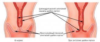
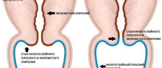
Ectopia, pseudo-erosion, endocervicosis are synonyms for erosion. The term “ectopia” is more suitable to describe a process in the external opening of the endocervix, which, in the absence of symptoms, does not require treatment.
When severe symptoms appear, ectopia should be treated, if the diagnosis does not reveal koilocytosis - evidence of moderate cervical dysplasia - cauterization is not necessary.
Forecast of the course of the disease
If koilocytes are detected during a cytological examination of a cervical smear, this does not mean that you have cervical cancer. In most cases, only monitoring of the condition is required - periodic examination by a gynecologist, taking smears. Often koilocytic atypia resolves after a course of anti-inflammatory treatment.
If the doctor considers the detected koilocytosis to be potentially dangerous, immediate diathermoelectrocoagulation, cryotherapy, and laser ablation are recommended.
To prevent the risk of HPV infection, practice safe sex.
If you are under 26 years old or have a child, you can talk to your doctor about preventing infection and creating immunity to this virus. Vaccines against some types of HPV have become available.
The use of the vaccine when planning or during pregnancy is contraindicated. There is no cure or remedy to get rid of papillomavirus forever.
The article has been reviewed by the site editors
Source: https://VashaDerma.ru/hpv/kojlotsitoz
Etiology of development
There are many reasons and factors influencing changes in the genome of a cell and its degeneration. Some of the most common are:
- Inflammatory pathologies of the genital organs. These diseases contribute to the development of necrobiosis processes in the stratified squamous epithelium and its scaly peeling, which causes the development of erosive lesions in certain areas of the mucous membrane of the cervix. Reparation of these lesions occurs due to hypertrophy of the cells of the cervical canal (cylindrical epithelium), which is not typical for the vaginal ecosphere. In the future, cracks may appear in the restored areas due to overly active sexual intercourse, leading to the penetration of papilloma virus DNA into the woman’s body and the development of koilocytosis.
Also, the combined progression of HPV and HIV significantly increases the risk of malignancy (development of precancerous conditions, in particular koilocytosis).
- Traumatization of the mucous membrane of the cervix after childbirth or abortion. The provoking factor is a violation of the blood supply to tissues, the use of barrier contraceptives (uterine devices) and tampons.
- Hormonal dysfunction.
- Disturbances in the functioning of the immune system.
- Early onset of sexual activity, promiscuous and unprotected sexual intercourse.
- Age-related changes in the tissues of the genital organs.
- Use of oral contraceptives.
- Hereditary predisposition to the development of cancer pathologies, in particular on the cervix.
Koilocytosis and HPV
As already mentioned, the identification of multiple koilocytes (abnormal cells) during a histological examination of the cervical canal of the uterus may be the cause of the development of HPV. Koilocytes are affected cells with varying degrees of disruption of the nuclear structure and perinuclear vacuolization. If this process is excessively expressed, then the cells are called “halo”; if the nucleus has pushed the environment closer to the periphery, then they are called “ballooning”.
Diskeratocytes are another type of cell that can be found in the cervix along with those described above. These cells also develop in the squamous epithelium layer and are characterized by their small size, excessively stained nuclei of varying diameters. With the development of tumor processes, it has been established that the number of this pair of epithelial units decreases in the active phase of pathological cell division.
The results of her study in 89% help to timely detect precancerous conditions (koilocytosis, dyskeratosis) and oncological changes in the cervix.
HPV effect koilocytic atypia
1. Persistence of HPV in the body (or latent course)
- the virus exists in episomal form without causing pathological changes in cells, there are no clinical manifestations, its existence can only be determined using molecular biological methods.
2. Mild neoplasia (dysplasia)
- HPV exists in an episomal form - in this case, changes occur in the structure of the cell, called koilocytic atypia - which occurs in the surface layers of the epithelium, while the nucleus takes on an irregular shape and becomes hyperchromatic, vacuoles appear in the cytoplasm. Lesions are most often localized in the transformation zone of the cervix and can progress. They are detected by cytological examination, as well as by histological examination of a biopsy specimen.
3. Severe neoplasia (dysplasia)
— HPV exists in an integrated form, with altered “atypical” cells appearing, characterized by an enlarged nucleus and reduced cytoplasm, indicating the malignancy of the process; the more such cells in the sample, the higher the degree of dysplasia. Lesions are most often localized in the transition zone of cervical transformation and can progress to invasive cancer (carcinoma). It is detected by cytological examination, as well as by histological examination of a biopsy obtained during a colposcopic examination.
4. Carcinoma
— HPV exists in an integrated form, with altered “atypical” cells indicating a malignant process (invasive tumor).
The target for the effects of oncogenic types of HPV is the transformation zone of the cervix, where precancerous changes in the cervix develop. Progression from cellular changes associated with HPV infection (koilocytic atypia) to the development of cervical cancer usually takes 10-40 years, but in rare cases can occur in 1-2 years. An analysis of the outcomes of CIN associated with HPV showed that with CIN1, regression is observed in 57%, persistence in 32%, progression in 11%, and the development of invasive cancer in only 1% of cases. At the same time, with CIN3, malignancy occurs in more than 12%, and regression occurs in only 32% of cases. The progression of cervical neoplasia depends primarily on the type of virus. The highest proportion of progression of cervical dysplastic processes associated with HPV infection is observed during infection with virus types 16, 18, 31, 33 and 45, the smallest - during infection with HPV types 51 and 56.
Diagnostics
The main methods for diagnosing all precancerous conditions of the cervix include:
- Collecting anamnesis data and conducting a gynecological examination. When visually examining the mucous membrane of the cervix, its relief, shape, condition of the external pharynx and the nature of the discharge, various changes can be identified, for example, hyperemia, foci of leukoplakia, polyps, etc. Conducting a bimanual examination and examination in mirrors is an informative first stage of diagnosis. Collecting medical history data can provide information about a possible infection and the route of its entry into the woman’s body. All data obtained play an important role in the course of koilocytosis and the tactics of its further treatment.
- Clinical and laboratory methods for koilocytosis: clinical blood test, glycemic level study, Wasserman reaction, reactions to HIV, clinical urine test, blood biochemistry and coagulogram, bacteriological culture of genital tract discharge, PCR test for sexually transmitted infections, including all types of HPV.
- Cytological examination. The resulting cervical biopsy is visualized using cytological dyes. This method helps to identify precancerous conditions and cancer of the cervix in the early stages of their development.
- Colposcopy, cervicoscopy, vulvoscopy are methods for examining the external genitalia and cervix for koilocytosis and other diseases. There are several types of colposcopy. A simple procedure is carried out without applying medications and removing mucous discharge; an extended procedure is carried out after treating the vaginal part of the cervix with a solution of acetic acid, adrenaline, hematoxylin (dye) and Lugol's solution. Colpomicroscopy is carried out for life and consists of applying hematoxylin to the cervical mucosa and further examining it under a microscope (magnification 160-280 times). Thanks to this study, koilocytosis can be detected in the initial stages.
- Histological diagnosis. The collection of the necessary material is carried out under the control of simple colposcopy. The required area of tissue is excised with a sharp scalpel in the area of pronounced changes in the cervix. The resulting biopsy is placed in a formaldehyde solution and then sent for diagnostics.
When epithelial dysplasia is detected, treatment of koilocytosis is carried out surgically with parallel correction of immunity and the prescription of antiviral drugs. The main techniques are conization (excision of a cone-shaped area of pathological cells and tissues in the cervical canal) or amputation of the cervix.
Koilocytosis: diagnosis and treatment | University Clinic
Although this condition is not cancer, the diseases that cause it require prompt treatment due to the high risk of malignancy.
Why does koilocytosis appear in a smear?
The cause of the pathology is damage to the epithelium covering the cervix by the papilloma virus (HPV) or other pathological processes. Because of this, cell division is disrupted, and incorrectly developed koilocytes are formed instead of epithelial cells:
- These cells are larger in size compared to healthy ones.
- Inside any cell there is a nucleus necessary for its division. Due to damage by a virus or inflammation, the nuclei of koilocytes are enlarged, deformed, have an irregular structure and structure, and can fall apart.
- In healthy cells, there is a fluid around the nucleus - cytoplasm. In koilocytes, it is preserved only along the edge, forming a “halo” - a zone of clearing.
A cell with such changes cannot divide normally, therefore, with koilocytosis, cell division disorders often occur - incorrect mitoses, leading to cancer.
Koilocytes
Koilocytes are diverse and can have varying degrees of deviation from the normal structure. The more pronounced it is, the more likely the development of a malignant tumor. Only a morphologist examining the smear can say exactly how dangerous a particular case is. But in any case, the presence of koilocytes is a dangerous symptom.
For what diseases is koilocytosis detected in a smear?
Cervical dysplasia (cervical neoplasia, CIN). This pathology is caused by damage to the cervical mucosa by the human papillomavirus (HPV). The pathogen disrupts cell division and leads to cellular changes. There are three degrees of pathology:
- The first (CIN I), in which 1/3 of the outer tissue of the cervix (epithelium) is affected. This is the mildest degree of the disease.
- Second (CIN II) – the lesion occupies 2/3 of the thickness of the epithelium. This form of the disease is of moderate severity.
- Third (CIN III) - with this pathology, the epithelium is affected by more than 2/3. The most severe form of the disease, followed by cervical cancer. The more severe the dysplasia, the more often koilocytosis is detected and the higher the content of koilocyte cells in smears.
Dysplasia
Cervical cancer. The initial stage of the oncological process in its cellular structure and other characteristics differs slightly from precancer - dysplasia.
Initially, the malignant tumor does not extend beyond the epithelium (carcinoma in situ) and koilocytes may be detected in the smear.
Soon, aggressive cancer cells will appear in the tissues, the tumor will grow deeper, penetrate into neighboring organs and metastasize.
Ectopia (erosion, cervicosis) of the cervix is also often accompanied by koilocytosis. With this disease, the columnar epithelium of the cervical canal, which runs inside the cervix, appears on its surface.
Columnar epithelial cells, which are unable to withstand the acidic environment of the vagina, become inflamed. This process is accompanied by the appearance of koilocytes.
Ectopia must be treated, as it leads to cancer.
Koilocytosis can be detected during infection with the herpes virus and cytomegalovirus. These viral infections are also considered cancer causing factors.
Sometimes single koilocytes may be detected in the smear. It's not scary. This condition indicates an inflammatory process. In women, inflammation of the cervix is detected - cervicitis, of the cervical canal passing inside it - endocervicitis, of the cervical mucosa - ectocervicitis, of the vagina - vaginitis (colpitis).
| Cervical disease | Cases of detection of koilocytes, % |
| CIN1 | 6,6% |
| CIN2 | 18% |
| CIN3 | 40% |
| Squamous cell carcinoma | 5% |
| Erosion (ectopia) | 26% |
Koilocytosis can occur not only on the cervix. Koilocytes are found on the mucous membrane of the stomach, esophagus, anal canal and other organs where HPV lesions and inflammatory processes occur.
Is it possible to determine koilocytosis by complaints and signs?
This lesion of the cervix does not give obvious symptoms. Existing discharge, spotting, pain and discomfort in the genital tract are caused by concomitant diseases. Therefore, in order to detect such a condition, you need to contact a gynecologist and undergo the necessary tests.
Methods for detecting koilocytosis
- You can suspect the presence of pathology when examining a woman on a gynecological chair using mirrors. There are pathological focal changes in the form of light or reddish spots on the cervix.
- In some cases, dense keratinized areas are found on the cervical mucosa. This condition is called keratosis. There are several variants of it: dyskeratosis, parakeratosis, hyperkeratosis, acanthosis. In this case, cells with signs of compaction (keratinization) are found in the smear along with koilocytes.
- The clinical picture can be clarified using colposcopy, a study performed with a colposcope. Extended colposcopy is more informative, during which the mucous membrane is lubricated with a solution of iodine or vinegar. This effect makes pathological lesions more visible and allows one to assess the size of the lesion.
- A smear is taken from the woman’s cervix for cytology. For this purpose, cytobrushes are used - devices that allow you to collect mucosal cells. The resulting material is transferred to the laboratory for cytological examination. This is where koilocytosis is detected.
- A biopsy is performed. Fragments of cervical tissue are taken from the patient for subsequent histological examination, which reveals cervical cancer, precancer and other pathologies. This study also reveals cellular changes consistent with koilocytosis.
- An analysis is carried out for papillomavirus, identifying the pathogen and determining its type (strain).
- In case of inflammation of the cervix and vagina, smears are taken for genital infections from the cervical canal, genital tract and urethra. An excellent option is the Femoflor study, during which various pathogens that cause the inflammatory process are identified. This examination replaces several tests at once.
Analysis for papillomavirus
How to treat cervical koilocytosis
Koilocytosis of stratified squamous epithelium is a symptom of diseases, therefore, in order to get rid of it, it is necessary to eliminate the pathologies that caused it. For this purpose the following is carried out:
- Antiviral and restorative therapy directed against papillomavirus. In the early stages of dysplasia, such treatment is often sufficient. Antiviral therapy is prescribed when herpes viruses and cytomegalovirus are detected.
- To treat inflammatory processes, antibiotics, anti-inflammatory and antifungal agents are prescribed orally and vaginally in the form of suppositories, ointments and vaginal tablets.
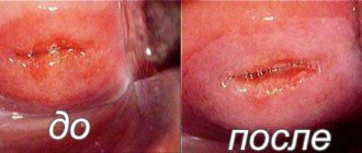
- Treatment with laser, radio waves, and photodynamic therapy is used to eliminate erosions and shallow dysplasia.
- In case of deep tissue lesions, conization is performed - removal of part of the cervix with capture of the cervical canal. The tissue obtained during the operation is sent to the laboratory for testing for malignant cells.
In order to timely identify diseases accompanied by koilocytosis, you need to go to the University Clinic, undergo an examination by a gynecologist and take tests. There is all the necessary diagnostic and treatment equipment and experienced specialists work here.
link:
Source: https://unclinic.ru/kojlocitoz-diagnostika-i-lechenie/