The uterus is the main reproductive organ of the female reproductive system. It consists of various elements, has a muscular structure and a free cavity inside. Its main function is the proper development of the fetus during pregnancy, as well as the regulation of menstrual flow.
Every woman knows approximately the structure of the uterus. For example, it is known that this organ is not a pair and is located in the pelvic area. In close proximity to it is the rectum and bladder. However, let’s take a closer look at the structure of a woman’s uterus, a photo with a description, and what changes may occur in it.
Essential elements
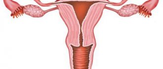
You can see what the uterus looks like in pictures, of which there are many on the Internet. But such a review cannot provide a clear understanding of the anatomical features of the organ. The following photo shows in close-up what a woman’s uterus looks like.
Parts of the reproductive organ. Source: ru.wikipedia.org
It can be studied in more detail through an ultrasound examination. The first part is the fundus of the uterus. It is located at the top and has a convex shape. In the middle part there is an expanded cavity - this is the body of the organ, the neck is in the lower part, and it is narrowed.
Walls
The walls of the uterus also consist of several layers. The first is the serous membrane, called the perimetrium. The tissues are called external because they face the cavity and are closely connected with the integument of the intestines and bladder. The main components here are connective fibers.
The myometrium is the next layer that covers a woman's uterus. It is thick and located in the middle part of the organ. It consists of three muscle structures, namely longitudinal, circular and internal. Here are the layers that the uterus has (photo, allows you to visualize the anatomical features as much as possible).
Localization of the layers of the uterus. Source: ikista.ru
The last layer is the endometrium, which has not only basal but also functional layers. They face the inside of the uterine cavity. The main component here are epithelial cells, due to which secretions are formed.
Neck
When wondering what the uterus is, we can say that it is a reproductive organ that has a rather complex structure, many constituent elements and is responsible for the normal intrauterine development of the fetus after the embryo attaches to its wall. The cervix is distinguished by the content of a large number of connective tissues, in which the content of collagen is increased.
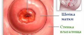
As for the muscle elements, there are significantly fewer of them here than in other parts of the organ. The structure of a woman’s uterus, in particular her cervix in the lower section, can be called unique in its own way. The size is within 3-4 centimeters, and according to topography it can be divided into vaginal and supravaginal parts.
The photo shows a close-up of a woman’s uterus, and also highlights the part where the cervix is located.
Where is the cervix located? Source: mioma911.ru
Outside the cervix is the entrance to the cervical canal, which is called the pharynx. If a woman has already gone through natural childbirth, then this element takes on a round shape; in nulliparous girls it is slit-like. This anatomical element is located in the central part of the small pelvis.
Depending on the period of the menstrual cycle, the position of the cervix will change, which is due to hormonal changes in the body. However, it is not recommended to carry out manipulations to determine the current period on your own, since the factor of infection cannot be excluded.
The walls of the cervix contain a large number of blood vessels. Blood circulation here is regulated due to the presence and operation of paired uterine arteries and internal branches of the iliac artery. Thanks to the tree-like base, small vessels are nourished, thereby enriching the entire organ as a whole with oxygen and nutrients.
The photo demonstrates as clearly as possible how the circulatory system of the female uterus works.
The circulatory system of the reproductive organ. Source: embryology.med.unsw.edu.au
Initially, blood passes through the capillaries, after which it accumulates in large vascular structures. They are the uterine veins, iliac vessels and ovaries. In addition, the organ also has lymph nodes. The endocrine system of the body produces certain sex hormones, thanks to which the normal functioning of the uterus and the vital activity of tissues occurs.
Also, the stability of the nervous system is of certain importance in the normal functioning of the reproductive organ. The fact is that on the walls there is a certain number of nerve endings that branch towards the pelvis and are also interconnected with the hypogastric plexus.
Picture at different periods of development of the disease
Genital prolapse, as a rule, extends over time and does not happen at once. Initial changes cannot be recognized externally; this requires a gynecological examination. The difficulty of diagnosis lies in the fact that a woman, being already sick, does not feel anything. Sometimes they have pain in the lower abdomen, discomfort, but such symptoms are not given much importance and people do not go to the doctor. That is why it is important to visit an obstetrician-gynecologist for preventive purposes and fight fears of not entirely pleasant manipulations. Doctors recommend checking annually so that a specialist can conduct a vaginal and rectal examination.
The first degree of uterine prolapse is established only after examination in a gynecological chair. The doctor observes a picture in which the cervix reaches approximately the middle of the vaginal canal. This diagnosis is still considered quite favorable, since the problem can be eliminated without surgery, using special Kegel exercises, gynecological massage, pessaries, bandages, etc. can be prescribed.
The second stage is easier to diagnose, since the woman may present a number of complaints. A common reason for seeking medical help is pain in the lower abdomen or pelvis, which gets worse when walking, exercising, or during sexual intercourse. A specific symptom is characteristic: the feeling of a “foreign” body in the vagina. Also at this stage, disorders of the urinary system and digestive tract may appear. During a general examination of the genital organs, the uterus may not be visible. But if you ask a woman to tense the muscles of the anterior abdominal wall (the same will happen when coughing, sneezing - increasing pressure inside the abdominal cavity of any etiology), then a similar picture will be observed:
If at the second stage of uterine prolapse the cervix extends beyond the genital slit only when intra-abdominal pressure increases, then at the third stage it is constantly outside. In this case, the body of the uterus is actually located in the vagina. This condition is characterized by an increase in all previous symptoms. Externally, the genitals at this stage of development look like this:
Unfortunately, it is not uncommon for women to see a doctor only in this condition. Conservative treatment will be completely ineffective and will be used rather in the form of symptomatic therapy. A woman can only be cured through surgery.
If treatment at this stage of the disease is ignored or surgery cannot be performed due to medical contraindications, uterine prolapse develops. Topographically, the body and cervix are located outside the genital tract. This is a serious condition that requires immediate treatment. Complete uterine prolapse looks like this:
The uterus is the main reproductive organ of the female reproductive system. It consists of various elements, has a muscular structure and a free cavity inside. Its main function is the proper development of the fetus during pregnancy, as well as the regulation of menstrual flow.
Every woman knows approximately the structure of the uterus. For example, it is known that this organ is not a pair and is located in the pelvic area. In close proximity to it is the rectum and bladder. However, let’s take a closer look at the structure of a woman’s uterus, a photo with a description, and what changes may occur in it.
Ligaments and muscles
The inside of the female uterus consists of ligaments that have connective tissue. Thanks to them, the reproductive organ is securely fixed in the pelvis. Wide or paired are characterized by an anatomical connection with the structures with which the ovaries are fixed, and they are attached to the walls of the abdominal cavity.
The round ligaments contain not only connective tissue, but also muscle tissue. It is localized along the entire uterine wall, and reaches the deep opening of the groin canal, and in the area of the labia majora it ends with fiber. With the help of the cardinal ligament, the organ connects to the lower part of the urogenital diaphragm, which helps prevent displacement to the left or right.
The presented photo shows a close-up of the uterus and its muscular-ligamentous apparatus.
Musculo-ligamentous apparatus of the organ. Source: lediveka.ru
The anatomy of the uterus is well thought out. Thanks to the described ligaments, her body is connected to the fallopian tubes and ovaries. But it is worth saying that such features are typical only for healthy women who do not have any gynecological pathologies. In addition to the ligaments, muscles are also responsible for the normal position of the organ.
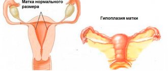
Organ sizes
Some girls may wonder how much a woman's uterus weighs. Experts say that in a completely healthy girl who has not yet given birth, the mass of the organ does not exceed 50 grams. If there has already been a pregnancy, then this figure reaches 100 grams.
The length of the organ ranges from 7 to 8 centimeters, and its width is no more than 5 centimeters. When the layers of the uterus, in particular the muscular structure, hypertrophy, which occurs during the intrauterine development of the child, these indicators definitely increase. Inside, the cavity is no more than 5-6 cm in size, since its walls are quite thick.
In the absence of congenital or acquired anomalies of structure and development, the reproductive organ is localized in the middle part of the small pelvis, parallel to the bone structures. Since the level of physiological mobility of the uterus is quite high, it can move freely to the areas of the most closely located structures. In this case, temporary bends occur.
If the bladder is not filled with biological fluid, then the fundus of the uterus is directed forward towards the peritoneum. When it stretches, when it is filled with urine, the organ temporarily bends backward and approaches the intestine.
Growth of the uterus by week of pregnancy
To find out how the belly grows during pregnancy, you need to focus on generally accepted norms for uterine enlargement.
By the twelfth week, the uterus will reach the size of a newborn’s head, while still being completely contained in the pelvis. The uterus can be clearly felt to the touch a little above the pubis; over time, its bottom will begin to rise.
By sixteen weeks, the fundus of the uterus will be located halfway from the pubis to the woman’s navel. At the twentieth week it will be at a distance of two transverse fingers below the navel.
At this time, most expectant mothers have a noticeable belly. Already at the 24th week, the fundus of the uterus will reach the level of the navel, and at 28 weeks it will rise above the navel by three fingers.
By the thirty-second week, a woman’s navel may completely smooth out, at which point the fundus of the uterus will be located between the xiphoid process of the sternum and the navel.
The thirty-eighth week is the peak for the fundus of the uterus to rise, and the navel may protrude outward. This time is considered a turning point; by the end of pregnancy, the child will strive to enter the pelvic area, and the uterus will descend accordingly.
At the onset of the fortieth week of pregnancy, the fundus of the uterus will drop to the middle of the distance between the xiphoid process and the navel. This condition can be compared to the level when the uterus is in the thirty-second week of pregnancy, but the volume of the abdomen will already be increased by eight or even ten centimeters.
Physiological changes
When considering the structure of the vagina and uterus, it is worthwhile to touch upon the topic of natural physiological changes that occur to the organ during ovulation or during pregnancy. Until the ovary ruptures and the follicle comes out, the cervix is quite elastic and dry.
Ovulation
Since before ovulation, the body begins to actively produce sex hormones necessary for pregnancy, as well as implantation of the embryo to the wall, under their influence the cervix softens. This indicates that the reproductive organ is ready for conception. A film of viscous mucus forms on the internal pharynx. Thanks to it, sperm have a greater chance of penetration, and the cervix itself lowers slightly.
Changes in the uterus during ovulation and pregnancy. Source: 28dnej.com.ua
If fertilization does not occur, regular menstrual bleeding begins. The internal pharynx expands, which makes it possible for the annealed endometrium and blood clots to come out. If during this period you do not follow the rules of intimate hygiene and do not use a condom during intimacy, the likelihood of infection of the reproductive organs is very high, which is due to the position of the cervix after ovulation.
On average, the duration of the menstrual cycle is 25-29 days. After the completion of menstrual bleeding, the organ returns to its natural position, the pharynx narrows, the cervix rises and the endometrial layer is restored. Before the next ovulation occurs, complete regeneration of the mucous membrane is noted so that the embryo can attach if fertilization occurs.
Pregnancy
Considering what the uterus looks like in girls and what changes may occur to it, experts draw attention to the fact that transformations of the organ are observed even after pregnancy. During the entire period of intrauterine development of the fetus, the uterus will constantly increase in size.
Also, along with this, there is a change in hormonal levels. If this does not happen, the process of endometrial cell rejection will begin, and pregnancy failure (miscarriage) will occur, after which menstrual bleeding will begin. During pregnancy, the following changes will be noted:
- By the middle of pregnancy, the thickness of the endometrium is 3-4 mm, as the spindle cells elongate and their diameter increases;
- Before labor, there is stretching and a decrease in the thickness of the myometrium;
- After conception, the cervix acquires a blue tint, and access to it is closed by a mucus plug;
- The ligaments grow rapidly, so the last three months of pregnancy are accompanied by pain with sudden movements, which is caused by tissue tension.
It is also worth noting that the anatomical location of the uterus changes in accordance with the timing of pregnancy. At approximately 13-14 weeks of gestation, an examination can determine the height of the fundus of the organ. The uterus itself gradually enlarges in the upper part, so it extends beyond the pelvis.
At about 24 weeks, the fundus of the uterus is localized in the navel area, and at 36-38 weeks its height is maximum, so the organ can be felt between the ribs. Subsequently, it descends, which allows the fetus to reach the birth canal. It is by the type of uterus that specialists determine the duration of pregnancy.
It is important to understand that the anatomical description given is the norm and can only be seen in completely healthy women. If there are any pathological abnormalities, this clearly affects the appearance and functionality of the reproductive organ.
This project was led by a 25-year-old woman. She has never given birth and has no history of STDs. Each photograph was taken at approximately 10:00 p.m., starting on the first day of the menstrual cycle. Throughout this project, she used condoms as a contraceptive method and also to prevent semen from being released during the photo shoot. She did not use tampons during her period.
This cycle is 33 days, which is the norm. The follicular phase of her cycle lasts until about 20–21 days. Favorable days for fertilization last several days from the 13th to the 21st day with ovulation on the 20th day. The luteal phase is 13 days (12–16 days is normal).
How the cervix changes depending on the physiological state of a woman
Depending on the physiological state of the woman, the cervix stretches or shortens. At a time when conception is impossible, it feels hard and short. These days it is a bit difficult to reach it with your fingers as it rises to the top. On the days of ovulation, on the contrary, the cervix drops down and swells. Therefore, it becomes soft and loose. At the same time, the depression in its center becomes deeper, and it is during this period that sperm can overcome the barrier of cervical mucus and fertilization will occur.
During pregnancy, the cervix rises high, becomes hard and closes. Therefore, during the examination, the doctor cannot reach the cervix with his fingers. In this regard, some women cannot feel their cervix when pregnancy occurs during a self-examination.
Based on these indicators, the onset of conception can be determined. During the examination, the gynecologist feels the cervix and presses with the other hand from above in order to determine some reflexes that confirm a change in the woman’s physiological status.
IMPORTANT! If you suspect pregnancy, it is not advisable to examine the cervix yourself, since this is a rather gross intervention in the body.
This can cause muscle contractions and lead to miscarriage. Even doctors during this period try to perform this manipulation a minimum number of times, touch the organ, insert and remove instruments into the genital tract. Therefore, to determine pregnancy, it is best to use a test and then confirm suspicions using an ultrasound examination.
Until 25 weeks of pregnancy, the cervix is normally long and firm. As a rule, its value is approximately 30 mm. Then, the length of the neck gradually begins to shorten. In pathological conditions, isthmic-cervical insufficiency (ICI) occurs. It leads to shortening occurring at an earlier date. In this case, the cervix softens ahead of time. In this case, the child puts pressure on the cervix from the inside, and the loose tissue of the pregnant cervix may not support the weight of the child and dilatation will occur. Accordingly, this condition can easily lead to early labor. With ICI, pregnant women experience pain in the lower abdomen and strong muscle tone is felt.
IMPORTANT! If you suspect untimely shortening of an organ during pregnancy, consult a doctor immediately!
When palpating the cervix, you should not insert foreign objects into the genital tract or touch it yourself. It is not advisable to fiddle with the organ, roughly palpate it, or climb inside with a mirror and other things. This can cause damage and injury to delicate tissues.
At first it is quite difficult to determine the consistency of the tissue and its height. With regular inspection, you can learn to establish these indicators quite easily.
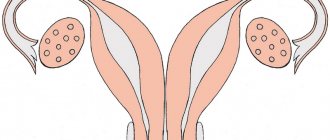
General structure of the uterus
This internal muscular organ of the reproductive system has a pear-shaped shape that is flattened in front and behind. In the upper part of the uterus on the sides there are branches - fallopian tubes, which pass into the ovaries. The rectum is located behind, and the bladder is located in front.
The anatomy of the uterus is as follows. The muscular organ consists of several parts:
- The fundus is the upper part, which has a convex shape and is located above the line of origin of the fallopian tubes.
- A body into which the bottom smoothly passes. It has a cone-shaped appearance. It narrows downwards and forms an isthmus. This is the cavity leading to the cervix.
- Cervix - consists of the isthmus, cervical canal and vaginal part.
The size and weight of the uterus vary from person to person. The average weight for girls and nulliparous women reaches 40–50 g.
The anatomy of the cervix, which is a barrier between the internal cavity and the external environment, is designed so that it protrudes into the anterior part of the vaginal vault. At the same time, its posterior arch remains deep, and the anterior arch, on the contrary.
How to evaluate the results?
Insert your finger deep into [email protected] #$%& and feel the cervix. During ovulation, the cervix will be soft, like lips, and the first days after menstruation, it will be hard, like the tip of the nose. With a low position, you will most likely be able to easily touch it with the middle of the fingertip, and with a high position, you can barely reach it with the tip. You will also learn to determine the degree of opening only with practice. Feel the depression in the center of the cervix. When closed, it rather resembles a small gap, and when open, it becomes rounded and deeper. It should be borne in mind that in women who have given birth, the cervix is always slightly open. When you learn to accurately detect the slightest changes in the cervix, the period of research during the cycle can be reduced to one week on presumably fertile days.
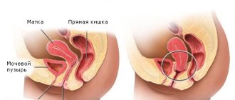
Where is the uterus?
The organ is located in the pelvis between the rectum and bladder. The uterus is a very mobile organ, which also has individual characteristics and shape pathologies. Its location is significantly influenced by the condition and size of neighboring organs. The normal anatomy of the uterus in terms of the place it occupies in the small pelvis is such that its longitudinal axis should be oriented along the axis of the pelvis. Its bottom is tilted forward. When the bladder is full, it moves back a little, and when emptying, it returns to its original position.
Anatomy of the uterus: photo and wall structure
The organ is three-layered. It consists of: perimeter, myometrium and endometrium. The surface of the uterine wall is covered by the serous membrane of the peritoneum - the initial layer. At the next – middle level – the tissues thicken and have a more complex structure. Plexus of smooth muscle fibers and elastic connective structures form bundles that divide the myometrium into three internal layers: internal and external oblique, circular. The latter is also called the average circular. It received this name in connection with the structure. The most obvious is that it is the middle layer of the myometrium. The term “circular” is justified by a rich system of lymphatic and blood vessels, the number of which increases significantly as it approaches the cervix.
Bypassing the submucosa, the uterine wall after the myometrium passes into the endometrium - the mucous membrane. This is the inner layer, reaching a thickness of 3 mm. It has a longitudinal fold in the anterior and posterior region of the cervical canal, from which small palm-shaped branches extend at an acute angle to the right and left. The rest of the endometrium is smooth. The presence of folds protects the uterine cavity from the penetration of vaginal contents that are unfavorable for the internal organ. The endometrium of the uterus is prismatic; on its surface there are uterine tubular glands with glassy mucus. The alkaline reaction they provide preserves the viability of sperm. During ovulation, secretion increases and substances enter the cervical canal.
Uterine ligaments: anatomy, purpose
In the normal state of the female body, the uterus, ovaries and other adjacent organs are supported by the ligamentous apparatus, which is formed by smooth muscle structures. The functioning of the internal reproductive organs largely depends on the condition of the muscles and fascia of the pelvic floor. The ligamentous apparatus consists of suspension, fixation and support. The combination of the properties of each of them ensures the normal physiological position of the uterus among other organs and the necessary mobility.
Composition of the ligamentous apparatus of the internal reproductive organs
Ligaments forming the apparatus
Connects the uterus to the walls of the pelvis
Paired wide uterine
Supporting ligaments of the ovary
Own ligaments of the ovary
Round ligaments of the uterus
Fixes the position of the organ and stretches during pregnancy, providing the necessary mobility
Main ligament of the uterus
Forms the pelvic floor, which is a support for the internal organs of the genitourinary system
Muscles and fascia of the perineum (outer, middle, inner layer)
The anatomy of the uterus and appendages, as well as other organs of the female reproductive system, consists of developed muscle tissue and fascia, which play a significant role in the normal functioning of the entire reproductive system.
Characteristics of the hanging apparatus
The suspensory apparatus consists of paired ligaments of the uterus, thanks to which it is “attached” at a certain distance to the walls of the pelvis. The broad uterine ligament is a transverse fold of the peritoneum. It covers the body of the uterus and the fallopian tubes on both sides. For the latter, the structure of the ligament is an integral part of the serous covering and mesentery. At the lateral walls of the pelvis it passes into the parietal peritoneum. The suspensory ligament arises from each ovary and has a wide shape. Characterized by durability. The uterine artery runs inside it.
The own ligaments of each of the ovaries originate from the uterine fundus on the posterior side below the branch of the fallopian tubes and reach the ovaries. The uterine arteries and veins pass inside them, so the structures are quite dense and durable.
One of the longest suspensory elements is the round ligament of the uterus. Its anatomy is as follows: the ligament looks like a cord up to 12 cm long. It originates in one of the corners of the uterus and passes under the anterior sheet of the broad ligament to the internal opening of the groin. After which the ligaments branch into numerous structures in the tissue of the pubis and labia majora, forming a spindle. It is thanks to the round ligaments of the uterus that it has a physiological inclination anteriorly.
Structure and location of fixing ligaments
The anatomy of the uterus should have suggested its natural purpose - bearing and giving birth to offspring. This process is inevitably accompanied by active contraction, growth and movement of the reproductive organ. In this connection, it is necessary not only to fix the correct position of the uterus in the abdominal cavity, but also to provide it with the necessary mobility. Fixing structures arose precisely for such purposes.
The main ligament of the uterus consists of plexuses of smooth muscle fibers and connective tissue, radially located to each other. The plexus surrounds the cervix in the area of the internal os. The ligament gradually passes into the pelvic fascia, thereby fixing the organ to the position of the pelvic floor. The vesicouterine and pubic ligamentous structures originate at the lower anterior part of the uterus and attach to the bladder and pubis, respectively.
The uterosacral ligament is formed by fibrous fibers and smooth muscles. It extends from the back of the cervix, envelops the rectum on the sides and connects to the fascia of the pelvis on the sacrum. In a standing position, they have a vertical direction and support the cervix.
Pathogenesis of the disease
In a healthy person, the uterus is located above the vagina in the pelvic cavity. Anterior to it are the bladder and urethra, posteriorly the rectum. This position helps the organs to be supported by powerful muscles and ligaments of the pelvis. The complex of these structures is otherwise called the pelvic floor. The normal anatomy of the pelvic organs is shown in the diagram below:
When the pelvic floor weakens, the topography of the pelvic organs changes, they move downwards under the force of the earth's extension. The reason for this may be factors related to lifestyle and work activity. Women who have given birth most often get sick. Any reason leading to an increase in intra-abdominal pressure leads to weakening of the muscles and ligaments of the pelvis. This could be a simple cough that lasts for a long time, or sneezing.
Weakening of the muscles and ligaments of the uterus leads to its descent. She begins to pass through the vaginal canal, “crushing” the vagina in front of her. Depending on the anatomical level of the uterus, there are several degrees of prolapse, the last of which is prolapse. Schematically, uterine prolapse looks like this: