Female body
Representatives of the fair sex differ in many ways from men. In addition to external signs, there are also internal structural features of the body. Thus, representatives of the weak half of humanity are able to reproduce their own kind and feed them. A woman’s uterus, ovaries, pituitary gland and other organs play a major role in this process. Men are structured more primitively and simply.
A woman's uterus: what is it?
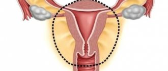
This organ is located in the small pelvis of every woman even before birth. Thus, the reproductive region is formed approximately at the 10th week of intrauterine life. Externally, the uterus resembles a small inverted pear or cone.
On the sides of a woman’s uterus there are two so-called processes. Doctors are more familiar with them under the name fallopian (uterine) tubes. Also under each of these processes there is a small oval-shaped organ. These structures are called ovaries.
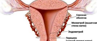
In addition to the internal structural features of the uterus in women, it has a cervix and a cervical canal, which opens into the vagina. The internal cavity of the reproductive organ has three layers. The main one is the endometrium - the inner lining.
Meaning of the word uterus
(uterus, metria), a special section of the genital ducts in female animals and women; is an expanded part of the oviduct. M. has a powerful muscle wall and is well supplied with blood.
Uterus in animals. M. is found in roundworms, arthropods, mollusks, most lower vertebrates (some cartilaginous fish and all viviparous bony fish), some amphibians, many reptiles, birds, and all mammals.
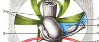
In oviparous vertebrates (reptiles and birds), mature eggs are temporarily placed in the membrane. In viviparous animals, embryonic development of the organism occurs in the mother due to either the nutrients of the egg or the nutrients of the mother’s body. In the latter case, communication and metabolism between the developing embryo and M. is carried out using the placenta. The structure of the muscle in different mammals is very diverse. In cloacals (platypus, echidna) the cloaca has a steam room and each opens separately into a cloaca. In marsupials (kangaroos and others) the vagina also has a steam room, but each of them opens into a special vagina. In placental mammals, all transitions from paired to unpaired M. are observed ( Fig. ).
Depending on the degree of fusion of the oviducts, there are 4 main types of uterus: double (uterus duplex) ≈ two uterus, each opening with an independent opening into the common vagina (in some rodents, elephants and others); bipartite M. (uterus bipartitus) ≈ also two M., but fused together by the posterior sections and opening with one common mouth in the vagina (in some rodents, ruminants, pigs and carnivores); two-horned (uterus bicornis) is the most common type of uterus in mammals: it consists of two uterine horns, which are connected to form an unpaired body of the uterus, which opens into the vagina (in many carnivores, insectivores, cetaceans, artiodactyls and odd-toed ungulates); simple (uterus simplex) ≈ consists only of an unpaired body into which 2 oviducts open (in most chiropterans, primates and humans).
Lit.: Kholodkovsky N. A., Textbook of Zoology, 6th ed., M. ≈ Leningrad, 1933; Course of Zoology, 7th ed., vol. 1 ≈ 2, M., 1966; Marshall's physiology of reproduction, ed. A. S. Parkes, 3 ed., v. 1≈2, L. ≈ NY ≈ Toronto, 1956≈58; Giersberg N., Rietschel P., Vergleichende Anatomieder Wirbeltiere, Bd 2, Jena, 1968.
K. M. Kurnosov.
The human uterus is a reproductive muscular hollow organ located in the woman’s pelvic cavity between the bladder and rectum. Weighs 40≈50 g in nulliparous women and 90≈100 g in multiparous women. M. has a pear-shaped shape. Most of it is made up of the body, the upper section of which is expanded and called the bottom; the lower, narrowed end of the M. (neck) is covered by the vagina. There are 2 parts in the cervix: facing the vaginal cavity - the vaginal part and the upper part - the supravaginal part. The M.'s bottom is inclined forward, and the body and neck form an angle open anteriorly. Inside the M. there is a cavity in the shape of a triangle, with two holes at the top leading to the uterine (fallopian) tubes. M.'s cavity passes into the cervical canal, which opens with its external opening (uterine os) into the vagina. The wall of the muscle consists of three membranes: outer (serous), middle (muscular), and inner (mucous). The serous membrane is represented by the peritoneum, which envelops the M. from the front, back and sides and passes to the bladder and rectum, limiting 2 recesses: vesicouterine and rectouterine; on the sides of the peritoneum, the layers of the peritoneum grow together and form the broad ligament of the peritoneum, which, together with the fascia and muscles of the pelvic floor, participates in its fixation. The middle shell of M. is the most powerful; it consists of three layers of smooth muscle mixed with elastic fibers. The mucous membrane is lined with columnar ciliated epithelium, is equipped with numerous glands and is subject to changes due to the menstrual cycle. M.'s arterial blood supply is carried out by branches of the uterine and ovarian arteries, which develop especially strongly during pregnancy. Venous blood flows from the lymph nodes through the veins of the same name, and lymph flows through drainage vessels to the aorto-abdominal, hypogastric, and iliac lymph nodes. M.'s innervation is carried out by the branches of the inferior mesenteric plexus and the pelvic nerves.
Ya. L. Karaganov.
Dimensions of the uterus and structural features
A woman's uterus has different sizes. It all depends on what phase of the cycle the body of the fair sex is in. Normal sizes after the end of menstruation are in the range of 4 to 5 centimeters. In this case, the length of the organ may be slightly greater than the width and cross section.
The cervix in women who have never given birth and have not undergone dilatation of the cervical canal has a rounded shape and the same tightly closed opening. If a representative of the fair sex has already become a mother, then her cervix may have a slit-like opening, which is somewhat expanded. All this is a variant of the norm. The length of the cervical canal in different women can vary from 2 to 5 centimeters. Moreover, special attention is paid to this figure during pregnancy.
The female reproductive organ has an interesting feature. The uterus is not anchored by any devices or bones. Her body is held together only by ligaments and muscles. One can only imagine the stress these components undergo while carrying a child. The female uterus may be positioned correctly or may have anterior or posterior deviation. This is not a pathology, but problems with conception may occur.
In Vasmer Max's dictionary
I uterus I., mother “fever”. Tabuistic name from mother (cf. kuma); see Zelenin, Taboo 2, 76. The leg (Μνῆμα 449) incorrectly brings the mother closer to the mother. II •• (II., dial., Belomorsk. “compass”, already in 1599 in a handwritten Russian-English dictionary. Possibly, tracing paper of the name of the part of the astrolabe - box, Latin. mater, Arab. umm “mother”; see Bogorodsky, “Dokl. history of the vocabulary of Eastern European languages, Leningrad, 1957, p. 60) sees here the influence of Finnish, Karelian matka “path.” - T.)
Functions of the female uterus
The female reproductive organ has many important functions. Let's look at the main ones.
- One of the main functions that a woman’s uterus has is childbirth. Every month, the inner layer changes and is exposed to hormones. Thus, the body prepares for conception. If fertilization has occurred, then the embryo is securely attached to the wall of the female organ and remains there until full development and readiness for life in the external environment.
- In addition, the female uterus performs a cleansing function. Each menstrual cycle, the organ contracts, pushing the unnecessary inner layer out. It is during this period that a woman menstruates.
- The female reproductive organ also has a protective function. The uterus reliably protects the fragile fallopian tubes from the penetration of pathogens and infections. The cervix, in turn, secretes mucus, which helps flush these bacteria out of the cervical canal and vagina.
- The function of promoting sperm is also inherent in the female organ. After sexual intercourse, the uterus actively contracts, helping male gametes penetrate the cavity and enter the fallopian tubes for fertilization.
- Also, the female uterus can be assigned the function of supporting organs and various systems. Due to being in its usual place, the uterus does not allow the intestines and bladder to move in different directions.
Diseases of the female organ
Many representatives of the fairer sex have to deal with pathologies that affect the reproductive system. These include endometritis, fibroids, uterine prolapse in women and other diseases. Some of them can be safely treated and have a favorable prognosis. Others lead to such a horrifying conclusion as hysterectomy. Women who have had to undergo this procedure feel depressed and inferior. Let's look at a few examples of pathologies of the female organ.
Uterine rabies (nymphomania)
Uterine rabies in women is a process in which the mental state is disturbed. This disease is often called hysteria. This name is now obsolete. Modern medicine does not recognize such a disease as uterine rabies in women. The symptoms of the pathology remained. Most often, the disease manifests itself as increased sexual desire, clouding of consciousness, laughter followed by tears. Now such women are called nymphomaniacs and are prescribed psychological correction.
In the dictionary D.N. Ushakova
UTERUS, uterus, female. 1. The internal part of the female genital organs, in which the embryo develops. Diseases of the uterus. 2. Female breeder in animals. Deer queen. The horse farm had many breeding queens. Queen bee. 3. transfer Designation of an object that serves to form others of the same kind or to transfer to them some properties or qualities (special). Womb bath (at the electrolyte plant). | Rock containing ore or precious stones (geol.). 4. Horse breeder, leader in games, for example. in lapta (reg.). 5. Mother, mother (simple region). He grew up as an orphan and became a helper for his mother. Mamin-Sibiryak. • Truth-uterus (simple) - truth, truth. “I’ll lay out the whole truth to you, right there in the palm of your hand.” Melnikov-Pechersky. Always cuts the truth.
Removal of the reproductive organ
There are several ways to remove the uterus. Depending on the capabilities of the medical institution and the qualifications of doctors, the most suitable option is selected. The most common procedure is laparoscopic surgery. However, there are times when a laparotomy is required. Let's consider both of these options.
Removal of the uterus using a laparoscope
If you have time to prepare for surgery, then it is preferable to perform this procedure. During the operation, the doctor makes several incisions in the patient's abdomen and inserts small manipulators into them. Using a video camera, the doctor sees everything that is happening on a large monitor. Small manipulators carefully cut the ligaments and muscles that support the uterus. After this, the organ is removed from the abdominal cavity.
Recovery after such an operation is quick. However, a woman may experience discomfort and pain during the first month after the procedure.
Laparotomy surgery to remove the uterus
If the procedure for removing an organ from the peritoneum is urgent, then laparotomy is performed. This method is also chosen when a woman has a large layer of fat in the pelvic area. During the operation, the doctor makes an incision in the lower abdomen. It can be horizontal or vertical depending on the situation. After removing the reproductive organ, the incision is sutured layer-by-layer.
Recovery after such an operation is much more difficult. The woman is incapacitated for one month after the procedure.
In Dahl's dictionary
and. French mask, in literal and figurative meaning, false face, for fun; | pretense, double-mindedness. | Okrutnik, mummer, disguised, disguised. To disguise, to rip someone off, to surround someone, to dress them up in unusual clothes for fun, and to put on a mask; | what, to cover, to hide from view: the fence is masked by bushes. Mask your actions and intentions, hide them under the guise of another, feigned action. -sya, they suffer. and return according to the meaning of speech. | To put on, assume a guise; put on a disguise; | to be dressed up; to be hidden behind something. Masking cf. duration valid according to verb. in all meanings Disguise g. about. valid in meaning a hidden matter, an object, behind another, deceptive matter. Masquerade m. entertainment gathering, congress, kind of ball, in unusual clothes and disguises: | comic bath, bathing. Masquerade, related to a masquerade. MASK or smear? and. north eastern blood, more about blood smeared, for example. from the nose, after a fight, struggle, or from killed game, as ore is more commonly used. about vein blood, bloodletting. Maskara perm. mask, blood. | Ichor. | Arch. a mixture of salt and fresh water, at river mouths.
What happens to the female body after surgery?
After removal of the uterus, a woman changes not only internally, but also externally. Most representatives of the fair sex note an internal emptiness in moral and physical terms. If a woman is of childbearing age, then in addition to depression, she feels helpless and useless.
After hysterectomy, many women are recommended hormone replacement therapy. This treatment allows you to maintain internal ligaments and muscles in tone.
Why do many gynecologists advise removing the uterus for fibroids?
The main reason for such a long dominance of therapeutic radicalism in the treatment of uterine fibroids is that for too long uterine fibroids have been considered, although benign, to be a tumor process, and the tumor, as the canons of surgery say, must be removed. Indeed, there is a list of organs without which a person can more or less exist. And from the point of view of many gynecologists, the uterus is almost in first place on this list.
For some reason, it is believed that having realized her reproductive function, a woman can part with the uterus completely painlessly, that is, a unique monofunctional relationship has been developed towards this organ. Wrong attitude. At the same time, it is quite obvious that there are no unnecessary organs in the body, and the uterus, in addition to the reproductive function, also has others, some of which are clear to us, and some of which are still not fully understood. To simplify, we can say that, being integrated into the whole organism, the uterus maintains a natural physiological balance.
A person can exist without one kidney, a lung, or part of the intestines, but everyone understands that this existence is no longer a completely full-fledged person, so why is a woman without a uterus considered healthy in the minds of a number of doctors? Indeed, it has been known for many years that removal of the uterus entails the development of the so-called posthysterectomy syndrome - a symptom complex of disorders of the endocrine, nervous, cardiovascular and other systems that occurs after removal of the uterus and is associated with this removal through a direct cause-and-effect relationship.
A special place is occupied by psychological consequences - the presence of a uterus is a subconscious element of femininity, involvement in the female gender. The presence of a uterus gives a woman constant inner confidence that she can give birth to a child. And even if she definitely does not want to have more children, permanent deprivation of this function may be emotionally unacceptable for her.
Some studies that examine the functions of the uterus show interesting results. For example, in 2021, scientists from Arizona State University (USA) conducted an experiment on rats: some animals had their ovaries removed, others had their uterus, others were not operated on, and their uterus and ovaries continued to function. Animals whose uterus were removed were less able to navigate mazes.
However, other studies show that women who have a hysterectomy have an increased risk of developing dementia. The uterus somehow influences the functioning of the nervous system, and these relationships remain to be studied.
In addition, during the experiment it turned out that the hormonal levels in the body changed in rats that had undergone hysterectomy. And this, as we know, can potentially create a number of health risks. This suggests that the uterus is needed not only during pregnancy. When a woman is not pregnant, the uterus does not “sleep” and is not at rest. It performs some functions that have yet to be understood in more detail.
The results of such studies provide a strong argument against hysterectomy in cases where there is no truly justified indication for it.