Functions of the uterus
The main function of the uterus is reproductive. This organ is continuously connected with the process of procreation. Directly in it the development of a small organism from two germ cells occurs. In addition, there are a number of other functions that the uterus performs:
- Protective. The organ is a barrier to the spread of pathogenic microorganisms and viruses from the vagina to the appendages.
- Cleansing – monthly, along with menstruation, self-cleaning of the cervical canal and vagina occurs during menstrual flow.
- Participation in the process of fertilization is a connecting link on the path of sperm from the vaginal cavity to the fallopian tube.
- Participates in the implantation process.
- Strengthens the pelvic floor along with its own ligamentous apparatus.
Woman's uterus - dimensions
It should be noted that such a parameter as the size of the female uterus has a special diagnostic value. Thus, based on an increase in the volume of an organ, the doctor can make the first assumptions about pathology or pregnancy already at the first stage of the examination, without the use of equipment. The size of the uterus can vary and depends on several factors:
- the presence of pathologies and diseases of the reproductive system;
- presence of pregnancy and childbirth;
- woman's age.
Normal size of the uterus of a nulliparous woman
Diagnosis of diseases of the uterus and determination of the size of the organ is carried out using ultrasound. This hardware method helps to accurately determine structural changes in an organ and establish its exact location. The normal dimensions of the uterus in a woman who does not have children are as follows:
- length – 7–8 cm;
- maximum width – 5 cm;
- weight – about 50 g.
Sizes of the uterus at different stages of pregnancy
Pregnancy is a complex and lengthy process, accompanied by the growth and development of the fetus. The immediate increase in the size of the unborn baby causes the growth of the uterus and its volume. At the same time, structural changes in the composition of the walls of the organ are observed: not only a qualitative, but also a quantitative increase in muscle fibers occurs. At the same time, the female uterus increases throughout the entire period of pregnancy.
In the first weeks of gestation, the reproductive organ retains its pear-shaped shape and practically does not change in size, since the embryo is still small. However, by the second month the organ acquires a rounded shape, and the size of the uterus during pregnancy by this time increases several times. The weight of the uterus itself also increases, and by the end of the gestation period it reaches almost 1 kg! At each examination of a pregnant woman, the doctor determines the height of the uterine fundus. The change in this parameter by week of pregnancy is shown in the table below.
RyzhikI
Hi all
I was looking for photos of ultrasound of follicles and VT for myself and came across this.
Maybe this information was already on the site, but I didn’t find it, I decided to post it in case it might be useful to someone, and then, for me, it’s easier to search here than to surf the internet.
Normal ultrasound of the uterus and ovaries: normal size of the uterus and ovaries
It has been established that the size of the uterus is influenced not only by previous pregnancies, but also by the stages of the menstrual cycle. During the so-called proliferative stage, in the first 7-11 days after the end of menstruation, it is reduced in size, and during the secretory phase, i.e. after ovulation and before the onset of menstruation, on the contrary, it is increased. Dimensions may also vary slightly depending on the examination technique. So for TAI, the thickness is slightly reduced due to the pressure of the bladder, but for TVI, on the contrary, it can increase due to increased muscle tone of the uterus.
The shape of the uterus is most often pear-shaped and only after several pregnancies it tends to become more rounded. The myometrium, the muscular lining of the uterus, has an average density similar to that of the parenchyma of the intact liver.
Endometrium in the early stage of the proliferation phase Endometrium in the middle stage of the proliferation phase Endometrium in the late stage of the proliferation phase
Ultrasound anatomy of the inner mucous membrane of the uterus - the endometrium should be considered in conjunction with the various phases of the menstrual cycle. For simplicity and convenience, we will consider the cycle to be a normal duration of 28 days with the onset of ovulation on the 14th day.
For early proliferation, i.e. in the period 5-7 days after the end of menstruation, the mucous membrane has a low density and a relatively uniform structure. Its thickness is on average 5 mm. In the central part, a thin line of increased density may be observed, arising from the contact of the anterior and posterior walls of the mucosa.
8 or 10 days after the end of menstruation in the stage of medium proliferation, the thickness of the mucosa gradually increases to 8 mm on average, while the structure remains almost unchanged.
Finally, closer to the onset of ovulation, on days 11-14 of the cycle, the thickness of the mucosa continues to increase up to 14 mm and at the same time increased echogenicity is observed.
Endometrium in the early stage of the secretory phase Endometrium in the middle stage of the secretory phase Endometrium in the late stage of the secretory phase
At the very beginning of the secretory, post-ovulation phase, on days 15-18 after menstruation, the thickness continues to increase, reaching a maximum of 16 mm, and the density increases from the edges to the center and thus takes on a drop-shaped shape with thickening towards the fundus of the uterus and elongation towards its cervix . During this period, the line of increased density already becomes fuzzy.
Uterus, 4th day of menstruation Postmenopausal uterus
In the stage of medium secretion, on days 20-23 of the cycle, the thickness of the mucous membrane reaches its maximum value. On average it is 14 mm, but thickening up to 18 mm is possible. The density continues to increase, and the clear thin line of increased density almost completely visually disappears.
Finally, in the last stage of secretion, days 24-28 of the cycle, the size of the endometrium may even decrease slightly - to an average of 12 mm. The main feature of this phase is the high density and very heterogeneous structure of the mucous membrane, which visually leads to the complete disappearance of the line of the walls of the shell petals.
Ultrasound of the ovary, early follicular phase Dopplerography of the ovary, early follicular phase
Continuation:
Uterine diseases - list
Uterine diseases in women can occur at any age. However, according to the observations of doctors, hormonal changes in the body are often the trigger for their development. This confirms the high frequency of development of pathologies of the reproductive system during puberty, after childbirth and during menopause. Most pathologies of the uterus are inflammatory and infectious processes in the reproductive organ. Among the common diseases of this organ are:
- Inflammatory processes: metritis, endometritis, adnexitis.
- Pathologies of the uterine cervix: erosion, ectopia, dysplasia, cervical cancer.
- Acute conditions associated with the uterus: ectopic pregnancy, ovarian apoplexy, spontaneous abortion.
- Tumor-like processes: myoma, fibroma.
Congenital pathologies of the uterus
Diseases of the uterus that occur during the embryonic development of the reproductive system and the formation of the genital organs are called congenital. Among the common pathologies of this kind, the following should be noted:
- Bicornuate uterus - formed as a result of non-fusion of parts of the Müllerian canals. There are different types of pathology:
- Saddle-shaped - a case when only the bottom of the organ is divided.
- A uterus with an incomplete or complete septum - the shape does not change externally, but a septum appears in the cavity, partially or completely separating it.
- Separate body with a common neck - formed by the fusion of the Müllerian ducts in the neck area.
- Duplication of the uterus - not only the body of the uterus is divided, but also the cervix.
Infectious diseases of the uterus
Infectious female diseases of the uterus are the most common type of pathology of this organ. They can arise due to banal non-compliance with the rules of intimate hygiene. Often the spread of an infectious agent occurs through sexual contact, so women of reproductive age are more often exposed to diseases. Pathology is almost always accompanied by changes in microflora, so additional symptoms appear that make it possible to identify the disorder (itching, burning in the perineal area, hyperemia). Common infections in women include:
Oncological diseases of the uterus
Women's diseases of the uterus, accompanied by tumor-like processes, are separate from all pathologies of the reproductive system. In most cases, the provoking factor for their development is chronic inflammatory and infectious processes, hormonal imbalances. The difficulty of diagnosing these pathologies lies in the absence of an obvious clinical picture, sluggish, latent course. Often a tumor is discovered during a random examination. Among the possible tumor-like diseases of the uterus, it is necessary to highlight:
Prolapse of the female uterus
With age, the female genital organs and the uterus may change their location. Often in older women, uterine prolapse is recorded, caused by a violation of the ligamentous apparatus and age-related changes. In most cases, the organ moves downwards, towards the vagina. The disease is accompanied by specific symptoms:
- feeling of pressure;
- discomfort in the groin area;
- pain in the lower abdomen;
- urination disorder (frequent urination, urinary incontinence).
The danger of the pathology lies in the possibility of complications by prolapse of the uterus from the vagina. This situation requires emergency medical care, so you should consult a doctor when the first symptoms appear. Treatment consists of surgical restoration of the integrity of the ligamentous apparatus of the pelvic floor and suturing of the vaginal muscles.
Documentary photo
Uterine fibroids
Stage of suturing the uterus. A seromuscular vicryl suture is applied. There is no active bleeding, the edges of the wound are well aligned.
Close-up of a subserous myomatous node on a broad base. The vessels are well defined. Ultrasound shows signs of active extranodular blood flow.
Body of the uterus with many myomatous nodes. One of them is subserous on a wide base along the anterior wall, more on the right, located on the uterine ligaments. On the back wall there is uterine fibroid, deforming the uterine cavity. The patient was admitted with complaints of heavy menstruation and pain for the last 3 months. Ultrasound shows rapid growth of nodes.
The incision on the uterus is closed with a vicryl suture. The edges are neatly matched. The uterus is of normal shape. The suture material will dissolve in 90 days.
Laparoscopy. Removal of a myomatous node that deforms the uterine cavity.
Laparoscopy. Conservative myomectomy. The stage of desquamation of the myomatous node. For this purpose, a special tool (corkscrew) is used, with the help of which adequate tissue tension is carried out.
The same myomatous node. Above it are 2 additional small subserous uterine fibroids.
Large subserous-interstitial uterine fibroid, located along the posterior wall of the uterus, more on the left. The patient complains of defecation problems (constipation), pain in the lower abdomen.
Uterine fibroids removed. The picture shows the bed of a myomatous node on the uterus. The next step is to stitch up this incision.
Stage of laparoscopy, removal of uterine fibroids. The myomatous node is desquamated. Uterine fibroids are almost removed.
Uterine fibroids of subserous localization along the anterior wall of the uterus of a low location. The patient complains of nagging pain in the lower abdomen and difficulty urinating.
Endometriosis
“Black” foci of endometriosis on the broad uterine ligament. This form is considered inactive.
Minor forms of endometriosis. Purple lesion on the left fallopian tube. There is no adhesive process.
Superficial foci of endometriosis on the wall of the bladder. They look like “rusty” spots. These are minor forms of endometriosis.
A deep focus of endometriosis on the left broad uterine ligament. Visible in the photograph as a bluish spot.
A focus of endometriosis on the right broad uterine ligament.
Retrocervical endometriosis with invasion into the right uterosacral ligament. The patient complains of pain radiating to the rectum, dyspareunia (painful sexual intercourse), and infertility.
Ovarian endometriosis. Lesions (red) are visible on the left ovary.
External genital endometriosis, small forms. Lesions on the broad uterine ligament. Laparoscopy, coagulation of endometriosis on the peritoneum.
Ovarian cysts and cystomas
Site of surgery after removal of a paraovarian cyst. Fallopian tube without visible changes. The pelvis is sanitized with antiseptic solutions.
The cyst has been removed. The capsule is not opened during enucleation. Preparations are underway for extraction in a special container.
In the photo there is a paraovarian cyst on the left, almost the size of the uterus. The patient complains of periodic pain in this area, radiating to the lower back. The vessels are clearly visible, the cyst capsule is well supplied with blood.
Laparoscopy. The ovary on the right is enlarged due to a cyst (the slide shows a bluish-colored formation in the ovary.
Laparoscopy. View of the left ovary after removal of the cyst.
Ovarian cyst removed. On the left, the cyst bed on the ovary is visualized.
The ovarian cyst is removed. Healthy tissue is preserved as much as possible, which will subsequently ensure the presence of ovulation.
Left ovarian cyst. The cyst has almost disappeared. The capsule is not opened, it is tense.
Left ovarian cyst. Laparoscopy stage – the ovary is opened, the cyst capsule is visible.
Infertility
Stage of surgery for hydrosalpinx. The fimbrial region is opened. The absence of folding of the mucous membrane is clearly visible. Since the probability of relapse is very high, the patient has been suffering from infertility for 10 years; by prior agreement with the patient, it is planned to remove the fallopian tube.
Left fallopian tube after plastic surgery. The fimbrial region is visualized.
Adhesions in the area of the pouch of Douglas in a patient with a history of chlamydia.
The patency of the left fallopian tube was restored. Chromohydrotubation is performed. The contrast agent (methylene blue) flows freely into the abdominal cavity.
Tubal-peritoneal factor of infertility. To get to the pelvis, it is necessary to separate the adhesion between the omentum and the posterior surface of the uterus.
Left fallopian tube after plastic surgery. The fimbrial section was formed and the paraovarian cyst was removed.
Right fallopian tube after plastic surgery, separation of adhesions. The fibrillary section is clearly visible.
The patient has been infertile for 5 years. Hydrosalpinx on the left. The fallopian tube is closed. A parovarian cyst (bluish in color) is present in the ampullary section.
The patient has been infertile for 4 years. Laparoscopy stage. Hydrosalpinx on the right side was diagnosed. The right fallopian tube is closed in the ampullary section. The fimbrial region is absent.
Adhesive process in the pelvis
The left fallopian tube and ovary are not visualized due to a pronounced adhesive process. The adhesions of the fallopian tube with the broad uterine ligament are clearly visible.
The right uterine appendages (fallopian tube and ovary) are in an intimate conglomerate with each other, as well as with the surface of the uterus. In the foreground there are yellowish liquid formations - closed cavities of adhesions filled with inflammatory fluid (serosocele).
Adhesions in the pelvis of the 4th degree. Thick adhesions with pronounced vessels between the ligaments and the intestine in the retrouterine space.
In the photograph there is a thick commissure located in the retrouterine space. Condition after suffering a chlamydial infection 10 years ago. Currently, she has been complaining of infertility for 2 years, and periodic pain in the lower abdomen.
An example of intestinal adhesion to the anterior abdominal wall. The patient suffers from constipation.
Dense adhesions between the left fallopian tube and the ovary. The stage of cutting adhesions with special scissors.
Adhesive process in the pelvis. The greater omentum is soldered to the posterior surface of the uterus.
Thick adhesion of the ovary to the fallopian tube on the right. The tube is not visualized because the ampullary section is fused to the bladder. The anatomy of the location of the uterine appendages is dramatically changed. The patient has been suffering from infertility for 6 years.
Adhesion between the liver and the anterior abdominal wall. "FiTz-Hugh-Curtis syndrome." The result of a previous infection in the pelvis (chlamydia).
Laparoscopy. Stage of coagulation of the adhesions, preparation for cutting off from the right fallopian tube. The patient has a history of chlamydia.
Laparoscopy. Membranous adhesion between the left fallopian tube and the uterus. Separation stage. The left fallopian tube is closed and impassable.
Laparoscopy. A commissure with numerous vessels is visible between the right fallopian tube and the broad uterine ligament.
Pathology of the uterine cavity
Hysteroscopy. The uterine cavity has a wide septum. The entrances to the right and left horns are visible. Incision of the intrauterine septum is planned.
Removal of the uterus - hysterectomy
The uterus has been removed. The stage of peritonization is when the cervix is “covered” by the anterior and posterior layers of the peritoneum.
The body of the uterus has been removed. Morcellation stage, i.e. removal of the uterine body from the abdominal cavity. A special tool (shown on the slide) is used to cut into pieces, which are removed in several stages from a small incision.
The body of the uterus has been removed. Close-up of the cervix. Coagulation of the cervical canal is performed.
The slide demonstrates the stage of cutting off the body of the uterus from the cervix. The cervix is healthy and there are no plans to remove it.
Laparoscopy. Hysterectomy stage. Coagulation of the ligaments and vessels connecting the left appendages with the body of the uterus is carried out.
The right appendages are cut off from the body of the uterus. It is planned to carry out the same manipulations on the other side.
Laparoscopy. Uterus removal. The stage of cutting off the right appendages of the uterus from the body of the uterus. The ovaries are completely healthy, so supravaginal amputation without appendages is planned.
On the back wall, bluish foci of adenomyosis (internal endometriosis) are clearly visible. This is only its external manifestation. The entire body of the uterus actually consists of similar foci. This is the so-called diffuse nodular adenomyosis.
The body of the uterus is enlarged until 11-12 weeks of pregnancy. On the back wall inside the uterus there is a large myomatous node. The patient suffers from bleeding. Supravaginal amputation of the uterus is planned.
Polycystic ovary syndrome
Laparoscopy. Completion of ovarian cauterization.
Beginning of ovarian cauterization in polycystic disease. Incisions are made on the cortex.
Characteristic appearance of the ovaries in polycytosis. The capsule is thickened, the vascular pattern is pronounced.
Emergency gynecology
The photo shows a very rare case of ovarian pregnancy. The diagnosis was made in a timely manner, at the progression stage, without bleeding. The ovary is completely preserved after removal of the fertilized egg. According to the histological report, the diagnosis was verified.
View of the right uterine appendages at the end of the operation. The right fallopian tube has contracted well. The place where the fertilized egg was removed is almost invisible.
The fertilized egg is removed from the fallopian tube by “washing out”. The photo shows a clot with a developing embryo.
View of the same tube after removal of blood from the pelvis.
In the right fallopian tube, a bluish-purple expansion is clearly visible - the fertilized egg (ectopic pregnancy). Progression stage (B-CG 2500 mU/ml). An organ-preserving operation is planned. In the pelvis - free blood - about 100.0.
Prices for services
Removal of a woman's uterus
Removal of the reproductive organ is referred to by doctors as a hysterectomy. This radical treatment method is used for diseases that cannot be treated, the presence of which can negatively affect the general condition of a woman. Doctors identify the following disorders as indications for hysterectomy:
- uterine fibroids with a size greater than 12 weeks;
- fibroids;
- rapid growth of myomatous nodes;
- malignant polyps;
- carcinoma;
- necrosis of myomatous nodes;
- suspicion of cancer;
- cancer of the body or cervix.
Pathogenesis of the disease
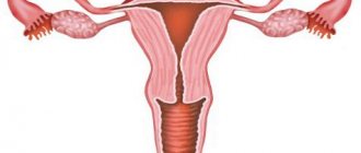
In a healthy person, the uterus is located above the vagina in the pelvic cavity. Anterior to it are the bladder and urethra, posteriorly the rectum. This position helps the organs to be supported by powerful muscles and ligaments of the pelvis. The complex of these structures is otherwise called the pelvic floor. The normal anatomy of the pelvic organs is shown in the diagram below:
When the pelvic floor weakens, the topography of the pelvic organs changes, they move downwards under the force of the earth's extension. The reason for this may be factors related to lifestyle and work activity. Women who have given birth most often get sick. Any reason leading to an increase in intra-abdominal pressure leads to weakening of the muscles and ligaments of the pelvis. This could be a simple cough that lasts for a long time, or sneezing.
Weakening of the muscles and ligaments of the uterus leads to its descent. She begins to pass through the vaginal canal, “crushing” the vagina in front of her. Depending on the anatomical level of the uterus, there are several degrees of prolapse, the last of which is prolapse. Schematically, uterine prolapse looks like this:
Structure of the uterus
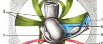
The uterus is a muscular, hollow organ shaped like a pear, slightly flattened from front to back. The upper largest part is called the body; downwards it narrows into the cervix. It has two parts: the supravaginal and the vaginal, which protrudes into the vagina itself. Surprisingly, the fundus of the uterus is the uppermost part of its body, located above the openings of the fallopian tubes (located in the corners on the sides). The cervical canal begins below the body, its inner part is called the internal uterine os, which gradually narrows and opens into the vagina with the external uterine os. In girls and young nulliparous girls, the opening of the pharynx is round, and after childbirth it looks like a transverse slit. The cervical canal is filled with thick mucus, which performs a protective function to maintain the sterility of the uterine cavity. During sexual intercourse, this mucous plug partially comes out, facilitating the penetration of sperm inside. Inside, the lumen of the canal is lined with a single-layer cylindrical epithelium; closer to the external uterine pharynx, it is replaced by a multilayered squamous epithelium. Interestingly, quite often in girls under 20 years of age, the cylindrical epithelium can move to the vaginal part of the cervix (which should not happen), creating a picture of erosion. This condition is called false erosion, which does not require any treatment.
The uterine wall is formed by three layers: the mucous layer (endometrium) lining the inside of the wall, the muscular layer (myometrium) and the serosa (perimetry) covering the outside of the organ. The thickness of the endometrium changes with each day of the cycle. In the endometrium itself, two layers are distinguished: the functional layer changes depending on the phase of the cycle and is rejected during menstrual periods; The basal layer of the endometrium is adjacent to the muscular layer of the uterus. The basal layer consists of secretory glands that ensure the restoration of a new functional layer with the beginning of each new menstrual cycle. Simply put, every month during a failed pregnancy, a completely new layer of endometrium is formed. And in the event of conception, this layer grows even more, and the embryo “immerses” in it.
The muscular lining of the uterus is very powerful, these are the strongest muscles in the female body, since they are designed for serious work: first, securely holding the growing fetus inside, and then expelling it out at the time of birth. The uterine muscles consist of three layers: the outer longitudinal, the middle circular (the most powerful) and the inner longitudinal. During pregnancy, muscle cells hypertrophy, in later stages the wall thickness reaches more than three centimeters, and after childbirth the uterus quickly returns to its original size.
The presence of an amazing organ, which serves as the first, most comfortable cradle for every person, makes a woman out of a girl, and a mother out of a woman. Therefore, you need to take special care of your female reproductive health.