Involution of the mammary glands is a natural aging process that is activated after 45 years. During it, the development of organs and tissues is suspended and a reverse process occurs - regression. As a result, the glandular lobules of the breast are replaced by adipose tissue. This process requires periodic monitoring by a specialist, as it can lead to the formation of cysts.
Appearance
Mammary glands are present not only in women, but also in men.
The anatomy of the female mammary gland is initially completely identical to the male anatomy, since the mammary glands are formed in both sexes during prenatal development. Before puberty, the difference is completely unnoticeable, and only during the period of hormonal changes do the size, shape and structure of the female breast change.
Form
The mammary glands in women look like two symmetrical convex hemispheres. The location of the mammary glands in women is at the level of the third to sixth pair of ribs. Just below the center of the roundness is the nipple, surrounded by the areola.
There is also a generally accepted classification of breasts depending on their shape:
- discoid - a small gland with a wide base;
- hemispherical - diameter and height are approximately equal;
- pear-shaped – the height significantly exceeds the base;
- mastoid - similar in parameters to the pyriform, but the gland itself is more strongly lowered, the nipples are located lower and directed downwards.
Size
You cannot take any specific size as the norm, since it develops individually for each woman.
The average is considered to be a girth of 80 to 85 cm. A slight asymmetry, when one gland is slightly larger, can be considered normal.
The size of the mammary glands depends on several factors:
- amount of adipose tissue:
- the size of the gland itself;
- full of milk.
The weight of the mammary gland in a nulliparous girl is on average 200 g; during breastfeeding it can reach 800-900 g. After the end of lactation, the gland decreases in size. Size does not affect the amount of milk and the possibility of lactation.
The hormonal background of the female body affects the size of the mammary gland; its appearance can change depending on the phase of the menstrual cycle and change with age.
Nipples
The areola (a pigmented round area of skin with a diameter of 3-5 cm) is located slightly below the middle of the breast gland, approximately at the level between the fourth and fifth pair of ribs. In its center there is a nipple, which has a flat-cylindrical or cone-shaped shape. The color of the areola and nipple varies from light pink in nulliparous and fair-skinned women to dark brown or brown in parous women or in women with darker skin. In nursing women with a large bust, the diameter of the areola can exceed 10 cm, and the pigmentation of the areolar-nipple area becomes more intense during this period.
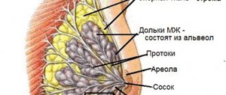
The structure of the nipple: the milk ducts come out and form the nipple, around which the areola is located. Under the skin of the areola are 10 to 15 vestigial areolar glands and a small number of sebaceous and sweat glands. There are small holes on the surface of the nipple, which are the exit of the milk ducts, through which milk flows.
The skin on the nipples and areola is very thin and covered with small folds that resemble wrinkles.
Bundles of smooth muscle cells located on the areola and nipple contract when exposed to cold or touch, causing the nipple to shrink and slightly increase in size.
Treatment
A woman’s body is a rather complex reproductive system that requires careful attention. Any deviations should be detected in time and appropriate measures taken. However, fibro-fatty involutional changes in the mammary glands, discovered after forty years, are a healthy course of the life of the female body. It is important to lead a healthy lifestyle and undergo a routine examination with a doctor on time.
For a young woman’s body, where involution of the mammary gland is considered a pathology, diagnostic actions of doctors are provided in order to select a treatment method. Typically therapeutic measures look like this:
- Taking medications that stabilize hormonal levels.
- Taking painkillers against the background of pain.
The choice of treatment is individual for each woman. If you follow the instructions of medical specialists, then you limit the possibility of losing the main function of the mammary gland - feeding the child.
Breast structure
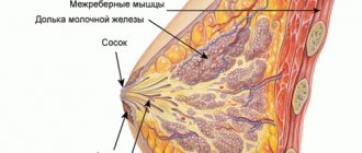
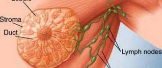
The mammary gland consists of adipose, glandular and connective tissue, and also contains milk ducts and glands, blood vessels and sensory nerve fibers. The mammary glands are attached to the pectoralis major muscle using connective tissue.
The soft tissues of the breast are supported by suspensor “Cooper's ligaments”. These ligaments begin deep in the body and connect to the subcutaneous area of the upper chest. They are not rigid and allow for natural breast movement. Ultimately, this leads to sagging as the Cooper's ligaments weaken over time.
The body of the mammary gland consists of 15-20 lobes, with their apex facing the nipple and separated by layers of connective tissue. The latter also pass between the anterior surface of the gland body and the deep layers of the skin and above the aponeurosis of the pectoral muscle, forming dense connective tissue cords in the form of a mesh, attached to the collarbone. Below, splitting along the entire length, connective tissue strands form a capsule in which the mammary gland is included. If the subcutaneous fat layer is not very well developed, when palpating the gland, granularity is determined. This or that form of the mammary gland largely depends on the strength and elasticity of the capsule.
Every woman feels changes in the mammary glands during the menstrual cycle. Depending on the phase of the cycle, the mammary glands of the same woman can significantly change their structure. In each menstrual cycle, a few days before ovulation, the amount of epithelium of the ducts and lobules begins to increase. In this case, there is an increase in the volume and density of glandular tissue due to blood supply to the organ and swelling. It is this circumstance that explains the feeling of engorgement, thickening, expansion and increased sensitivity. In a normal gland this process is moderately expressed. In preparation for lactation, the mammary glands also enlarge due to the proliferation of glandular tissue. If pregnancy does not occur, atrophy of the newly formed structures occurs over several months. At the end of menstruation, the described phenomena decrease or disappear. Throughout life, tissue proliferation and regression occur in parallel.
The weight of the breast, in which the amount of adipose tissue significantly exceeds the amount of glandular tissue, directly depends on the overall change in body weight. This is explained by the fact that fat in the human body is burned and added evenly throughout the body. The size and weight of breasts in which breast tissue predominates is not affected by changes in body weight.
The size of the mammary glands themselves is greatly influenced by hormones, which explains the change in breast size during menstruation or menopause. Due to the effects of hormones, slight breast enlargement is possible in women who are sexually active.
In addition to hormones, another positive side of sex is the rush of blood into the breast tissue, the development of the capillary system, which leads to breast enlargement.
Based on the above, it is clear that breast size is determined by the size of the mammary gland, adipose tissue, the size of the pectoral muscles, and the development of the circulatory system of the breast. Since women's muscles are not very developed, it is impossible to greatly influence breast size through training, but minor increases can still be achieved. It should be noted that with weak pectoral muscles, they may “sag”. Weak pectoral muscles, as a rule, also determine weakness of the Cooper's ligaments, which leads to even less elasticity of the chest.
Since adipose tissue predominates in the breast, with each new kilogram gained, the weight of the breast will increase by approximately 20 g. If you gain 5–10 kg of “extra” weight, the breast will increase by at least one size.
The mammary glands play a decisive role in the elasticity of the breast; they have a rather dense structure compared to fatty tissues. With exhausting diets, the amount of fat in the breasts decreases, the breasts become smaller and more elastic. With an abundance of nutrition, as the total body weight increases, the amount of fatty tissue in the breast increases, the breasts become enlarged, the firmness decreases, and breast sagging is possible.
The main reason for small breast size is most often a lack of estrogens - female sex hormones that determine the size of the mammary glands.
With age, another factor that affects the shape of the breast appears - this is the stretching and aging of the skin, which plays a fairly important role in maintaining and giving the shape of the breast. Frequent weight gain and loss, and any means for breast enlargement also contribute to stretching of the skin.
Symptoms
Before age 50, the internal structure of the breast changes; after age 50, external changes also occur.
Internal ones usually do not cause disturbances in well-being. Sometimes there is a burning sensation, tingling, nagging unilateral or bilateral pain, muscle pain, and a feeling of discomfort. They most often occur a few days before the end of the cycle in menstruating women.
When hormone levels remain high during menopause, mastodygenia sometimes occurs, manifested by painful sensations of varying intensity, engorgement, and increased sensitivity of the mammary glands. The pain syndrome often imitates osteochondrosis.
Involution proceeds slowly. Changes occur gradually. Initially, the breasts increase slightly, remaining painless. Then its appearance deteriorates - there is a decrease in size, a change in shape, a decrease in tone, and wrinkling of the skin. A woman sees that her breasts have sagged, become flabby, wrinkles have appeared on the skin, the nipple has sank downwards, and the contours of the bust “float”. By old age, the mammary glands turn into flabby, wrinkled sacs.
Sagging occurs due to the characteristics of FGI - fat accumulates first in the lower lobules, the breasts become heavier and sag under their own weight. At the same time, there is a thinning of Cooper's ligaments and stroma - the fibrous frame of the breast. It becomes weak and ceases to perform its functions. There are no muscles in the mammary glands, and they cannot restrain the process.
The disease most often does not manifest itself in any way, but there are also pronounced manifestations that require treatment. Everything is individual.
Complications arising on an involutive background
- Mastodigenia (see above).
- Mastoptosis is sagging breasts. Considered as an aesthetic defect. With a large size and significant ptosis, maceration occurs - in the fold under the breast, the skin loosens, becomes warm, and itching and burning occur.
- Cancerophobia is the fear of cancer as a result of negative experiences or iatrogenic words and actions of medical personnel.
- Depression. It manifests itself as depressed mood, fears, irritability, and sleep disturbances.
Stages and degrees
The process of involutional changes with pathological processes is called fibrous mastopathy.
It occurs in several stages:
Fat involution. The stage of reverse development begins from the end of the first lactation period. It can last until menopause or re-pregnancy. The adipose tissue actively grows, and the mammary gland fades - it becomes lighter, connective soft tissues appear between the fibers. Milky ducts and blood vessels become thinner over time
During this period, it is important to undergo an annual examination with a mammologist to observe endocrine changes. In nulliparous women, a similar process is accompanied by hormonal imbalance.
Fibrocystic mastopathy is manifested by disturbances in the area of hormonal imbalance and developing pathological processes
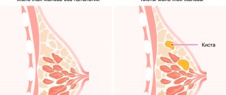
In the tissues of the glandular layer, zones of fibrosis are formed, which are filled with cystic compounds. These are benign capsules that contain cystic fluid. The cyst is detected by palpation. If the secretion becomes inflamed, an infection joins the pathology - pus mixed with blood is released. In severe cases, the glandular tissue decreases and the breast is destroyed from the inside.
Fibrous involution means the process of degeneration of the glandular layer into zones of fibrosis. The cells are replaced by connective and fatty layers. At the age of 45 years, this process is the norm, and for nulliparous girls it is a violation of hormonal and endocrine processes. This pathology is called fibrofatty involution.
The latter type of disease can occur in several stages of development. The stages of tissue replacement are different, depending on the nature of the manifestations and the characteristics of the symptoms.
When a cancerous tumor is detected, it is worth separately highlighting the stages of development of such a process:
Fibrofatty involution degenerates into cancer
| Form/stage of malignancy | Manifestation | Development |
| Nodal form | It occurs in young nulliparous women who have had one or more abortions. Age – from 30 to 45 years. The formation is local and does not affect the lymph nodes. At the first stage, its presence can be studied and established by palpation. Externally, the breast is covered with small dots, the contours of the areola become unclear, the nipple “looks” to the side. Low temperatures may appear in the evenings. | Upon examination, the woman does not have any pain, but there is discomfort due to the immobility of the nodes. During the transition to the second and third stages, the nipple is retracted or fixed without the possibility of maintaining mobility. In some places, lymphedema appears, similar to an orange peel. It may indicate the development of the last stage of the disease. |
| Edema form | This form of cancer occurs in women aged 16 to 68 years. Body temperature is not observed. This is due to the proliferation of adipose and connective tissue. The predominance of connective tissue in the structure can occur in women 25 years of age, as well as among those over 50. Edema and “swollen” forms of the mammary gland indicate a characteristic sign of diffuse cancer. | The tissues of the structure are compacted, heavy, sometimes the chest hurts when touched even at an early stage. An x-ray will reveal a node around which the walls will be significantly thickened. Transverse heaviness and microcalcifications are diagnosed. |
| Erysipelas cancer | This form of cancer is difficult to miss due to changes in the skin. It is covered with erosion points that look like craters. Skin hyperemia, damage to the nipples and areola are established and diagnosed. | Cancer of this form is severe and painful, body temperature rises to 39 degrees, and health worsens. The course of the disease does not occur without metastasis to the lymph nodes and neighboring organs. Metastases can also form in neighboring lymph nodes with the risk of affecting individual organs. |
| Hidden cancer | The most complex and dangerous form. Temperatures can range from 36 to 39 degrees. The neoplasm cells initially affect the lymph nodes, only then they spread to the tissues. When nodes become infected, the disease develops within 1-2 weeks until the final stage. | As a rule, the lymph nodes located above the chest are the first to be affected, then the nodes of the cervical spine, and only at the end - the nodes of the mammary gland. This can be noticed when the lymph nodes are enlarged and the cells mutate into metastases - diseased areas that cannot be diagnosed. Education cannot be determined by clinical methods. |
Prevention of involution
The premature manifestation of involution can be prevented and its course can be alleviated using preventive methods. First of all, a woman needs to undergo regular preventive examinations, even if there are no complaints, so as not to miss the onset of pathology. Preventive examinations are especially necessary for older women.
Every woman should have a mammogram over the age of 45-50 years. If a woman has just finished feeding her baby, she needs to monitor the condition of her breasts and go to the doctor. During the incomplete lactation period, no treatment is required. It is started only after finishing feeding for special indications.
In order not to have health problems, you need to eat right, walk regularly, lead an active lifestyle, and give up bad habits.
Involutional changes in the breast are a natural process of aging of the mammary gland, to which all women are susceptible when they reach the age of over 45–50 years, as well as older ladies. To avoid complications, you need to have an idea of what this process is, when it occurs, what features it has, and in what cases the alarm should be sounded.
Diseases of the glandular tissue of the mammary gland
Pathological changes in the layers occur with such ailments as:
- Endometriosis
- Uterine fibroids
- Polycystic ovary syndrome
- Tumors of the uterus or ovaries
- Endocrine disorders
Blast hyperplasia develops with regular stress, breast trauma, and prolonged lactation. The most common pathology is the proliferation of glandular tissue of the mammary gland. Often the mammary glands undergo mastopathy. Cysts, tumors, benign and malignant formations can develop inside the mammary gland.
Hyperplasia of glandular tissue
The pathology is characterized by the proliferation of thoracic structures and imbalance of hormones. The onset of the disease is caused by genetic predisposition and a number of negative factors. The disease can develop after trauma to the mammary gland, nervous exhaustion, stress, or taking hormonal medications. Sometimes pathology is differentiated when a person works with hazardous chemicals. The thickness of dense tissue can increase due to hormonal age-related changes in the body.
Main signs of the disease:
- Breast pain
- Presence of nodes in glandular tissue
- Breast swelling
- Secretion from the nipple
The diffuse form of hyperplasia occurs without pronounced symptoms, so detecting the disease is extremely difficult. The disease is usually diagnosed during a routine examination of a woman. Glandular hyperplasia can affect boys and men, but the incidence rate among males is significantly reduced.
In pathology, neoplasms are sometimes diagnosed in the form of nodes of varying mobility and size. There is active growth and thickening of the tissue. Pain with hyperplasia can moderately radiate to the armpit or shoulder. There are menstrual irregularities and changes in the structure of the gland.
In medicine, hyperplasia is divided into several types:
- Dishormonal – occurs against the background of hormonal imbalance.
- Epithelial – accompanied by damage to the epithelium.
- Nodular – nodes are present.
- Diffuse – characterized by the presence of granular small tumors.
- Fibrous - dense affected areas of tissue are fused with surrounding tissues.
- Cystic - multiple and single cysts are diagnosed.
- Ductal - tissue growth is observed in the milk ducts.
- Stromal – a benign fibromuscular tumor is present.
- Atypical - proliferation is observed in glandular, as well as adipose and epithelial tissues. Diagnosed as a precancerous condition.
Glandular mastopathy
The disease manifests itself as severe pain inside the mammary gland. Discomfort and heaviness appear in the chest. The pain sometimes radiates to the armpit. There is discharge of secretion or pus from the nipple. Mastopathy is also characterized by the proliferation of glandular layers. Photos show what changes in glandular tissue look like. To the touch, you can detect a lump of various sizes in the thickness of the chest.
In medicine, the following forms of the disease are distinguished:
- Glandular-fibrous – benign seals are diagnosed
- Glandular-cystic – cysts are present in the thickness of the breast
- Fibrous-adenomatous – the tumor is characterized by the proliferation of organic structures
With glandular mastopathy, formations are palpated that are densely localized in the lobules of the mammary gland. Their predominance, as well as increased breast tenderness, indicate a serious pathology. These are benign cystic neoplasms or small tumors. With the adenomatous form, characteristic tuberous formations are found deep in the thickness of the chest. The disease usually does not affect the lymph nodes.
In the diffuse form of mastopathy, the seals are small in size and distributed over the surface of the entire gland. Cystic mastopathy is a pathology with the formation of small capsules filled with liquid. As the cysts grow, they compress the nerve endings and cause pain in the chest. There is also a mixed form of the disease, in which cysts may predominate.
Diagnostics
Diagnosis of involutional manifestations must be confirmed by laboratory and instrumental methods.
- External examination is palpation and assessment of the shape and color of the mammary glands.
- An X-ray examination using contrast agents will allow you to assess the condition of the milk ducts.
- Ultrasound and mammography will allow you to assess the condition of the mammary gland tissues and identify the focus of the pathological process.
- An additional diagnostic method is MRI and CT, which will allow you to accurately determine the source of pathology.
- Blood test to check hormone levels in the body.
In the early stages of involution, it is almost impossible to make a diagnosis, especially if you try to do it yourself. As a rule, it is discovered during a preventive examination, which takes place annually at the workplace. When a professional mammogram or ultrasound examination is performed, a decrease in the amount of iron tissue in the lower part of the breast can be easily detected.
Nowadays, mammography is the most effective and informative method for detecting breast pathology. The efficiency of the technology helps to detect everything with a probability of about 97%. Ultrasound examination also helps to make a diagnosis, but in the initial stages it does not recognize altered areas that are less than 1 cm in size.
When a woman reaches 40 years of age, she should undergo a mandatory breast examination once a year. When she reaches 50 years of age, the examination should be done 2 times a year. This should be done not only to monitor the progress of involutional changes, but also to monitor other pathological conditions and diseases that may occur against the background of this process.
· inspection and palpation;
· Ultrasound examination;
X-ray examination;
· biopsy;
· study of hormonal status.
By examination and palpation, it is possible to determine a decrease in the elasticity and firmness of the mammary glands, their sagging, and the presence of fine-grained lumps in the thickness of the breast. Special instrumental and laboratory methods allow for more accurate diagnosis.
· 20-40 years – the normal ratio of glandular and connective tissue components is determined, milk lobules have normal sizes;
· 41-50 years – early signs of involution are detected in the form of fibrous mastopathy (nodules and cords in the form of hyperechoic formations of different sizes and shapes, hypoechoic areas of adipose tissue);
· 51-55 years – milk lobules are significantly reduced, fibrous and fatty changes are expressed;
· over 55 years – glandular tissue is practically absent or there are residual areas, the structure of the glands is almost uniform, there are no obvious focal compactions.
The main radiological method for determining fibrofatty degeneration is mammography. If there is no indication, mammography is performed on all women annually, starting at age 40. If there are symptoms of any pathological changes in the breast, then a study is prescribed regardless of the woman’s age.
The mammary glands are placed between two holders and, as it were, spread out to give them the optimal thickness for examination. Then a photo is taken. An involution mammogram reveals diffuse darkening of the tissue. Individual lobules, cords and small nodules of fibrous tissue and a vascular pattern are clearly visible.
Mammography is a more diagnostically informative method compared to ultrasound, but it cannot be prescribed to pregnant women, while ultrasound has virtually no contraindications. Ultrasound scanning should be preferred in young patients, especially under 35 years of age. The structure of their mammary glands is most optimally suited for studying with ultrasound, and not with X-rays.
Among laboratory methods, enzyme-linked immunosorbent or immunochemical studies of hormonal status are most often performed. The level of the most significant hormones is determined: progesterone and estradiol, follicle-stimulating, luteinizing and lactotropic hormones. If indicated, the content of thyroxine and triiodothyronine (thyroid hormones), thyrotropin and some others are examined.
The early stages of fibrofatty involution of the mammary glands cannot be detected during self-examination. Most often, it is detected during an annual preventive examination during a mammogram or ultrasound examination.
At the moment, mammography is the most informative method for detecting breast pathologies. The effectiveness of the method is 97 percent.
Ultrasound also makes it possible to detect involution of glandular tissue, but its capabilities do not allow recognizing the process in the initial stages when the size of the changed areas is less than 1 cm.
When a woman reaches 40 years of age, a preventive breast examination must be performed once a year, after reaching 50 years of age - 2 times a year. Such measures are necessary not only for monitoring involutive changes, but also for the prevention of other pathological conditions of the breast.
What is it and what causes it?
Glandular mastopathy of the mammary gland is a benign formation. Despite the fact that mastopathy is not on the list of malignant breast tumors, it can transform into cancer.
This occurs under the following conditions:
- the glands are regularly exposed to electromagnetic waves (for example, X-ray radiation);
- the patient’s living and working conditions are unfavorable;
- constant stress;
- with decreased immunity;
- when carcinogens accumulate in the body.
The occurrence and development of the disease is associated with changes in hormonal levels. Characterized by hyperplasia of glandular tissue cells. Glandular tissue is the main structure of the paired organ.
After the process of cell division has begun, the glandular tissue of the mammary gland grows. Then small nodules and compactions form.
Adenosis is often diagnosed in teenage girls towards the end of puberty. The appearance of a neoplasm can also be caused by pregnancy and lactation. In these cases, the disease often goes away on its own - as soon as hormone production returns to normal. The main risk group is women between 30 and 40 years old. In this age category, the incidence ranges from 30 to 70%.
The main cause of the disease is an imbalance in hormone production. If the hormone progesterone is produced in insufficient quantities, neoplasms may develop. If estrogen is produced in excess, the risk doubles. The following factors lead to hormonal imbalance:
- Obesity. Fat cells take part in the synthesis of estrogens. The more fat, the higher the production of these hormones.
- Termination of pregnancy artificially or naturally. Abortion is stress; it causes hormonal changes, against the background of which adenosis develops.
- Pregnancy in adulthood (after 35 years). At this age, the activity of the ovaries fades away, and pregnancy reactivates them. The result is hormonal imbalance.
- Lack of milk production after pregnancy. This violation indicates a decrease in the amount of progesterone and an excess of estrogen.
- Refusal of breastfeeding. Milk stagnates in the ducts, clogs them and dilates them. As a result, the structure changes and cysts form.
- Taking hormonal drugs without medical supervision (especially contraceptives). Incorrect selection of medications or their incorrect use.
Also, as a concomitant disease, mastopathy develops with diseases of the uterus and ovaries. The reasons for the development may lie in problems with blood pressure, stomach and pancreas. Chronic stress, smoking and alcohol abuse, sexual dysfunction, and routine violations leave their mark on health.
Risk group
Premature onset of involution is not such a common occurrence, but there are still certain risk groups that are most susceptible to such changes. These include women:
- Those who have terminated their pregnancy prematurely, as during an abortion there is a sharp hormonal surge;
- Have had an abortion more than once;
- Smoking;
- With pathology of the thyroid gland;
- nulliparous;
- Those who have suffered a mammary gland injury, regardless of its duration;
- Having an irregular sex life;
- Sunbathing without a swimsuit, both in the open sun and in a solarium;
- With pathology of the pancreas;
- For diseases of the ovaries, especially in the chronic stage;
- Aged 40 years or more.
All this requires regular mammography, as well as visits to specialists. Self-examination usually does not help identify any problems until it is too late. It is worth understanding that involution is often accompanied by other, more serious problems, such as mastopathy, etc., therefore, its progress should be monitored with professional doctors.
Quite often, involution occurs before its physiological deadlines, and in this case, doctors say that the following women are most often at risk:
- If you have had abortions repeatedly, mechanical injuries and hormonal imbalances lead to pathological processes.
- Smoking and alcohol abusers.
- If pathological changes in the thyroid gland are diagnosed.
- Previously nulliparous and having irregular sex life.
- If a woman has suffered a chest injury, she often visits the beach or solarium, where she sunbathes without a bra.
- For pathologies of the pancreas or ovaries, especially in the chronic stage.
- Age 40 and older.
All this obliges a woman to undergo regular examinations and diagnostics by a doctor.
Reasons for appearance
Fat involution is considered normal for women who go through all stages of maturation in their lives. Fibrous involution depends on the criteria through which the “life” of the mammary gland passes. These criteria include abortion, pregnancy, lactation and menopause. At these moments, the glandular tissue changes depending on the operating factors.
Sometimes they do not represent the norm, but a violation accompanied by a number of aspects:
- A decrease in the level of sex hormones - during menopause, this moment is considered normal. In other cases, it is a pathology caused by disorders of the endocrine and genitourinary systems. Their precursors can be STDs, infections inside the urinary system, diseases of the pelvic organs (polycystic disease, cystitis, etc.).
- Poor nutrition affects the functioning of individual organs of the endocrine system. Subsequently, the hormones produced by these organs affect those human systems that are responsible for premature aging and withering of the reproductive system. For example, an increase in subcutaneous fat as a result of obesity can lead to the replacement of fibrous tissue with adipose tissue.
- Infections detected during the lactation period. Often women experience stagnation of milk and blockage of the mammary glands. This all affects hormonal levels, the elasticity of the breast skin and changes that occur during the post-lactation period. The glands lose volume, become thinner, and the infection spreads to neighboring tissues.
- The phases of the menstrual cycle after and before childbirth also affect the involution of the mammary gland. If before the start of the cycle a woman experiences pain and lumps in the breasts, the culprit is the hormone estrogen. After menstruation stops, progesterone is produced by other organs that are responsible for preparing the uterus for a subsequent possible pregnancy. If processes in these phases are disrupted, changes in the area of connective tissue proliferation can be diagnosed.
Fibrofatty involution may be observed every month after bleeding has stopped. The period is short and is considered the norm for women in labor and those who do not yet have children.
Restoring breast size and shape
It happens that, despite all efforts, a woman’s breasts lose their shape and sag. Modern medicine is no longer powerless against this problem, which can be solved surgically. Augmentation mammoplasty is combined with breast lift.
There are different types of breast surgeries. To obtain the best cosmetic result, the choice of plastic surgery must be approached taking into account individual characteristics.
Plastic surgery can be performed with or without endoprostheses. The choice of method depends on the degree of preservation of glandular tissue and the degree of breast ptosis. Despite all the achievements in the field of plastic surgery, the current state of surgical correction of the mammary gland cannot be considered complete.
How to remove fat from breasts
Women are more prone to breast enlargement than men, but the methods for eliminating the pathology are similar in both sexes. If the volume of the bust has increased, it is necessary to exclude the possibility of hormonal imbalance - visit an endocrinologist and undergo the necessary tests. After treatment, the mammary glands can take on their natural shape.
If your health is good, excess weight may be the cause of breast enlargement.
In this case, you should reduce the calorie intake, increase physical activity, and evaluate the effect of medications and cosmetics taken on the body. It’s not just hormonal medications that can cause weight gain.
In special cases, they resort to surgical removal of fat - liposuction. During the operation, 3–4 mm incisions are made on the rib near the mammary gland, through which excess fat is pumped out using special instruments. Depending on the size of the operated area, plastic surgery is performed under local or general anesthesia.
Ways to reduce chest girth in men
Men are naturally given a wide chest, so even a slight increase in the bust becomes noticeable. If the cause of the pathology is a disease, it can only be eliminated by treating the underlying disease or by surgery.
When breast growth in a man is due to excess weight, intensive training in the gym will help. Exercises with weights and barbells are effective. Regular training helps strengthen all muscle groups and reduce fat, including in the ribs.
Strength exercises for breast reduction lead to a significant loss of energy reserves in the body. To restore balance, a man is recommended to eat a protein diet. Exercising and eating a balanced diet will not give immediate results. The older a man is, the more difficult it is for him to lose weight.
How to lose weight in a woman's chest
If a healthy woman's weight increases by 1 kg, on average her breasts become 20 g heavier. And vice versa - the mammary gland decreases in proportion to the decrease in body weight. Simple calculations show that if you lose 10 kg, your breasts will lose 200 g of weight.
A sharp decrease in the fat layer often leads to another problem - the mammary gland loses its shape, stretches, sag, and breast asymmetry occurs. This is especially noticeable if the body experiences age-related changes. When losing weight, you should take care of the elasticity of the mammary gland and prevent sagging skin.
Regular breast massage gives tangible results. Light stroking, pinching, patting, squeezing increase blood circulation in the mammary gland area and prevent congestion. At home, using a warm shower is easy to perform hydromassage. A contrast shower has a good effect on the shape of the bust - it effectively tones and strengthens blood vessels.
Women use cosmetics and medications more often than men. This creates an additional risk of weight gain. Even vaginal cream can provoke breast growth if it contains hormones. It is necessary, if possible, to exclude those medications that can lead to obesity.
If you have a large bust, you need to choose only high-quality underwear - it supports the breasts and does not allow them to sag under their own weight. You need to wear compression garments while doing fitness or any other sport.
Invasive form
Depending on the symptoms, oncologists distinguish several types of invasive breast cancer.
- Infiltrative-edematous .
With this type of disease, the mammary gland increases in size, swells, the skin turns red, and an “orange peel” appears on it. The pain is moderate. - Mastitis-like.
Accompanied by elevated temperature, there are signs of inflammation of the mammary gland. - Erysipelas.
Invasive cancer spreads to the lymphatic and blood vessels and skin. The chest shrinks, becomes wrinkled, and looks like a shell covering. - Preinvasive ductal carcinoma.
At this stage of the disease, the tumor does not spread beyond the milk ducts. But if treatment is not started in time, invasion develops - germination into surrounding tissues, which is accompanied by pain and heaviness in the chest.
10 more proven methods
Let’s not forget to consider other methods of tightening the problem area, which have shown high effectiveness:
- Strength exercises. They will strengthen the pectoral muscles and make the bust firmer and lifted.
- Gymnastics. An excellent opportunity for physical activity and aesthetic pleasure. It prevents bust sagging.
- Special creams. An important component of the bust care complex. Actively fight skin sagging.
- Masks. Masks with natural ingredients are the basis for tightened and beautiful breast skin.
- Wraps. They have the ability to exfoliate, moisturize, renew skin cells and increase its elasticity.
- Massage. Improves blood circulation, removes sagging skin and tones.
- Diet and food. A balanced diet is the basis for youthful and beautiful skin. Controls weight and helps you stay slim.
- Pull-up bra. Makes the silhouette slender and fit, lifts the bust and prevents it from sagging.
- Special stickers. A fashionable part of women's clothing that allows you to wear clothes with an open back and a large neckline.
- Threads and mesothreads. A breast lift using threads makes it firmer and prevents it from sagging.
It is a mistake to think that only by eating certain foods you can increase this part of the body in size. The breast consists of glandular, fatty and connective tissue, which is located on a cushion of pectoral muscles. By pumping them up through intense physical exercise, you can make your bust rise and become taller. Fat is most often not deposited locally in one place, for example, in the bust area. It is also distributed evenly in other roundness of the female body, for example, buttocks and thighs. By adding breast size, you are likely to increase other parts of your body as well.