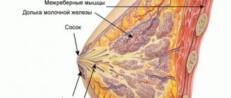
Breast ductal ectasia is a condition that is diagnosed in women aged 37 years and older. It occurs for several reasons, in most cases it is due to natural hormonal changes in the patient’s body, which are associated with menopause and a decrease in estrogen levels in the blood. The condition is not considered pathological and in some cases there is no treatment. But the cause of ectasia can be not only an imbalance of hormones, but also a tumor of both benign and malignant origin.
Total information
The described condition as an independent disorder is detected extremely rarely - in fact, it is a complication of a number of pathological processes, which is their unique marker.
The normal lumen of the milk ducts ranges from 1.5 mm at the narrowest part to 2 mm at the widest. It is considered that ectasia of the milk ducts has occurred if the lumen of the ducts increases to at least 3-5 mm or more .
note
The described pathology is most often diagnosed in women over 40 years of age. This fact indicates that age-related changes that occur in a woman’s body are of greatest importance in the development of lacteal duct ectasia.
Breast duct ectasia itself is not a critical condition. But it often occurs against the background of diseases of the mammary glands, the prognosis for which is serious or questionable.
Causes
Ectasia of the mammary gland ducts can be equally provoked by both physiological and pathological factors.
Normally, moderate ectasia develops under conditions such as:
- second phase of the ovulatory cycle;
- pregnancy;
- lactation.
In the first two cases, ectasia means normal physiological preparation of the mammary glands for lactation. During lactation itself, the milk ducts are able to expand most often when:
- large volumes of produced mother's milk;
- lactostasis - stagnation of milk in the mammary glands, which indicates disturbances in the evacuation of milk from the glands.
If ductectasia of the mammary gland is observed during the development of involutive processes (reverse development against the background of withering and aging), then it can be regarded as both physiological and pathological - this depends on the degree of its manifestations. Normally, this is a slight dilation of the mammary gland ducts, which develops against the background of:
- hormonal changes in mammary tissue;
- their structural changes.
Pathological expansion of the milk ducts often develops against the background of diseases and pathological conditions such as:
- dyshormonal disorders;
- hyperprolactinemia. This is an increase in the production of prolactin, a hormone whose function is to stimulate the development of breast tissue;
- secerating mammary gland - this is the name of the gland from the nipple of which pathological secretion is constantly secreted;
- inflammatory processes;
- deforming transformations.
Dyshormonal conditions most often lead to the development of ectasia of the subareolar canals - those that are located under the areola of the mammary gland. This happens when there is a violation of the ratio between the amount of estrogens and gestagens. The reasons for this condition are:
- pathologies of female genital organs;
- disorders of the hypothalamic-pituitary system, a complex of brain structures that plays a regulatory role.
Of all the pathologies of the female genital organs, the most common cause of a dyshormonal state that leads to ductal ectasia of the mammary gland is:
- oophoritis - inflammatory lesion of the ovaries;
- adnexitis - an inflammatory process in the uterine appendages (ovaries and tubes);
- neoplasms are most often voluminous. They can be either benign or malignant;
- Endometriosis is a disease in which endometrial cells (the inner layer of the uterus) appear in extragenital areas.
Hyperprolactinemia plays the following role in the occurrence of ductal ectasia of the mammary gland. Since prolactin is produced in greater quantities, it further stimulates the breast tissue, and this provokes a constant secretion of milk, which puts intense pressure on the milk ducts from the inside and thus contributes to their expansion. Hyperprolactinemia can be caused by diseases and pathological conditions such as:
tumors of the pituitary gland - an endocrine gland that is located at the base of the brain and affects growth, development and metabolism in the body;- neoplasms of the hypothalamus - a small area in the diencephalon, which consists of groups of cells that regulate neuroendocrine activity of the brain and homeostasis (stable state) of the body;
- hypothyroidism – lack of production of thyroid hormones;
- chronic renal failure - severe impairment of kidney function;
- liver cirrhosis – replacement of its parenchyma with connective tissue;
- hyperestrogenism - an increase in the production of female sex hormones estrogen.
With a secerated mammary gland, the milk ducts begin to expand due to the fact that pathological secretions enter their lumen, which puts pressure on the walls of the ducts. Most often, this pathological process develops in diseases and pathological conditions such as:
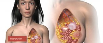
- secretory mastopathy - a change in the tissues of the mammary gland, which develops against the background of hormonal imbalance and is accompanied by the release of pathological secretion from the nipples;
- intraductal papillomatosis of the mammary glands - the formation of small short growths of tissue in the ducts;
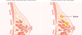
- malignant tumors of the mammary glands. Most often these are intraductal cancer (a cancerous tumor develops inside the milk ducts) and Paget's disease (a cancerous lesion of the nipple and areola).
Inflammatory processes in the mammary glands lead to changes in the normal structure of their tissues, which entails a change in the structure of the milk ducts. The following components of the inflammatory process play a role in the development of ductal ectasia:
- swelling of tissues during an acute process;
- proliferation of connective tissue during a chronic process.
Ectasia of the mammary gland ducts against the background of its deformation develops practically according to the same “scenario” as ectasia against the background of inflammatory processes. This deformation most often occurs against the background of such pathological conditions as:
- traumatic injury to the mammary gland suffered in the recent past;
- medical manipulations - diagnostic (puncture) and therapeutic (surgical intervention for a particular disease of the mammary gland);
- tissue germination by neoplasms – benign and malignant.
Ectasia - what is it
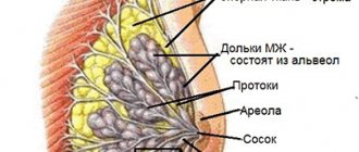
Mammary duct ectasia is a change in the mammary glands associated with dilation of the ducts that cannot be explained by lactation or breastfeeding.
Normally, milk is constantly being produced in the female breast, but the amount is so small that it is absorbed quite quickly without being released from the nipples. With ectasia of the mammary glands, there is a pathological expansion of the passage through which milk is excreted, which leads to its accumulation and, as a consequence, subsequent release.
The pathology, as doctors note, does not pose a danger if it is not associated with the presence of a malignant neoplasm in the mammary gland.
Development of the disease
The process of formation of milk duct ectasia consists of several parts, the development of which can be provoked by various factors. The following changes are observed:
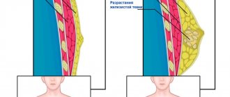
with dishormonal disorders, the layer of epithelial cells that line the milk ducts hypertrophies (grows and thickens, blocking the lumen of the ducts - therefore, in order to “push through” the fluid, they expand compensatoryly;
- the entry of physiological (milk) or pathological (excretion) secretion into the lumen of the duct also provokes its compensatory dilatation (expansion). With increased viscosity of the intraductal secretion or the formation of clots in it, the outflow worsens even more, the milk ducts become blocked. Because of this, the pressure in the terminal sections of the milky passages increases even more, and their subsequent persistent expansion develops.
The described changes can occur with local (local) tissue swelling, which develops in such pathological conditions as:
- acute inflammation;
- adhesive process;
- formation of scar tissue after traumatic injuries to the mammary gland;
- invasion of the gland by tumors.
In addition to the processes described, in women during menopause, the development of ectasia of the milk ducts is played by the fact that the breast tissue is stretched due to its sagging, which, in turn, develops due to a decrease in elasticity, characteristic of involutive processes.
Diagnosis of the disease
The further success of treatment of the pathology depends on how quickly diagnostic measures are carried out. It is much easier to get rid of ectasia at the beginning of its development, without allowing the disease to worsen, so you should not start the pathological process, hoping that the problem will solve itself.
If a woman suspects she has ductectasia, she should contact specialized specialists:
- Gynecologist.
- Mammologist.
To confirm the diagnosis, you will need to undergo laboratory and hardware diagnostics.
Laboratory examination of the body involves the following tests:
- Blood test for prolactin or other hormones.
- Analysis of nipple discharge (smear examination allows you to determine the presence of inflammation or tumor).
- A biopsy is prescribed if lumps of suspicious origin are detected.
In order to accurately diagnose the present disease, an instrumental examination is prescribed:
| Hardware examination | What allows you to find out |
| Mammography | Evaluates the condition of the milk ducts. Determines the presence of tumors, polyps and papillomas. |
| Breast ultrasound | The results of mammography are being clarified. |
| Ductography (X-ray) | It is performed using a special contrast agent, which is injected into the milk canals. It is considered one of the informative methods of mammography. |
If the echo signs identified by ultrasound are not enough to make a final diagnosis, and ectasia has a complex course, the following is prescribed:
- Establishment of tumor marker CA 15-3.
- Radioisotope scintigraphy of the breast.
- Computed tomography of the chest.
- Magnetic resonance imaging.
Additional instrumental studies make it possible to differentiate the disease from other serious diseases:
- Paget's disease.
- Malignant formations.
- Subareolar abscess.
- Galactophorite.
In addition, the woman will need to consult an endocrinologist, surgeon and oncologist.
If the diagnostic results confirm the presence of the disease in both mammary glands, then in this case a diagnosis of bilateral ectasia is established.
Symptoms
Clinical signs of breast duct ectasia often develop gradually and often appear only with significant progression of the described disease. The main signs of the described disease are:
- itching or burning in the areola area;
- soreness there;
- tissue compaction;
- changes in the nipple;
- discharge;
- skin irritation.
Characteristics of pain:
- by localization - in the area of the affected milk ducts;
- in terms of distribution – irradiation as such is not observed;
- by nature – oppressive;
- in terms of intensity - moderate, with the progression of pathology - increasing;
- by occurrence - they mainly appear with a significant expansion of the milk ducts.
Often there is no pain, the patient complains of discomfort.
note
With ductal ectasia of the mammary gland, tissue often becomes denser in the subareolar area.
Changes in the nipple appear as:
- offsets;
- retractions;
- deformation.
Characteristics of discharge:
- in quantity - scant or in moderate quantities;
- by frequency – regular;
- by nature - serous or sanguineous;
- by color - yellowish or whitish;
- The consistency is liquid.
Skin irritation occurs due to contact with the described pathological discharge from the nipple. It manifests itself in the form of skin changes such as:
- swelling;
- redness;
- maceration - wet corrosion.
note
If ectasia of the milk ducts occurs due to dyshormonal disorders, dilation of the ducts is observed in both mammary glands.
The ducts are affected in only one mammary gland, most often in such pathological conditions as:
- inflammatory processes;
- consequences of traumatization;
- volumetric processes (formation of tumors).
Symptoms of the disease
Breast ectasia is characterized by severe symptoms; the most common signs of the disease are:
- Unpleasant discomfort in the breast.
- Itching and burning in the nipples.
- Pain in the chest area.
- Swelling of the nipples.
- Redness of the nipple tissues.
- Nipple displacement.
- Retraction of the nipple into the inner part of the chest.
- On certain days of the menstrual cycle, one or both breasts increase in size and become firm.
- Discharge from the nipples of unknown origin.
- When palpating the breast, moderate compactions are felt in the area of the areola.
The release of foreign fluid from the nipples is the main sign of the development of breast ectasia, and the watery substance may have a different color. A particularly alarming symptom is discharge interspersed with blood fluid, which indicates the development of a benign or malignant tumor.
Diagnostics
The first task of the diagnostic process is to determine whether breast ductal ectasia is physiological or pathological. Hasty conclusions should be avoided, since sometimes physiological ectasia can develop into pathological one. The main thing in the diagnostic process is to identify not only the pathology being described, but the reasons that provoked its appearance. To do this, it is necessary to use all available information - the patient’s complaints (if any), age characteristics and features of the obstetric and gynecological history, the results of additional examination methods (physical, instrumental, laboratory).
A physical examination of the breast takes into account:
- upon examination - size, symmetry, visible changes (or lack thereof) of the mammary glands, skin changes, presence of discharge;
- upon palpation (palpation) - consistency of breast tissue, soreness, presence or absence of lumps.
Instrumental research methods used in the diagnosis of ductal ectasia of the mammary gland are as follows:
- mammography – a set of methods for examining the breast;
- ductography is an x-ray examination of the mammary gland ducts with the preliminary introduction of a contrast agent into them. Allows you to evaluate the milk duct system, identify areas of dilatation (expansion) and analyze its severity. The method is valuable in identifying intraductal (intraductal) neoplasms;
- radioisotope scintigraphy of the mammary gland - the patient is intravenously injected with pharmaceuticals containing radioisotopes; when examining the mammary gland with a special tomograph, they create a color image. It is used to determine changes in the mammary gland - in particular, areas of dilated milk ducts;
- biopsy - removal of breast tissue for subsequent examination under a microscope.
Mammography includes examination methods such as:
- X-ray mammography is a survey radiography of the mammary glands in two or three projections;
- ultrasound mammography - using ultrasound, areas of dilatation (expansion) are identified in the thickness of the mammary gland - this is a sign of ectasia of the milk ducts;
- tomosynthesis - using a two-dimensional image of the mammary gland, which is created during this method, the structure of the gland tissue is assessed;
- magnetic resonance (MRI) mammography – a method of tomographic examination of the mammary gland;
- optical mammography – breast tissue is studied using optical equipment.
Laboratory methods used in the diagnosis of the described disease are as follows:
determination of the content of estradiol, progesterone, follicle-stimulating hormone (FSH), luteotropic hormone (LH). If necessary, a study of thyroid hormones is carried out;
- histological examination - a biopsy specimen is examined under a microscope to exclude cancer;
- cytological examination - smears of nipple discharge are examined under a microscope to identify atypical cells;
- determination of tumor markers - specific compounds that appear during the development of a malignant neoplasm.
Since ductal ectasia of the mammary gland in the majority of cases develops as a complication of other diseases, consultations with related specialists - a gynecologist, endocrinologist, oncologist, dermatologist, and surgeon - may be necessary.
Diagnostics and its principles
Diagnosis of ectasia or dilation of the mammary gland ducts is carried out in several stages. The following examinations may be prescribed to the patient:
- X-ray of the chest area, a study is carried out if mammography or ultrasound is impossible for one reason or another. X-rays may also be part of a comprehensive examination.
- Collecting anamnesis - making a diagnosis begins with interviewing the patient and collecting complaints. On the basis of which the doctor draws up a clinical picture and makes a presumptive diagnosis for the patient. This also includes a medical examination and palpation of the breast.
- Ultrasound of the ducts is a fairly accurate study; it helps to determine the presence of not only pathological changes in the breast area, but also tumor formations.
- Mammography is an examination that is most often performed on women over 40. It helps to diagnose pathological changes using ionizing radiation.
- Biopsy - helps determine the nature of the tumor. The tissue is collected using a needle and then sent for further examination. Carried out if there are suspicions about the nature of the changes.
- Collecting secretions for histology - by examining the secretion under a microscope, a specialist can detect cancer cells in it and suspect the patient has cancer. The analysis helps determine the nature of the tumor and diagnose cancer at an early stage of development.
You will also have to donate blood for hormones. The analysis is carried out in several stages, if necessary. Hormones are gynecological and endocrinological. For this reason, the doctor often advises you to additionally consult a gynecologist and endocrinologist, which will help identify the cause of the disease.
Complications
Physiological ectasia of the mammary gland ducts does not pose any risk to the health and life of a woman. But in some cases it may be accompanied by complications such as:
- galactophorite;
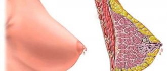
- deformation of the mammary gland - can develop with a significant expansion of the milk ducts.
With the development of the pathological form of the described pathology, complications such as:
- dermatitis – inflammatory lesion of the skin (most often in the alveolar zone);
- dermatosis is a non-inflammatory lesion (most often in the alveolar zone).
Both of these complications develop with ectasia of the lacteal ducts in patients with secerated mammary glands - their development is provoked by the constant secretion of secretions from the dilated ducts.
It is also possible to develop another pathological condition, which is not a complication of lacteal duct ectasia, but develops against its background - this is cancerophobia (fear of getting cancer).
Treatment of milk duct ectasia
If breast duct ectasia has formed due to physiological reasons, treatment is not required. However, regular monitoring by a mammologist is indicated.
If the dilation of the mammary ducts is determined to be pathological, treatment is based on the elimination of the underlying disease that led to the appearance of the described pathology. Treatment methods can be conservative and surgical.
Conservative therapy is based on the following:
- for dishormonal disorders - hormonal drugs;
- for inflammatory processes - antibacterial drugs taking into account the sensitivity of the pathogen and anti-inflammatory drugs (non-steroidal anti-inflammatory drugs, or NSAIDs, are often used);
- for malignant neoplasms - cytostatics.
The following purposes are common:
- immunocorrectors;
- sedatives;
- vitamins and minerals (often in the form of complexes).
If ductal ectasia of the mammary gland occurs due to hormonal disorders, patients are recommended to:
- reduce weight;
- adjust your diet - in this case, the consumption of fatty foods and simple carbohydrates should be reduced (buns, pastries, cakes and other sweet products are rich in them).
Surgical treatment for breast duct ectasia is indicated in the following cases:
- lack of effect from conservative therapy;
- steady progression of pathology and development of complications;
- severe pain that is difficult to relieve;
- severe deformation of the mammary gland;
- cancerous lesion.
The type and extent of surgical intervention depends on the type of pathology and the degree of its development. The following operations are carried out:
- selective ductolobectomy – excision of part of the mammary gland with affected milk ducts;
- desquamation of a benign neoplasm from its bed;
- sectoral resection of the mammary gland - removal of a section of it;
- mastectomy – complete removal of the mammary gland.
In the presence of oncopathology that provoked the development of ductal ectasia of the mammary gland, radiation therapy is indicated.
Treatment methods
Treatment directly depends on the cause of the pathology. May have several varieties:
1. Conservative therapy. 2. Surgery. 3. Taking hormonal medications.
Conservative therapy includes taking anti-inflammatory drugs. Treatment is long-term and can last for several months.
Medicines help stop inflammation and reduce the risk of complications. If necessary, the doctor can prescribe the following drugs to the patient: • Indomethacin; • Fusidine sodium; • Cefuroxime.
Important: The course of treatment is supplemented with vitamin therapy and medications that strengthen the human immune system.
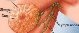
The operation is performed if conservative or hormonal therapy has not had the desired effect. And also if there are tumor formations in the area of the ducts (ranging from a cyst, polyp and ending with a malignant neoplasm). The essence of the operation is that the mammologist excises a certain part of the duct that has undergone deformation. In rare cases, lymph nodes and breast tissue are removed.
Hormonal drugs are prescribed if the root cause of the disease is an imbalance in the patient’s body. One drug may be prescribed or a whole course of hormonal therapy may be prescribed, which will help overcome the menopause period and stop the progression of the disease. In most cases, medications are prescribed to help compensate for the lack of estrogen in the body.
Treatment with folk remedies is also carried out, but in most cases it acts as a supplement to the main therapy.
It is recommended to make warm compresses and use baths with a decoction of herbs that have a calming and anti-inflammatory effect (chamomile, oak bark, calendula, plantain, etc.).
Ectasia is a disease that requires constant monitoring. Even if the cause of the disease is a banal hormonal imbalance, it is important to ensure that the condition does not continue to progress. To do this, you need to see a doctor and undergo a series of diagnostic examinations.
Prevention
The set of preventive measures to prevent the development of duct ectasia of the mammary gland is very wide, since ectasia can occur against the background of many diseases of the mammary gland. To reduce the risk of morbidity, important measures are:
- prevention of diseases and pathological conditions that can provoke the development of the described disease, and if they occur, timely diagnosis and adequate treatment. This is especially true for diseases that can arise against the background of hormonal imbalance;
avoiding trauma to the mammary glands;
- competent choice of the volume and method of surgical operations on the mammary gland (in particular, when performing plastic correction);
- at the age of over 35-40 years – a preventive visit to a gynecologist and mammologist once a year;
- medical control of the balance of sex (and not only) hormones;
- wearing comfortable bras;
- regular weight control;
- balanced diet;
- physical education classes;
- normalization of work, rest, sleep, sexual intercourse;
- giving up bad habits - smoking, drinking alcohol and drugs.
Treatment of ductectasia
If diagnostic studies have confirmed the initial diagnosis of ectasia of two or one breast. If the pathology affects both breasts, then a diagnosis of bilateral ductectasia is made. The treatment regimen prescribed by the mammologist is aimed at eliminating the symptoms of the disease and the causes that led to the disease.
- More often, therapeutic treatment is prescribed for ectasia, depending on the reasons that caused the ectasia. If inflammation of any form is detected during the diagnosis, the patient is prescribed special anti-inflammatory drugs. Additionally, the doctor prescribes complex medications that should strengthen the immune system.
- In case of hormonal disorders, the disease is treated in such a way as to normalize the level of hormones in the body. The goal is achieved by taking hormonal medications. The choice and dose of medications is prescribed by a doctor, who will take into account the patient’s age, her state of health, and the presence of concomitant diseases. It is dangerous to prescribe hormonal drugs on your own; they are taken only after consultation with a doctor.
- If therapeutic treatment does not bring the desired result, the disease is treated with surgical intervention. The operation is especially often used when polyps or large papillomas are detected in the ducts.
Modern medicine offers two methods of performing operations for ductectasia.
- Only pathological areas of the canals and cellular material are removed. The material obtained during the operation is necessarily sent for histological examination to confirm or refute oncology.
- Complete removal of the ducts - this technique is usually chosen if the patient’s tumor is confirmed to have a malignant nature.
Both types of surgery are performed under general anesthesia. Today's surgical techniques are so exquisite and precise that they leave virtually no traces. For some patients, surgical treatment is contraindicated: usually due to heart disease or the desire to give birth to a child and feed him breast milk.
Only the attending specialist can make a decision about surgery or refuse it. In complex cases of oncology, the decision is made at a medical consultation. The results and outcomes of treatment depend on timely diagnosis and the causes of the disease. In the vast majority of cases, recovery is not difficult to achieve if the patient came to the doctor on time and the examination did not reveal cancer.
Forecast
The prognosis for pathological ductal ectasia of the mammary gland is quite different - it depends on the underlying disease against which the described pathology arose. Critical complications occur relatively rarely - they develop mainly in advanced forms of one or another pathology, against the background of which ectasia of the mammary gland ducts has arisen.
Kovtonyuk Oksana Vladimirovna, medical observer, surgeon, consultant doctor
10, total, today
( 57 votes, average: 4.53 out of 5)
Intraductal papilloma of the mammary gland: signs, treatment
Breast asymmetry: causes and correction