Uterine perforation: what is it?
Perforation of the uterus is through damage to its wall with surgical instruments during intrauterine interventions. The complication occurs in 0.03 - 1% of cases of intrauterine procedures, but in recent years there has been a tendency to increase the percentage of pathology, which is explained by the expansion of diagnostic intrauterine methods and the increase in the number of diseases of the female genital organs.
Important
The most dangerous case of uterine perforation is damage to the organ wall by a curette (sharp edges of the instrument) or an abortion forcemer (during an abortion), which can lead to the capture of abdominal organs and their removal into the uterine cavity.
Perforation of the fetal sac can occur at any stage of the operation, including its probing, dilatation of the cervical canal, curettage of the walls of the organ and extraction of tissue of the ovum.
Causes
Rupture of the walls occurs as a result of a doctor’s violation of the technique of performing various medical procedures, namely:
- during abortion;
- installation of an intrauterine device;
- examination of the uterus using a hysteroscope;
- during curettage due to a non-developing pregnancy.
There are certain risk factors that significantly increase the likelihood of perforation:
- progressive infection;
- scar formed after surgery;
- operations on the uterus;
- abortion in the second trimester of pregnancy;
- hypoplasia;
- changes in the reproductive organs that appear during menopause;
- retroversion of the uterus;
- cervical tumor;
- uterine cancer.
According to statistics, wall punctures most often occur during an abortion. This can happen at any stage of the operation.
The damage caused by the probe usually does not cause severe bleeding. Complications develop when the cervical canal expands. Due to the inept actions of the doctor, tears appear in the internal pharynx, isthmus, and lower part of the uterine body. Damage to the uterus with a curette and similar instruments poses a great threat to a woman’s life. The rupture will be large and localized in the area of the bottom or walls. In this case, large blood loss occurs and neighboring organs are affected.
Classification
The complication can be complete, when the entire thickness of the uterine wall is damaged, or incomplete (the perforation reaches the serous membrane, but does not damage it). Complete perforation of the uterus can be complicated (damage to the pelvic organs: colon, bladder, ovaries, omentum) and uncomplicated (abdominal organs are not damaged).
note
Perforation of the uterine wall is considered a serious surgical complication and can lead to massive bleeding, peritonitis, and, if medical care is not provided in a timely manner, death.
Signs
Symptoms of uterine perforation are determined by its characteristics (uncomplicated/complicated, incomplete/complete) and location. If the perforation is incomplete or the hole that appears is closed by a certain organ (for example, an omentum), the signs may be weakly expressed or absent altogether. You can suspect perforation of the uterus during an abortion only when, after undergoing manipulation inside the organ, a woman complains of severe bleeding from the vagina, sharp pain in the lower abdomen, weakness and dizziness. With severe internal bleeding, tension in the peritoneal wall, pale skin, decreased blood pressure, and tachycardia appear.
Causes and risk factors
An organ is injured during manipulation performed in its cavity. Most often, uterine perforation occurs during surgical termination of pregnancy. Despite the presence of predisposing factors in the patient, the complication is caused by a violation of the technique of performing the intrauterine procedure and/or the rude actions of the operating gynecologist. Perforation of the uterus can also be performed during the removal of a frozen pregnancy (often after 12 weeks) and the remains of the ovum, diagnostic curettage, during hysteroscopy and intrauterine excision of myomatous nodes or polyps, hysterography, dissection of intrauterine adhesions (Asherman syndrome), during laser restoration of the uterus cavity, insertion of an intrauterine device.
Percentage of uterine perforation during surgical abortion:
- organ probing (2 – 5%);
- dilatation (expansion) of the cervical canal (5 – 15%);
- curettage (curettage) or extraction of fetal parts with an abortion force (reaches 90%).
It is characteristic that puncture of the uterus with a probe is not as dangerous as gross expansion of the cervical canal with metal dilators, which may be accompanied by rupture of the cervix, perforation of the isthmus and lower uterine segment. If the wall of an organ is damaged by a curette or abort forcemer, the size of the perforation hole is quite serious, which causes massive bleeding and further damage to the internal organs.
The following factors suggest the occurrence of complications:
- bending of the uterus posteriorly or bending the organ anteriorly;
- acute and chronic endometritis;
- fibroids or (especially) uterine cancer;
- existing scar on the uterus (after cesarean section, conservative myomectomy);
- age-related involution of the uterus (reduction in its size after 50);
- interruption of gestation at 12 weeks or more;
- criminal abortion;
- rude and fussy actions of the doctor;
- low qualification of a specialist;
- underdevelopment of an organ or its malformations;
- numerous organ scrapings;
- lack of visual control (ultrasound or hysteroscopy).
Causes of injury
The greatest danger from the point of view of injury to the uterine wall are the abortion forceps and curettes, which have a sharp edge. At the same time, neighboring organs may be injured. The Hegar dilator is rounded at the end and has a large thickness, so it is much more difficult for them to perforate the organ. In 0.3% of cases, perforation of the uterus is possible when an IUD is inserted.
The leading cause of injury is considered to be technically incorrect performance of intrauterine interventions. Perforation of the organ wall can occur during the following operations:
- medical abortion;
- separate therapeutic and diagnostic curettage;
- hysteroscopy;
- insertion of an intrauterine device.
It is believed that it is almost impossible to pierce the wall of a healthy organ: it is quite elastic and durable. And with various diseases, the structure of the tissues is loose and fragile, so their damage becomes possible.
The risk of uterine perforation increases in the following cases:
- acute or chronic inflammation – endometritis;
- myomatous nodes of various locations;
- scar after artificial childbirth or surgical interventions;
- frequent intrauterine interventions, including abortions and diagnostic curettages;
- recent surgery, less than six months have passed since;
- abortion after 12 weeks of gestation;
- uterine hypoplasia;
- age-related characteristics during menopause;
- posterior deviation of the organ (retroversion);
- endometrial cancer.
Injuries with a uterine probe are rare and do not lead to heavy bleeding. The Hegar dilator is dangerous only in case of rough manipulation and with a pronounced bend of the uterine body anteriorly or posteriorly. If it is used to perforate the wall, a large hole is formed with profuse bleeding. But the greatest danger is posed by the curette and abortion forceps, which account for up to 80% of traumatic perforations.
The curette (above) and the abortion forceps are the most dangerous surgical instruments from the point of view of injury to the uterine wall.
Spiral perforation
The insertion of the intrauterine device is performed blindly; the accuracy of the procedure depends on the doctor’s technique and his tactile sensations. The reason for the perforation of the wall is that the uterine cavity does not always coincide along the axis with the cervical canal. Sometimes in the lower segment the wall is very thin, which serves as a risk factor. An additional risk arises when installing an IUD earlier than 6 months after childbirth and immediately after an induced abortion.
Perforation by means of a spiral occurs both immediately after its installation and as spontaneous consequences some time after insertion. Sometimes this is discovered when trying to remove the coil. In this case, the threads will be lost or the removal of the IUD will be difficult.
Injury from the coil can occur at the injection stage if there were active contractions of the myometrium - expulsion aimed at expelling the drug. In this case, the cervix is perforated, since the axis of the cervical canal does not coincide with the axis of the uterus.
Symptoms of uterine perforation
The clinical picture of the complication is determined by its nature (complete or incomplete perforation of the organ wall, injury and/or extraction of abdominal organs into the uterine cavity) and the location of the perforation. In case of incomplete perforation of the fetal sac or covering of the puncture site with some organ (most often the perforation hole is closed with an omentum), the symptoms of the pathology are absent or mild.
Perforation of the uterus can be suspected by the occurrence of sharp, stabbing pain in the lower abdomen during intrauterine manipulation and the persistence of severe pain after the procedure. The doctor should also be alerted to the appearance of heavy bleeding from the uterus during the operation and/or after it, increasing weakness, lethargy and dizziness. Significant bleeding into the abdominal cavity is accompanied by pallor of the skin and mucous membranes, increased heart rate, hypotension, tension in the abdominal muscles and the appearance of peritoneal symptoms.
Perforation of the uterus: types, causes, symptoms and treatment – GYNECOLOGICAL HOSPITAL OF OMSK REGION
Many intrauterine operations and manipulations are performed by the doctor almost blindly. In 1% of cases of all interventions, perforation of the uterus can occur - this is a through wound of its wall with a surgical instrument.
Causes of injury
The greatest danger from the point of view of injury to the uterine wall are the abortion forceps and curettes, which have a sharp edge. At the same time, neighboring organs may be injured. The Hegar dilator is rounded at the end and has a large thickness, so it is much more difficult for them to perforate the organ. In 0.3% of cases, perforation of the uterus is possible when an IUD is inserted.
The leading cause of injury is considered to be technically incorrect performance of intrauterine interventions. Perforation of the organ wall can occur during the following operations:
- medical abortion;
- separate therapeutic and diagnostic curettage;
- hysteroscopy;
- insertion of an intrauterine device.
It is believed that it is almost impossible to pierce the wall of a healthy organ: it is quite elastic and durable. And with various diseases, the structure of the tissues is loose and fragile, so their damage becomes possible.
The risk of uterine perforation increases in the following cases:
- acute or chronic inflammation – endometritis;
- myomatous nodes of various locations;
- scar after artificial childbirth or surgical interventions;
- frequent intrauterine interventions, including abortions and diagnostic curettages;
- recent surgery, less than six months have passed since;
- abortion after 12 weeks of gestation;
- uterine hypoplasia;
- age-related characteristics during menopause;
- posterior deviation of the organ (retroversion);
- endometrial cancer.
Injuries with a uterine probe are rare and do not lead to heavy bleeding. The Hegar dilator is dangerous only in case of rough manipulation and with a pronounced bend of the uterine body anteriorly or posteriorly.
If it is used to perforate the wall, a large hole is formed with profuse bleeding.
But the greatest danger is posed by the curette and abortion forceps, which account for up to 80% of traumatic perforations.
The curette (above) and the abortion forceps are the most dangerous surgical instruments from the point of view of injury to the uterine wall.
Spiral perforation
The insertion of the intrauterine device is performed blindly; the accuracy of the procedure depends on the doctor’s technique and his tactile sensations.
The reason for the perforation of the wall is that the uterine cavity does not always coincide along the axis with the cervical canal. Sometimes in the lower segment the wall is very thin, which serves as a risk factor.
An additional risk arises when installing an IUD earlier than 6 months after childbirth and immediately after an induced abortion.
Perforation by means of a spiral occurs both immediately after its installation and as spontaneous consequences some time after insertion. Sometimes this is discovered when trying to remove the coil. In this case, the threads will be lost or the removal of the IUD will be difficult.
Injury from the coil can occur at the injection stage if there were active contractions of the myometrium - expulsion aimed at expelling the drug. In this case, the cervix is perforated, since the axis of the cervical canal does not coincide with the axis of the uterus.
How to recognize the condition?
Based on the volume of damage, the classification of uterine perforation was determined:
- Complete when the hole is formed over the entire thickness of the wall. It can be uncomplicated or complicated. In the latter case, the omentum, appendages, intestines, bladder and other neighboring organs are additionally injured.
- Incomplete, with it the serous cover remains intact.
In many cases, the doctor can recognize the signs of perforation immediately at the moment it occurs.
Committed during curettage, perforation of the uterus is accompanied by an unexpected failure of the instrument to a greater depth than that which was determined during the probing process.
The instrument does not encounter obstacles in the form of organ walls. Sometimes such a sign is observed with sudden atony, then it is necessary to carry out a differential diagnosis with perforation.
If perforation occurs as a complication of an induced abortion, then increased bleeding is observed, and parts of the fertilized egg cease to appear in the discharged substance.
The onset of severe bleeding at the very beginning of curettage, when the fertilized egg is still attached to the wall, is especially suspicious.
Severe bleeding during abortion, when the removal of parts has already been completed, free movements in the abdominal cavity of the surgical instrument, which is accompanied by signs of shock, confirm the presence of injury.
But atony, especially in women who have given birth frequently or with repeated abortions, is also accompanied by massive bleeding and a flabby uterus. Therefore, differential diagnosis is necessary.
Perforation of the uterus: a - with a probe, b - with a dilator, c - with a curette, d - with an abortion forcem
The greatest difficulties will be caused by incomplete perforation during abortion. It may go unnoticed by the gynecologist and the patient. In case of severe injury to the uterine wall with prolapse of the omentum and intestinal loops, making a diagnosis is not difficult.
In some cases, symptoms of uterine perforation appear after the operation, when the woman has recovered from anesthesia. Then the following complaints appear:
- acute pain in the lower abdomen;
- bleeding from the vagina;
- weakness, dizziness, loss of consciousness;
- tachycardia with a simultaneous drop in blood pressure;
- pale skin.
If the perforated wound is covered by a neighboring organ, then the symptoms of the pathology are less noticeable or may not appear. It all depends on the amount of bleeding and the flow of blood into the abdominal cavity.
Diagnostic techniques
Signs of perforation appear at the time of injury. If this does not happen, then additional diagnostics are necessary when symptoms of an acute abdomen appear.
Careful medical history collection if a woman comes with complaints from home. If she is still in a hospital setting, it is much easier to assume this complication after the intervention.
A gynecological examination is performed. In most cases, there is bleeding from the vagina. If the spiral is pierced, the threads will not be visible from the opening of the cervical canal. In some cases, the threads are visible, but the spiral cannot be removed.
Injury may occur during hysteroscopy. In this case, the diagnosis is established based on the following criteria:
- the expander of the device goes to a greater depth than it should;
- it is impossible to create increased pressure inside the organ cavity;
- bowel loops or omentum are visible.
Sharp pallor and tachycardia indicate significant blood loss. Its severity is determined by the Algover shock index. To do this, the pulse rate must be divided by the systolic pressure. If the numbers are 0.8 or less, this indicates slight bleeding, up to 50 ml, with no obvious symptoms yet.
A 20% decrease in circulating blood volume is manifested by a shock index of 0.9-1.2. In this case, up to 1250 ml of blood is poured out. A loss of 30% of the bcc increases the index to 1.3-1.4. In this case, the bleeding pattern is accompanied by tachycardia up to 120 beats per minute, shortness of breath, and sweating.
Loss of 40% of blood leads to stupor, the pressure is critically low or undetectable. Shock index 1.5 or higher.
Blood leaking into the abdominal cavity causes irritation of the peritoneum, resulting in peritoneal symptoms. Abdominal pain worsens with movement. Injury to adjacent hollow organs leads to the release of their contents into the abdominal cavity, which also leads to the development of peritonitis.
Ultrasound diagnostics allows you to determine free fluid in the abdominal cavity. If coil perforation has occurred, in most cases it can also be detected using ultrasound.
Hysteroscopy can be used as a diagnosis. With its help, damage visible to the eye and the location of the IUD are identified.
Sometimes the spiral is not detected by ultrasound. In such cases, an X-ray examination is performed. Metal parts of the device will be visible on the overview image of the pelvic organs.
Using a general blood test, the level of red blood cells, hemoglobin and hematocrit is determined. This data is necessary to make a decision about the need for a blood transfusion. The blood type and Rh factor must also be determined.
Treatment methods
If perforation occurred at the stage of probing the uterus, then further manipulations are stopped.
Emergency care for uterine perforation, which was diagnosed during surgery, consists of administering uterotonics and applying a heating pad with ice to the abdomen.
If the hole was made with a small instrument (probe), and ultrasound shows no free fluid in the abdominal cavity, then a wait-and-see approach is chosen. The woman remains under medical supervision in the hospital.
Treatment of uterine perforation is individual in each case and depends on the severity of the damage and general condition. A through hole in the wall of an organ with massive bleeding is subject to surgical treatment.
A relatively small defect is sutured through a laparoscopic approach.
This is a minimally invasive operation that is performed under the control of a video camera inserted into the abdominal cavity through a small hole in the anterior abdominal wall (for more information about laparoscopy, read our article at the link).
If damage to the IUD has led to its release into the abdominal cavity, it must be removed as quickly as possible, especially for copper-containing IUDs. Copper ions lead to an inflammatory response.
The manipulation is performed laparoscopically. But if necessary, it is expanded to laparotomy.
The patient is informed before the operation that in the event of a large number of adhesions in the abdominal cavity or injury to other organs, the course of the operation will be changed.
Injury to other pelvic organs - intestines, bladder - requires the work of a surgeon, not a gynecologist.
In case of multiple major injuries to the uterus and if suturing the defects does not stop the bleeding, they resort to an extreme method - amputation of the organ. Bleeding due to injury to the vessels of the uterus is massive and often leads to disseminated intravascular coagulation syndrome. Therefore, in order to save the patient’s life, doctors have to take extreme measures.
Treatment of acute blood loss depends on the severity of the condition. Antishock therapy is carried out, as well as restoration of circulating blood volume.
For this purpose, colloidal and crystalloid solutions are used, which compensate for the deficiency with liquid and also restore the ionic composition. Depending on the clinical situation, plasma is used and blood transfusions are performed.
If bleeding has just occurred, then reinfusion of your own blood collected from the abdominal cavity is possible.
Antibiotics are required in all cases of perforation. Broad-spectrum drugs are selected from the group of cephalosporins (Cefotaxime, Ceftriaxone), Gentamicin, and Metronidazole for the prevention of anaerobic infection.
Source: //gb8-omsk.ru/zabolevaniya/perforatsiya-matki-vidy-prichiny-simptomy-i-lechenie.html
What is the complication?
Untimely detection of uterine perforation can lead to serious complications, which can subsequently affect the patient’s reproductive function. Early complications of pathology include:
- intestinal injury;
- bladder damage;
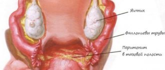
- massive intra-abdominal bleeding;
- the formation of hematomas between the layers of the uterine ligament, under its serous membrane;
- peritonitis turning into sepsis.
Long-term complications include:
- formation of ICN (damage to the muscle layer of the cervix);
- miscarriage;
- formation of intrauterine adhesions;
- infertility due to the development of Asherman's syndrome or hysterectomy.
Diagnostics
Possible perforation of the uterine wall during the intrauterine procedure is indicated by the sign of the instrument being immersed more than the expected depth, a feeling of “falling through” it. In this case, it is necessary to palpate the anterior wall of the abdomen without removing the instrument from the uterus to determine its end in the abdominal cavity. Complicated perforation is indicated by the removal of an intestinal loop, ovary or omentum from the organ cavity (most often). During the installation of the IUD, perforation of the wall of the fetal sac is indicated by the disappearance of the contraceptive threads from the cervical canal, and upon visualization, an attempt to remove the intrauterine device by pulling the “antennae” is unsuccessful, while the patient feels sharp pain, and the doctor resists.
If the wall of the uterus is injured during hysteroscopy, the doctor notes that it is impossible to maintain stable pressure in the cavity of the organ, there is no outflow of fluid poured into its cavity, intestinal loops and other contents of the small pelvis are visualized on the monitor screen.
If uterine perforation is suspected, but there are no reliable signs of a complication or a sharp deterioration in the woman’s condition, she is transferred to a ward under the supervision of medical personnel to further determine the patient’s management tactics. If the woman’s condition is satisfactory, a pelvic ultrasound with a vaginal sensor is performed. A complication is indicated by the presence of free fluid in the pelvic cavity and a perforation hole in the wall of the organ.
Treatment
Therapeutic tactics for perforation of the uterine wall are determined by the nature of the pathology:
- In case of incomplete perforation and minor trauma, absence of symptoms of internal bleeding and peritoneal manifestations, a conservative approach is used. The patient is administered uterotonic medications that promote uterine contractions and antibiotics, and the patient must be under the supervision of specialists.
- In case of a serious perforation with an extensive and acute clinical picture, emergency surgical intervention is required. Surgical tactics are based on laparoscopic or laparotomy access. In this case, minor traumatic lesions are simply sutured, and in case of massive injuries, a hysterectomy is performed, in which the uterine body is completely removed.
During surgical treatment, the patient is compensated for blood loss by administering fresh frozen plasma or special solutions.
After the operation and during the rehabilitation period, antibiotics and NSAIDs are indicated to prevent possible complications. In general, the rehabilitation and recovery period lasts about 8 weeks.
Treatment for uterine perforation
The tactics for managing a patient in the event of a complication is determined by the time of diagnosis, the size, location and mechanism of the injury (abortion, curettage of an organ or installation of an IUD) and trauma to neighboring organs. It is extremely rare that conservative management of the patient is possible with the prescription of bed rest, cold on the lower abdomen, contractions and antibacterial drugs. Indications for conservative therapy:
- incomplete perforation of the uterine wall;
- no injury to the abdominal organs;
- absence of hematoma in the periuterine tissue;
- no intra-abdominal bleeding occurred.
The choice of conservative observational tactics should be confirmed by transvaginal ultrasound data with subsequent dynamic monitoring.
All other cases of perforation of the fetal sac are subject to surgical intervention, before which intensive preoperative preparation is carried out: infusion therapy (blood transfusion if indicated), stabilization of blood pressure (glucocorticoids), hemostatics, uterotonics.
The extent of surgical intervention depends on the number of perforations, their size, damage to large uterine vessels and neighboring organs. If there is a small defect in the uterine wall and there is no bleeding, it is sutured with preliminary curettage of the uterine cavity through the perforated hole (when installing an IUD and perforation of the organ, the contraceptive is removed from the defect or pelvic cavity). Indications for supravaginal amputation of the uterus are multiple defects or one, but large rupture of the uterine wall, injury to the uterine artery. If the isthmus or lower segment of the uterus is damaged, gynecologists tend to perform hysterectomy. If the intestines or bladder are injured, their damage is sutured, and extensive operations on the abdominal organs are performed by an abdominal surgeon. In the postoperative period, antibiotics, uterotonics, and infusion therapy are prescribed.
Reviews
The consequences of uterine trauma depend on the number of injuries and their volume. Patients note that large holes heal, but a scar forms. After such an injury, a woman must register with the antenatal clinic.
The consequences of perforation can be different. Patients say that when intervening in the abdominal area, adhesions often form. Injury can be avoided with proper prevention.
Women also note that they have to seriously plan their pregnancy. A preliminary examination of the scar is required. It is best to become pregnant at least two years after the perforation. The main thing that is noted in the reviews is the need to pay close attention to your health status and contact trusted doctors.
Prognosis and prevention
Timely diagnosis and surgical intervention for uterine perforation provides a favorable prognosis. Subsequently, problems may arise regarding conception and pregnancy. Prevention of organ perforation includes adherence to the technique and stages of intrauterine manipulation, including the insertion of an IUD, insertion of instruments into the uterine cavity with caution, visual control of all intrauterine interventions (ultrasound at the end of the procedure, hysteroscopy during it). Women are advised to refuse abortions, plan a pregnancy, regularly visit a gynecologist and treat inflammatory processes in the genital organs.
Sozinova Anna Vladimirovna, obstetrician-gynecologist
just today
( 55 votes, average: 4.04 out of 5)
Atypical endometrial hyperplasia: symptoms and treatment
Estradiol: norm in women, causes of deviations, treatment
Related Posts
Consequences and complications
Cavity curettage (curettage) is a gynecological mini-operation that allows you to expand the cervix and clean out its cavity with a special surgical instrument.
Such manipulation can be caused by several reasons:
- To clean the cavity to remove fetal tissue, in time, after an abortion or spontaneous miscarriage. Sometimes after childbirth to completely remove placenta particles. In such cases, curettage helps prevent bleeding and infection.
- For the diagnostic treatment of uterine bleeding. This mini-operation will help in the diagnosis and treatment of such pathologies: endometriosis, polyps, fibrous tumors, uterine cancer and hormonal imbalances. During cleaning, a piece of collected tissue is examined for the presence of idioblasts.
The main purpose of such an operation is to remove the top layer of the uterine cavity, which should be shed during menstruation. Curettage is performed in the operating room using a gynecological chair.
As a rule, the operation is performed under anesthesia, since dilatation of the cervix is a rather uncomfortable and painful procedure. Intravenous drugs are used for anesthesia. Sometimes, if, for example, curettage needs to be performed immediately after childbirth, then it is performed without anesthesia.
Before the operation begins, the doctor places a dilator in the vagina, which straightens the walls so that the cervix becomes visible. Curettage is carried out with a specially designed device - a curette. The tool is similar to an ordinary spoon only with a long handle. The doctor carefully removes the surface layer and collects it in a test tube, which is sent for histological analysis. The operation lasts about 40 minutes.
Initial complications:
- perforation of the uterus - severe through damage to the organ with surgical instruments;
- hematometra - accumulation of blood in the cavity;
- incomplete abortion – incomplete removal of fetal or placenta particles;
- abortion that did not take place - the rejected fetus remains completely in the uterus;
- complete rupture of the cervix - tears and complete ruptures can occur;
- great blood loss.
Late: chronic adnexitis, salpingoophoritis and others.
Delayed diagnosis of uterine perforation can cause life-threatening and serious consequences and complications. These include intestinal wounds or bladder injuries, extensive hematomas, sepsis, peritonitis, and bleeding. Damage can cause the formation of isthmic-cervical insufficiency, as well as miscarriage during pregnancy in the future.
Uterine perforation itself is a complication of a therapeutic or diagnostic procedure. Damage to the walls of the reproductive chamber can be aggravated by the following circumstances:
- perforation of organs located nearby (when the integrity of the intestines, bladder, ovary, fallopian tubes is disrupted);
- peritonitis (the inner lining of the peritoneum becomes inflamed due to the penetration of pathogenic flora);
- hematoma (a blood clot forms in organs located near the uterus);
- death (occurs due to large blood loss and late diagnosis of uterine perforation).
To avoid damage to the thickness of the uterus, it is necessary to carefully prepare for any type of intervention
Before the procedure, it is important to exclude the inflammatory process. You should first perform an ultrasound and a gynecological examination to assess the size and position of the organ being examined.
Perforation can be prevented if any intervention is carried out under the control of an ultrasound scanner.
If the position of the reproductive organ is incorrect, before intrauterine intervention, forceps are applied to the cervix to eliminate the refraction angle.
The danger of the consequences of uterine perforation increases many times if this complication is not recognized in a timely manner. Meanwhile, the doctor’s attentive attitude to all manipulations during the abortion almost completely eliminates the possibility of not noticing a perforation of the wall or its consequences.