Here are the circumstances under which the chances of uterine subinvolution are high:
- Incomplete exit of the placenta from the cavity. In almost all cases of cobinvolution, particles of the placenta or amniotic sac and blood clots are found in the uterus. In such a situation, infection penetrates into the organ more easily, inflammation and endometritis occur, and the contractile function of the uterine muscles is disrupted.
- Pathological course of pregnancy . A predisposition to subinvolution is observed in women in labor with polyhydramnios, multiple pregnancies, and large fetal sizes. Also at risk are women who, during the period of bearing a child, had one or another inflammatory disease (cystitis, pyelonephritis, colpitis).
- Disorders of the birth process . Complications often arise when labor proceeds rapidly or, conversely, labor is too weak and the process is delayed.
- Features of body structure . Some researchers hypothesize that the contractility of the uterus may be influenced by certain hereditary factors and genetic changes in the female body. If they are present, the chance of insufficient myometrial activity after childbirth increases significantly.
There is also a connection between co-involution of the uterus and the presence of fibroids. The age of the woman giving birth matters; girls under 18 years of age and women over 40 years of age are at risk. Lack of breastfeeding for 4-5 days after the birth of a child is harmful, because the process of lactation and breastfeeding by the baby triggers the hormonal mechanism of stimulating uterine contractions. A certain role is played by the pathological location of the uterus, problems with the ligamentous apparatus, anemia, and chronic somatic diseases.
Physiological amenorrhea
Women who breastfeed have a longer period of amenorrhea. In women who do not breastfeed, the first ovulation usually occurs after 70-75 days; in 60% of women giving birth, the first menstruation occurs 12 weeks after birth.
In women who breastfeed, the duration of anovulation correlates with the frequency of breastfeeding, the duration of each feeding, and the presence of additional nutrition for the newborn.
If a woman exclusively breastfeeds her newborn, on demand, without a night break, ovulation earlier than 6 months after birth is possible only in 1-5% of cases (lactational amenorrhea). To maintain lactational amenorrhea, the interval between feeding a newborn should not be more than 4 hours a day and 6 hours at night, additional nutrition for a newborn should not be more than 5-10% of the total nutrition.
Contraindications to breastfeeding include the following conditions:
- maternal use of alcohol or drugs;
- galactosemia in a newborn;
- HIV infection in the mother;
- active tuberculosis in the mother without treatment;
- treating the mother for breast cancer;
- maternal use of drugs such as bromocriptine, cyclophosphamide, cyclosporine, doxorubicin, ergotamine, lithium, methotrexate, phenycilidine, radioactive iodine, etc.
Suppression of lactation is carried out by using modulators of prolactin receptors antagonists of the action of prolactin bromocriptine (Parlodel) 2.5 mg per day or more until cessation of lactation or carbegoline (Dostinex).
Ovulation suppression occurs due to increased prolactin levels in lactating women. Prolactin levels remain elevated for 6 weeks after birth, whereas in non-lactating women they return to normal within 3 weeks. Estrogen levels, on the contrary, remain low in lactating women, while in those who do not breastfeed, they rise and reach normal levels 2-3 weeks after birth.
Symptoms of the disease
The presence of uterine subinvolution is determined by the following signs:
- No pain in the lower abdomen, similar to contractions, in the first days of feeding the baby. When the uterus contracts, similar pain sensations occur for some time.
- Excessive bleeding or dark-colored discharge with an unpleasant odor.
- Presence of elevated temperature. It lasts for a long time, but does not exceed 37.5. Usually indicates the development of an inflammatory process in the body.
- During a routine gynecological examination, the doctor notes the looseness of the uterus and its dimensions that do not correspond to the norm.
The presence of the symptoms mentioned should alert the woman and force her to immediately seek medical help.
Postpartum contraception
Women in labor are usually advised to have sexual rest for 6 weeks, until the first postpartum visit. But some women begin sexual activity earlier than this period, so the issue of contraception should be discussed before the woman in labor is discharged from the hospital.
If a woman prefers hormonal methods of contraception and is breastfeeding, she is recommended progestin-only contraceptives: mini-pill, Norplant or Depo Provera. They do not affect the quality of breast milk and may even increase its volume. Asosa recommends starting to take progestin-only contraceptives 2-3 weeks after birth, Depo Provera (medroxyprogesterone acetate) - 6 weeks after birth. Combined estrogen-gestagen oral contraceptives affect the quantity and quality of milk to a greater extent, so they are recommended for patients who are not interested in breastfeeding.
If the patient is interested in non-hormonal methods of contraception, the use of a condom is recommended, which also allows for the prevention of sexually transmitted diseases. Diaphragms and cervical caps can be used no earlier than 6 weeks after birth (after completion of uterine involution).
Useful recommendations from doctors
To maintain muscle tone, doctors strongly recommend consuming herbal teas based on birch or nettle leaves. The following plants also have beneficial properties: horsetail, shepherd's purse, yarrow. To enhance the positive therapeutic effect, you can do vaginal douching with a decoction of the listed herbs.
For the entire period of treatment, it is better for a woman to refrain from lifting weights and playing sports. In your free time, you can take relaxing walks. Restoring the contractile activity of the uterine tissue is facilitated by attaching the baby to the breast. At the same time, the body begins to intensively produce the hormone prolactin. It helps the uterus return to its natural shape.
Postpartum care
Maternity hospital stays in the United States are limited to 2 days after a vaginal birth and 4 days after a cesarean section, although many hospitals shorten this period to 1 and 3 days, respectively. After vaginal birth, the issue of caring for the perineum, mammary glands and contraceptive methods are discussed with the patient. The doctor must provide psychological support and give recommendations on how to help the patient and newborn at home.
Post-cesarean section patients are given advice on wound care and physical activity. Patients are advised not to lift heavy objects (“nothing heavier than a child”) and excessive activity, including driving, is prohibited.
Care for women in labor after vaginal birth
Routine care for women in labor after vaginal birth consists of monitoring body temperature, uterine involution and the nature of postpartum discharge (lochia), caring for the condition of the perineum, supporting breastfeeding in the absence of contraindications, and reducing pain. Non-steroidal anti-inflammatory drugs are usually used for analgesia. Pain relief may be required for women in labor with grade III-IV perineal tears.
The wound after episiotomy is cared for, the presence of edema or hematoma is monitored (applying ice to relieve pain and reduce swelling, sitz baths, treating sutures with disinfectant solutions). Toilet of the external genitalia and perineal sutures is carried out after each act of urination and defecation, with warm water and soap or antiseptic solutions (pale pink solution of potassium permanganate) with movements from front to back, from the pubis to the perineum. If there are sutures in the perineum, it is recommended to regulate bowel function with the help of mild laxatives and reduce the load on the pelvic floor muscles. In the presence of severe pain, the possibility of hematoma of the vulva, vagina and retroperitoneal space should be excluded.
In patients suffering from hemorrhoids, ice application, a diet with sufficient dietary fiber, mild laxatives, and hemorrhoidal suppositories are used.
If body temperature increases > 38 ° C with two or more measurements during the first 10 days after birth, with the exception of the first 24 hours (puerperal fever), the patient is further examined (blood tests, urine tests, ultrasound) to identify possible causes of infectious complications.
Caring for patients after caesarean section
Management of patients after cesarean section includes adequate pain management, wound care, prevention of wound infection, control of uterine involution and vaginal discharge. Analgesics are used for pain relief, which can contribute to the development of postoperative intestinal paresis. Laxatives are prescribed. To reduce pain as a result of postpartum uterine contractions, non-steroidal anti-inflammatory drugs are used. Antibiotic prophylaxis includes the administration of I-II generation cephalosporins during the perioperative period (2 g intraoperatively, then 1 g twice daily).
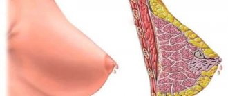
Breast care
Breast care is provided to all women in labor, regardless of their desire to breastfeed. Nipple preparation should be carried out during pregnancy (massage, treatment with tannins - oak bark tincture, cognac). The onset of lactation is accompanied by bilateral enlargement, pain, hardening of the mammary glands, an increase in their local temperature and the release of colostrum approximately 24-72 hours after birth. There may be an increase in body temperature to 37.8-39 ° (“milk fever”). When body temperature rises, it is important to exclude other causes of fever (mastitis, endometritis, thrombophlebitis). To reduce the pain associated with breast engorgement, ice is applied to the mammary glands, a supportive bra, analgesics and anti-inflammatory drugs.
Women who are breastfeeding may experience problems with nipple soreness and erosions. Residues of milk in the ducts of the glands are a breeding ground for opportunistic bacteria and contribute to nipple erosion. Patients are advised to wash their hands with soap and toilet before and after breastfeeding, and toilet the mammary glands (washing the nipples with soap, wiping with a clean dry towel).
Diagnosis of uterine subinvolution
Patients at risk of delayed recovery of prenatal parameters of the uterus require constant medical supervision. If detected in a timely manner, this complication is not dangerous; it can be quickly stopped and a complete recovery can be achieved.
The following techniques help to identify subinvolution:
- physical examination, clarification of the volume and nature of vaginal discharge;
- gynecological examination and bimanual examination;
- Ultrasound of the uterus through the vagina or through the abdominal wall;
- MRI of the pelvic organs;
- hysteroscopy is the most informative study that allows you to detect retention of the placenta in the cavity and clarify the parameters of the uterus;
- general blood analysis;
- culture of postpartum secretions on a nutrient medium.
Attention! The condition can cause a significant deterioration in a woman’s well-being and provoke the development of dangerous diseases. Treatment must be started immediately.
Complications of the postpartum period
The most common primary postpartum complications include postpartum hemorrhage, postpartum infectious complications (wound infection, endomyometritis, mastitis, etc.) and postpartum depression.
Postpartum hemorrhage usually occurs within 24 hours after birth, while the patient is still in the obstetric hospital. But these complications can develop several weeks after birth due to retention of fertilization products (remnants of the placenta or membranes). Endomyometritis and mastitis usually occur 1-2 weeks after birth. Postpartum depression can develop any time after childbirth, but is usually undiagnosed.
Postpartum hemorrhage
Postpartum hemorrhage is blood loss of more than 500 ml after vaginal delivery or more than 1000 ml after cesarean section. Domestic obstetricians and gynecologists define postpartum hemorrhage (pathological postpartum blood loss) as blood loss of >0.5% of a woman’s body weight.
Bleeding of more than 20% of the volume of blood volume (> 1-1.2 l) is considered massive. Massive postpartum hemorrhage, the main cause of maternal hypotension during the gestational period, is one of the leading causes of maternal mortality.
The possibility of sudden massive postpartum hemorrhage is determined by the speed of uteroplacental blood flow (600 ml/min). Limitation of blood loss after childbirth is ensured by adequate contraction of the myometrium at the site of placental attachment after childbirth, which leads to occlusion of open vessels of the placental plane.
Early postpartum hemorrhage is postpartum bleeding that occurs within 24 hours of delivery. Late postpartum hemorrhage occurs later than 24 hours after birth.
The most common causes of postpartum hemorrhage are atony (hypotonia) of the uterus, retention of conceptus products (parts of the placenta and membranes), and trauma to the birth canal. Less common causes are low placental implantation (in the lower uterine segment, which has less contractility) and coagulation defects. The use of obstetric forceps and vacuum extraction increases the risk of injury to the cervix and vagina.
While the cause of bleeding is being determined, the patient is given intensive infusion therapy and preparation for blood transfusion. If blood loss exceeds 2-3 liters, the patient may experience consumption coagulopathy - DIC syndrome, which requires transfusion of blood clotting factors and platelets.
In rare cases, which are accompanied by significant hypovolemia and hypotension, pituitary infarction (Sheehan syndrome) may develop. These patients may further develop agalactia (lack of lactation) due to a sharp decrease or absence of prolactin, or secondary amenorrhea as a result of insufficiency or absence of gonadotropins.
Pathogenesis
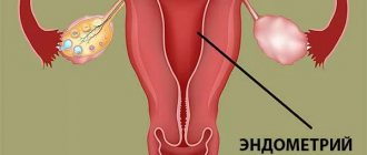
The main links in the pathogenesis of uterine subinvolution are: low activity of the uterine muscles, a decrease in its contractility; severe and prolonged postpartum tissue swelling; slow resorption of collagen fibers in the uterine wall. The myometrium contracts poorly if during pregnancy it was too stretched (large fetus, polyhydramnios, multiple pregnancy) or the neurohumoral regulation of the smooth muscles of the uterus is impaired. This mechanism is the main one when the complication is primary. Low uterine contractility affects not only recovery after childbirth. In such a situation, the infection penetrates more easily into its cavity, causing inflammation that ends in endometritis.
The second mechanism that contributes to the rapid reduction in the size of the uterus is a decrease in tissue swelling. Immediately after childbirth, the inner layer of the uterus resembles a continuous wound. But after 4-10 days it heals, the lumen of the blood vessels narrows, and tissue swelling goes away. If there are foreign bodies in the cavity or it is infected, swelling persists for a long time, blood flow in the capillaries, arterioles and small veins is disrupted. Delayed lysis of collagen is also noted, but this phenomenon cannot yet be fully explained.
Ruptures of the genital tract
Vaginal lacerations and hematomas
Immediately after birth, the mother's birth canal (perineum, labia, periurethral area, vagina, cervix) is examined in the speculum; Any tears found are sutured. Deep vaginal tears (up to the fornix) may be difficult to visualize, impinge on arterial vessels, and may cause noticeable bleeding or hematoma. To ensure adequate restoration of injuries to the birth canal, suturing is performed under adequate anesthesia (regional anesthesia).
Large hematomas are opened, injured vessels are found, stitched, and damaged vaginal tissue is restored. In some cases, extensive hematomas may form in the retroperitoneal space.
Clinical signs of such hematomas include back pain, anemia, and decreased hematocrit. The diagnosis is confirmed using ultrasound and, if necessary, computed tomography (CT). For small hematomas, a wait-and-see approach is chosen and anemia is treated. If the patient’s condition is unstable, surgical evacuation of the hematoma and ligation of injured vessels are performed.
Cervical ruptures . Cervical ruptures can lead to significant postpartum bleeding. The cause of these ruptures may be the rapid dilatation of the cervix in the first stage of labor or the beginning of the second stage of labor before the cervix is fully dilated. Immediately after birth, the cervix is examined in speculums using sequential application of fenestrated forceps following the movement of the clock hand. Sutures are repaired under adequate anesthesia (epidural, spinal or pudendal) with a continuous or interrupted suture using suture materials that are absorbable (absorbable).
Atony (hypotony) of the uterus
Uterine atony is the leading cause of postpartum hemorrhage. Increased risk of postpartum hemorrhage in patients with chorioamnionitis, when using magnesium sulfate, in multiple pregnancies, fetal macrosomia, a history of postpartum hemorrhage, in multiparous women (> 5 births). Developmental abnormalities and uterine fibroids can also affect the contractile function of the uterus and lead to atony and bleeding.
The diagnosis of uterine atony is determined by palpation of the uterus, which is soft, enlarged and weak. The fundus of the uterus may be contracted, and the lower segment may be relaxed. If uterine atony (hypotony) is suspected, Asos recommendations include the following:
1. Massage the uterus to stimulate its contractions and introduce an infusion of oxytocin (10-20 IU/l), which is usually also administered prophylactically; if ineffective, 20 IU of oxytocin is added intramuscularly. You should make sure that the bladder is stool (a full bladder prevents full contraction of the lower uterine segment). If bleeding continues, administer methylergonovine 0.2 mg IV or IM.
Maternal hypertension is a contraindication for the administration of methylergonovine. If bleeding continues, 250 mcg of PCP2a (contraindicated in bronchial asthma) is injected into the myometrium (transabdominal or transcervical) or administered no more than after 15 minutes with a maximum dose of 2 g. If ineffective, 1000 mg of misoprostol is administered rectally.
2. Examine the mother’s birth canal. If there are no visible ruptures, a careful manual examination of the uterine cavity is performed to remove retained products of the concept or to detect a uterine rupture (by the scar from a previous cesarean section). At the same time, an intensive infusion of colloidal (refortan) and isotonic solutions is carried out, and tests are performed before transfusion of fresh frozen plasma and blood.
3. If the bleeding continues, but is not rapid and massive, as an alternative to hysterectomy, if appropriate conditions are available, uterine artery embolization under radiological control can be performed. If delay is impossible or embolization is ineffective, laparotomy, ligation of the pelvic vessels (hypogastric, uterine, ovarian arteries) or hysterectomy are performed.
Remains of placenta and membranes
Immediately after birth, the placenta and membranes are thoroughly examined (integrity, presence of broken blood vessels, which may indicate an additional placental lobe). But with vaginal birth, it is often difficult to assess the retention of small parts of the placenta and membranes in the uterus. Typically, scraps of placental tissue and membranes emerge from the uterine cavity during postpartum contractions along with lochia. But the remains of concept products in some cases can lead to the development of endomyometritis and postpartum hemorrhage.
If remnants of the placenta and membranes are suspected in the postpartum period, a manual (if the cervix has not contracted) or, more often, instrumental inspection of the uterine cavity is performed. If after instrumental revision (curettage of the mucous membrane) of the uterus bleeding continues, placenta accreta is suspected.
Placenta accreta
Placenta accreta, as well as placenta accreta and placenta accreta, occur due to abnormal attachment of the placenta to the uterine wall, which can extend into the myometrium, leading to incomplete separation of the placenta from the uterine wall and the development of postpartum hemorrhage. Risk factors for placenta accreta include placenta previa and previous uterine surgery (cesarean section or myomectomy).
Clinical signs of placenta accreta may include a slowdown in the third stage of labor and fragmented separation of the placenta. If the duration of the third stage of labor exceeds 30 minutes, and there are no signs of separation of the placenta, perform manual separation and release of the placenta under adequate anesthesia. If the placenta is separated in fragments, a diagnosis of “placenta accreta” is made.
With placenta accreta, bleeding does not stop after uterine massage, the use of oxytocin, ergonovine and prostaglandins. If placenta accreta is suspected, treatment includes explorative laparotomy and surgical cessation of bleeding, which usually involves a hysterectomy. There are reports of cases of preservation of the uterus when fragments of the placenta are left in the uterus and subsequent successful treatment with methotrexate.
Uterine rupture
Uterine rupture may occur in 0.5-1% of patients with a previous uterine scar and in 1: (15,000-20,000) of women with an intact uterus. Uterine rupture can be traumatic (complicated childbirth, surgical vaginal delivery) and spontaneous (along a scar). This complication occurs during childbirth, but bleeding can develop in the postpartum period.
Uterine rupture is rare in nulliparous women (the uterus of primiparous women is “resistant” to rupture). Risk factors for uterine rupture include prior uterine surgery, fetal extraction in breech presentations, clinically narrow pelvis (disproportion between the fetal head and maternal pelvis), and an increased number of births in history. The classic clinical symptoms of uterine rupture are acute abdominal pain and a feeling of a “tear in the abdomen.” Treatment consists of urgent laparotomy, repair of the rupture, and, if surgical correction is not possible, hysterectomy.
Inversion of the uterus
Uterine inversion occurs when the fundus of the uterus is “born” through the cervix. Postpartum uterine inversion is rare (1:2000-1:2500 births). Risk factors for the underside of the uterus may include attachment of the placenta to the fundus of the uterus, uterine atony, placenta accreta, excessive traction on the umbilical cord in the third stage of labor. The diagnosis is determined by identifying the underside of the uterine fundus through the cervix, possibly with the placenta attached, at the birth of the placenta. Urgently perform manual separation of the placenta. In response to uterine inversion, the patient may experience a vasovagal reflex.
The doctor’s algorithm of actions after separation of the placenta in case of uterine incision includes stabilizing the patient’s condition, administering adequate anesthesia and restoring the position of the uterus (uterine reduction). To facilitate the reduction of the uterus, its relaxation is carried out using an infusion of beta-adrenergic agonists (terbutaline, ritodrine), magnesium sulfate or nitroglycerin. If it is impossible to manually reduce the uterus, a laparotomy is performed to surgically restore the position of the uterus using traction on the round ligaments. Sometimes, to restore the position of the uterine fundus, it is necessary to make a vertical incision of the myometrium.
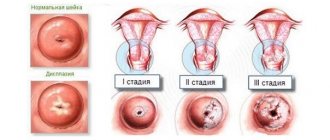
Types of disease
For the reasons that caused the complication, the following types of subinvolution are distinguished:
- True - the result of birth trauma after the birth of a large baby or several babies. Preeclampsia, cesarean section and some diseases can negatively affect the process of uterine restoration. For example, cervical fibroids or adenomyosis.
- Infectious - occurs when an infection occurs as a result of inflammation caused by the remains of the placenta in the uterus. Subinvolution of an infectious nature is promoted by an inflammatory process in the kidneys or anemia.
The exact cause will be determined by the doctor after the woman in labor undergoes an ultrasound examination.
Surgical treatment of postpartum hemorrhage
During vaginal birth, after taking conservative measures to stop bleeding, performing manual exploration and curettage of the uterus, if they are ineffective, the patient is transferred to the operating room for laparotomy and surgical stopping of bleeding.
During laparotomy, the presence of hemoperitoneum is assessed, which may indicate uterine rupture. In the absence of coagulopathy and the patient's stable condition, the first stage of surgical treatment is bilateral ligation of the uterine arteries. The second step is to ligate the hypogastric or internal iliac arteries. If the cause of bleeding is uterine atony, hemostatic compression circular sutures are applied to the body of the uterus to achieve hemostasis. If these measures are ineffective, a hysterectomy (postpartum hysterectomy) is performed.
If placenta accreta is found during cesarean section, the first step (after separation of the placenta) is to place hemostatic sutures on the placenta site. If the bleeding does not stop and there are no other causes of bleeding, the second step for an open uterus is to place circular sutures on the body of the uterus. If ineffective, the next step will be suturing the uterus (with or without tamponade) and ligation of the hypogastric arteries. If bleeding continues, a hysterectomy is performed.
If the bleeding is not massive, there is a reserve of time; if the patient’s condition is stable and there is a desire to preserve reproductive function, temporary uterine tamponade and further embolization of the uterine arteries under angiographic control can be performed.
With the development of consumption coagulopathy (DIC syndrome), a hysterectomy is performed with simultaneous rapid restoration of BCC and coagulation factors (fresh frozen plasma, cryoprecipitate, platelets, red blood cells, refortan, albumin, colloidal and isotonic solutions) under the control of hemostasis and coagulogram parameters.