Atrophic colpitis is a type of inflammatory process on the walls of the vagina associated with a decrease in the level of female hormones estrogen.
Insufficient levels of these hormones or their complete absence leads to thinning and dryness of the vaginal walls, resulting in complete tissue atrophy. Most often, this disease develops within a period of two to five years after physiological menopause.
- It manifests itself as degenerative changes in the vagina, in which the mucous membrane becomes thinner and the amount of vaginal discharge decreases.
- In addition, muscle tone drops sharply. All this together leads to an increase in the organ’s vulnerability to infectious agents. For this reason, inflammatory processes may occur and the disease may become chronic.
- If the disease occurred at an earlier age, then the first sign by which a woman may suspect it is discomfort and pain during sexual intercourse. The hormone estrogen, being one of the most important for the female body, has its greatest effect on the genitals.
Due to a decrease in its level, less vaginal secretion is produced, which causes discomfort. Subsequently, a decrease in the amount of vaginal discharge entails a decrease in the level of lactobacilli, which maintain a healthy balance of vaginal flora. Hence the increased vulnerability of the organ to infections.
This type of disease is not contagious. It is characteristic of age-related changes associated with menopause, but can also occur in women of childbearing age.
Epidemiology
About 58% of women experience symptoms of vulvovaginal atrophy in postmenopause, while only 25% receive appropriate therapy, and only 4% attribute their occurrence to menopause.
The symptoms of atrophic colpitis, in contrast to vegetative symptoms (classical “hot flashes”), only increase over time, becoming more and more intense.
About 80% of women note that the symptoms that arise disrupt not only their sexual life, but also their daily life, 68% talk about a decrease in self-esteem and loss of sexuality.
Atrophic colpitis changes a woman’s quality of life, affects her self-perception and relationships with her partner.
At the same time, only ¼ of the corresponding contingent goes to the doctor with complaints. Most women are embarrassed by intimate discussions with strangers or consider it a natural part of aging.
In both cases, an important factor is the poor awareness of women about the possible manifestations of menopause syndrome, postmenopause, and methods for their correction and elimination.
Causes of atrophic colpitis
All symptoms of atrophic colpitis have one single cause - a lack of estrogen in the body of a postmenopausal woman.
As is known, estrogens in a woman’s body cause:
- 1Female pattern hair;
- 2Pigmentation of nipples and genitals;
- 3Rejection of the functional layer of the endometrium, manifested by menstruation;
- 4Bone density;
- 5Formation of coagulation factors in the liver, due to which menstruation does not turn into bleeding;
- 6Increased blood concentrations of high-density lipoproteins, which have an antiatheromatous effect;
- 7Ensuring the affinity of receptors for progesterone, that is, ensuring the process of onset and gestation of pregnancy;
- 8Transition of fluid into the interstitium from the intravascular bed;
- 9Ensuring and maintaining normal vaginal microflora, maintaining local immunity.
Thus, all manifestations of menopausal syndrome, as well as the postmenopausal period, are caused by a lack of estrogen.
2.1. The role of vaginal microflora in a woman’s body
Due attention to the role and composition of the vaginal microflora began to be paid relatively recently. Its influence has already been proven not only on the “cleanliness” of the genitourinary tract and ensuring the normal course of pregnancy and childbirth, but also on maintaining the health of women in general.
During life, the composition of the vaginal biocenosis correlates with the hormonal background of a woman, undergoes cyclic changes, facultative flora and pathogenic microorganisms are added and eliminated, but, one way or another, its basis is lactobacilli.
They provide an acidic environment in the vagina by synthesizing lactic acid from glycogen, thereby suppressing the growth of facultative flora and preventing the development of the infectious process.
Of the 18 species of lactobacilli known to mankind, 1-4 species are present in a woman’s vagina, and their combination is individual; no pattern of their interaction has yet been identified.
In total, up to 400 species of various microorganisms can live in the genitourinary tract of a woman, but normally 90-95% should be lactobacilli.
Taken together, all microorganisms that normally form the vaginal biocenosis provide colonization resistance of the genital tract.
Estrogens, in turn, ensure the normal functioning of the stratified squamous epithelium of the vagina and the production of glycogen in superficial cells.
Constantly sloughing surface epithelial cells undergo breakdown (cytolysis) with the release of glycogen, which is the material for the synthesis of lactic acid by lactobacilli.
The pathogenesis of atrophic vaginitis involves a violation of the maintenance of local vaginal immunity against the background of atrophic changes occurring in it.
In a state of hypoestrogenism, the formation of glycogen in the vaginal epithelium is critically reduced, therefore, an acidic environment is not provided. In addition, the vaginal epithelium becomes significantly thinner.
All this leads to a significant decrease in colonization resistance of the genital tract, active reproduction of facultative flora and the occurrence of atrophic vaginitis.
It does not matter what causes estrogen deficiency, the mechanism of development of the disease remains the same, with possible variations in the severity of clinical manifestations.
Why does this disease occur?
The main reason that provokes atrophic colpitis is the reduced production of female sex hormones. They are the decisive factors influencing the condition of the vaginal epithelium. Estrogens most actively maintain stability in the vagina, because they determine the acidity of the vaginal environment, which is the norm for women. In such an environment, only beneficial bacteria live in the vagina, and the growth of other microorganisms that can upset the balance is not provoked. Estrogens also ensure stable blood circulation in the epithelial layer.
Age-related changes and the onset of menopause are the main markers that the vaginal environment will undergo certain changes. But if at first, even after the end of menstruation, the level of hormones can still provide the minimum norms for vaginal acidity, then already in postmenopause women begin to experience all the “delights” of a deficiency of female sex hormones.
Lack of estrogen leads to thinning of the vaginal epithelium and narrowing of its lumen. And microbes, previously restrained by the acidic environment, receive favorable conditions for development. Most often, microbes provoke the chronic course of the disease, and with mild symptoms, a woman may not be aware of the presence of pathology.
Another important reason is the influence of external factors. In some cases, atrophic colpitis is caused by taking hormonal drugs for a long time. And the course of the disease is aggravated by stress factors: hypothermia, previous genital infections, previous radiation therapy, removal of the ovaries, weakened immunity. Risk factors include excess weight, thyroid disease and diabetes.
Classification
There are two types of atrophic vaginitis depending on the etiology of hypoestrogenism:
- 1Postmenopausal;
- 2 Associated with artificial menopause.
The main difference in this case is age, since artificial menopause can be triggered at any period of a woman’s life.
This classification is quite sparse and reflects only the voluminous causes of the onset of hypoestrogenism. If we take a closer look, among the pathological conditions leading to a lack of estrogen in a woman’s body, we can additionally highlight:
- Polycystic ovary syndrome;
- Ovarian wasting syndrome;
- Resistant ovarian syndrome;
- Condition after extirpation of the uterus and appendages, oophorectomy, resection of the ovaries;
- The use of certain medications, in particular hormone-releasing hormone agonists;
- Excessively long protocols for stimulating superovulation in the in vitro fertilization program;
- Tumor, infectious process, hemorrhage in the area of the pituitary gland or hypothalamus - hypoestrogenism of central origin.
Characteristics of the disease
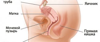
Uterine atrophy is a gradual thinning of the endometrium due to a decrease in estrogen synthesis. In the female body, these hormones restore the epithelial cells of the mucous membrane and promote the normal functioning of the glands.
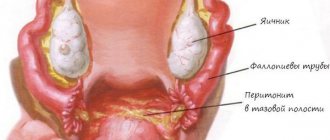
The uterine mucosa becomes pale and thins so that the boundaries of the fallopian tubes are visible. The thin endometrium is able to show through varicose veins in the muscle layer of the organ. As the disease progresses, intrauterine synechiae are formed.
With uterine atrophy, there is a significant decrease in the size of the reproductive organ as a result of age-related involution. The uterus is reduced to the parameters of the tonsil, and during a gynecological examination, the doctor palpates only the cervix. During menopause, the ratio of the size of the body of the uterus and cervix changes, which corresponds to children's sizes.
Basically, atrophic changes occur during menopause. A decrease in hormone production leads to the disappearance of mucous secretion and a change in the bacterial flora. As a result, a gradual reduction in the thickness of the epithelium occurs, which leads to tissue irritation and injury.
Standards for endometrial thickness during menopause have been established. The size of the mucous membrane is determined using ultrasound examination.
The thickness of the normal endometrium during menopause should not exceed 5 mm. When the mucosa thickens, endometriosis is diagnosed.
In addition to menopause, the reasons that provoke uterine atrophy are:
- artificially induced menopause after removal of the ovaries;
- chemotherapy for oncological formations in the female reproductive system;
- hormonal treatment of breast cancer;
- diseases of the endocrine system;
- disruption of the menstrual cycle, amenorrhea;
- taking medications that have an antiestrogenic effect (Tamoxifen, Danazol);
- long-term use of oral contraceptives, antidepressants.
In young women, the detected pathology indicates dysfunction of the endocrine glands. Cervical atrophy is observed in primary infertility, ovarian failure, and menstrual irregularities.
In adulthood, many representatives of the fair sex consider it unnecessary to regularly visit a gynecologist. Unfortunately, in such cases significant complications are often identified:
- complete atrophy of the body of the uterus, cervix;
- hypoxia of the vaginal walls, which provokes the formation of ulcers.
Atrophic changes are an unpleasant consequence of age-related involution of a woman’s body, which is associated with estrogen deficiency.
Clinical manifestations
As mentioned above, the symptoms of genitourinary menopausal syndrome are more persistent the more time a woman has been in menopause.
The clinical picture of vaginal atrophy is characterized by a continuously relapsing course.
So, atrophic vaginitis can manifest itself with the following symptoms:
- Feeling of dryness, persistent, severe itching, burning in the genital area;
- Painful sensations during intimacy (dyspareunia), often accompanied by minor capillary bleeding.
The painful sensations themselves, as a rule, are associated not only with vaginal dryness, which is natural for this period of a woman’s life, but also with thinning of the mucous membrane, as a result of which the nerve endings are “exposed”, providing hyperesthesia (increased sensitivity).
Bloody discharge also occurs due to the thinning of the vaginal epithelium and the exposure of most of the small blood vessels, which are often injured during any intervention, be it sexual intercourse or examination in the mirrors at a doctor’s appointment.
- Leucorrhoea, pathological vaginal discharge. This symptom depends on the severity of the inflammatory reaction and the type of causative agent of vaginitis.
Discharges can be:
- 1Scanty/moderate/abundant;
- 2Watery/mucous/mucopurulent/purulent.
Symptoms of atrophic vaginitis with estrogen deficiency may be accompanied by a decrease in libido, as well as urinary disorders (dysuria, frequent urination, night trips to the toilet, urinary incontinence, a feeling of incomplete emptying of the bladder).
We can say that the main clinical manifestations of atrophic colpitis are not very different from other types of vaginitis, but it is very important to correctly diagnose the atrophic nature of the process, since further treatment tactics depend on this. Let's move on to diagnostics.
Colpitis through the eyes of a specialist
The woman’s discomfort is supported by gynecological examination data. The doctor notes the following changes in the vagina:
- severe vaginal dryness and thinness of its surface;
- mucosal atrophy, pallor, presence of local hyperemic areas;
- sometimes you can find areas without epithelium or loose adhesive zones;
- bleeding even when taking a smear for examination;
- weak expression of the vaginal vault, lack of folding on the walls;
- with the rapid development of pathogenic microorganisms, areas emitting purulent contents may be noticeable.
After the doctor collects an anamnesis, conducts a visual examination and receives data from laboratory tests of a vaginal smear, he will be able to assess the condition of the vaginal lining and give the woman an accurate diagnosis of atrophic colpitis during postmenopause.
Diagnostic methods
It includes both methods of objective examination with elementary tests (Schiller test), and methods of instrumental and laboratory diagnostics: colposcopy, colpocytology, smear analysis for flora and PCR diagnostics of discharge.
5.1. Objective examination
When examined in the mirror, attention is drawn to the pale, thinned mucous membrane with translucent blood vessels and areas of small hemorrhages.
Sometimes the mucous membrane is injured during examination, which is manifested by minor capillary bleeding. The examination itself is quite unpleasant for the woman and causes pain.
In the presence of an infectious process, leucorrhoea is visualized during examination; its quantity and nature depend on the severity of inflammation; hyperemia of the vaginal mucosa is not excluded.
5.2. Flora smear
A smear is taken during a speculum examination from the posterolateral vaginal vault. The nature of the flora (rod, coccal, mixed), the number of leukocytes, desquamated epithelium are assessed; microscopy may detect red blood cells, mucus, and fungal mycelium (concomitant vulvovaginal candidiasis).
5.3. pH-metry of vaginal discharge
One of the express methods for determining the acidity of vaginal fluid using special indicator test strips. The method is very simple, convenient, quick to implement and, most importantly, informative.
By changing the color of the sensory part of the strip after applying vaginal discharge, the acidity of the vaginal environment is analyzed. Normal vaginal pH levels are 3.7-4.5 . Deviations from these figures (towards the alkaline side) indicate the course of the pathological process (colpitis).
5.4. Colposcopy
The examination is performed with a colposcope - a special device with which it is possible to examine the entrance to the vagina, its mucous membrane itself, and the vaginal part of the cervix under high magnification and with high-quality lighting. Colposcopy is distinguished:
- Simple (without special tests). When it is carried out, the character, color of the mucous membrane, and vascular pattern are only visually assessed;
- Extended (with special diagnostic tests using medications).
For atrophic vaginitis, the Schiller test with 3% Lugol's solution is very often performed. It is based on the ability of glycogen to absorb iodine and at the same time change the color of the epithelium.
To carry it out, the doctor, during colposcopy, clears the required areas of mucus and secretions, after which he applies Lugol’s solution and observes the reaction and color change.
5.5. Colpocytology
This study is aimed at assessing the saturation of a woman’s body with estrogen based on the nature of the discharge.
To do this, when examining a woman in the mirror (strictly before conducting a bimanual examination!), using a special instrument (preferably a Papanicolaou pipette, or a spatula or tweezer branches), secretions are collected from the posterolateral vaginal vaults, which are applied in a circular motion to a glass slide.
There are 4 types of colpocytological smears:
- 1Deep type. The epithelium in the smear is represented by cells of the deepest layers (basal and parabasal), the presence of leukocytes is possible. This type of reaction is characteristic of severe estrogen deficiency.
- 2 Mixed-deep type. In addition to the basal ones, a small number of intermediate cells are also found in combination with a large number of leukocytes. The result is also observed with severe estrogen deficiency.
- 3Medium mixed type. Against the background of single basal and parabasal cells and a small number of leukocytes, a fairly large number of intermediate cells are found. This picture characterizes a moderately reduced estrogen level.
- 4 Superficial type, in which keratinizing cells are found in a predominant number against the background of a small number of leukocytes. Basal and parabasal cells are not detected. The result confirms the normal saturation of the woman’s body with estrogen.
5.6. PCR diagnostics of a smear using the Femoflor test system
The Femoflor test system has already become firmly established in clinical practice and allows you to qualitatively and quantitatively assess the composition of the vaginal flora and identify the causative agent of the disease using real-time PCR diagnostics.
The scope of the study can be either the most minimal, including the study of the most typical representatives of the facultative and obligate flora of the vagina (Lactobacillus spp, Gardnerella vaginalis/Prevotella bivia/Porphyromonas spp, Candida spp, Total bacterial mass), or the most extensive with additional identification of pathogenic microorganisms and viruses (gonococci, trichomonas, herpes viruses, cytomegalovirus).
To carry out the analysis, a scraping is required from the posterolateral fornix, which is taken with a disposable probe, placed in a special tube with a nutrient medium, rinsed and the probe is disposed of. The test tube is delivered to the laboratory.
The most common results of examination of a patient with atrophic vaginitis are presented in the table below.
| Study | Deviations |
| Inspection | 1) mild trauma during intervention; 2) minor petechial hemorrhages, possibly capillary bleeding; 3) hyperemia of the mucous membrane, pathological leucorrhoea. |
| Flora smear | 1) leukocytes in the amount of more than 10 in the field of view; 2) unchanged red blood cells; 3) the predominance of coccal flora over rod flora, often the complete absence of rods. |
| pH-metry of vaginal contents | Alkaline environment above 6.0 |
| Colposcopy | 1) thinning, slight trauma to the mucous membrane, areas of hemorrhage; 2) when performing the Schiller test - weak and uneven coloring |
| Colpocytology | Deep type of smear (basal and parabasal epithelial cells predominate) |
Table 1 - Possible results of examination of a patient with atrophic colpitis
Diagnosis of pathology
The first thing to do when unpleasant symptoms appear is to visit a doctor. To make a diagnosis, the patient will be prescribed and undergone the following procedures:
- standard gynecological examination;
- colposcopy (examination of the vagina using a video camera with an image displayed on a monitor screen);
- measuring the acidity level in the vagina;
- smear for infections;
- cytological smear (Pap test for cellular changes that provoke cancer);
- Ultrasound diagnostics of the pelvic organs.
Usually the picture becomes clearer during a gynecological examination, when the doctor sees a thinned, smoothed, as if stretched, surface of the vagina. It can diagnose areas of erosion, hyperemia, minor hemorrhages and purulent foci. Most often, the vaginal mucosa is swollen, has a serous coating and can bleed even with a slight touch. The chronic stage of the disease does not produce such clear symptoms, but they are all slightly present.
After receiving the results of laboratory tests and conducting additional studies, there is no doubt about the diagnosis. The doctor begins to formulate a treatment strategy for the disease.
Drug therapy
The only proven effective treatment for vulvovaginal atrophy is estrogen replacement therapy..
To avoid the development of striking clinical manifestations and profound changes in the vaginal mucosa, therapy should begin no later than 18-36 months after menopause.
If there are signs of an infectious process, the first stage of treatment will always be antibiotic therapy. The choice of drug will depend on the type of pathogen isolated that caused the inflammatory reaction.
6.1. Hormone replacement therapy (HRT)
HRT at the initial stage includes a combination of local and systemic therapy, and then only systemic therapy for 5 years or more (as indicated).
Local therapy consists of local exposure to estrogens to eliminate the identified symptoms of atrophic colpitis.
Preparations in the form of suppositories and ointments containing estriol are used intravaginally; Treatment regimens depend on the drug.
Systemic therapy is aimed at restoring the hormonal imbalance caused in a woman’s body by turning off ovarian function.
These drugs are used for a long time (on average about 5 years), the dosage calculation depends on the individual characteristics of the woman and the intensity of the manifestations of menopausal syndrome.
Principles for prescribing HRT:
- 1It is preferable to prescribe natural estrogens;
- 2The use of minimally effective doses of the drug is indicated;
- 3Start HRT no later than 5 years after menopause;
- 4The drug for HRT is prescribed individually by the attending physician, after an appropriate examination.
Contraindications:
- 1Presence of bad habits (smoking) - with caution;
- 2 Tendency to thromboembolic complications: heart attacks, stroke, history of thrombosis;
- 3Severe arterial hypertension;
- 4 Severe varicose veins;
- 5 Suspicion or presence of cancer, especially breast cancer;
- 6Uterine bleeding of unspecified origin;
- 7 Individual drug intolerance;
- 8Severe liver damage;
- 9 Autoimmune diseases (systemic lupus erythematosus, scleroderma, etc.).
Menopausal hormone therapy was discussed in more detail in another article; to recall the information, follow the internal link.
Table 2 - HRT for atrophic vaginitis. To view, click on the table
6.2. Phytohormone therapy
One of the common options for hormonal correction during menopause is the use of products containing substances that can have an estrogenic effect due to their structure, similar to the chemical formula of estradiol.
The most common drugs:
- 1Klimadinon – available in tablet form, used once a day for a long time (2-5 years with a positive effect);
- 2Cliofit - available in the form of syrup or elixir, used three times a day. Before use, the drug is diluted in an amount of 10-15 ml in 100 ml of water. The course of therapy is no more than 21 days, which can be repeated after 14 days if necessary.
- 3Qi-Klim - available in tablet form, used once a day for a long time (acceptable for up to 5 years).
- 4This group also includes drugs: Climaxan, Remens, Menopace, Estrovel, Feminal and many others.
6.3. Unconventional methods
Therapy with medicinal herbs is carried out only against the background of the use of estrogen preparations.
Herbs only potentiate the effect of hormone replacement therapy, but are in no way suitable for independent prescription and treatment.
The following have a positive effect on partially eliminating the manifestations of atrophic vaginitis:
- Rhodiola rosea – decoction for taking sitz baths for 30 minutes twice a day until acute symptoms disappear;
- Baths with juniper infusion for 40 minutes daily;
- Apply a gauze pad with aloe juice every night until the acute symptoms subside.
Treatment of the disease
Pathology should never be ignored, so treatment of the disease is a key point for every patient. It is very important not only to receive prescriptions from a doctor, but to strictly comply with all his requirements, without hoping that pathological changes can magically disappear. Competent treatment of colpitis and compliance with all the doctor’s requirements is the key to successful and quick relief from atrophic colpitis.
The basis of treatment for the disease is the prescription of hormone replacement therapy. After hormone levels increase, the vaginal mucosa will begin to renew itself in the same way as it did before menopause.
Hormonal drugs are prescribed in the form of tablets or suppositories. It is necessary to take the drugs for a fairly long period of time - from a year to three years, but the first positive changes become noticeable after three months. It is impossible to interrupt treatment of the disease, since this will lead not only to a relapse of the disease, but also to the possible addition of a secondary infection.
Most often, for atrophic colpitis, suppositories Estriol and Ovestin . The main active ingredient of these drugs is the estrogen component, which effectively eliminates vaginal itching, genital dryness, soreness and frequent urge to urinate.
Gynoflor E has a good effect . With the help of lactobacilli acidophilus, the vaginal microflora is normalized, the blood supply to the vaginal epithelium is improved, the formation of new cells is stimulated, and normal vaginal acidity is maintained due to the development of lactic acid bacteria in the woman’s vagina.
Among other equally effective drugs, Elvagin , Orthoginest , Estrocard , Estrovagin , Ovipol Clio .
To reinforce local treatment, systemic drugs are also prescribed - Climodien, Cliogest, Divina, Pauzogest. The drugs are prescribed for early signs of atrophic colpitis, but after the complete end of menstruation, and Kliogest can be used as a preventive measure for pathology. Doctors also recommend continuing to take standard medications that are indicated for menopause - Activel, Cliofit, Eviana, Klimadinon, Menopace and others.
Contraindications
In some cases, women are not prescribed hormonal drugs. Hormone replacement therapy should not be used in patients who suffer from breast cancer, endometrial cancer, bleeding, or vascular thromboembolism. It is not recommended for those who have liver problems or pathologies of the cardiovascular system (myocardial infarction, angina).
In this case, therapy is replaced by other drugs that do not contain hormonal components. These can be douches and baths with decoctions and infusions of herbs, vaginal suppositories with antibacterial and anti-inflammatory effects.
Atrophic colpitis, unfortunately, is a familiar phrase for many women who have entered menopause. However, such changes in the body should not be taken with a negative connotation. The natural aging process cannot be delayed, but degenerative changes can be slowed down. This will not only prolong a woman’s healthy period, but will also help her endure the changes that occur to her body during menopause as easily as possible.
Interesting and educational video on this topic:
About
Monitoring the effectiveness of therapy
When a woman is treated with hormonal drugs, especially at the very beginning of therapy, regular visits to an obstetrician-gynecologist are necessary to monitor the effectiveness.
This is achieved using:
- 1pH-metry, as a result of which the doctor should note a gradual shift in pH to the acidic side. Naturally, you should not expect indicators typical for a woman of reproductive age. A result in the range of 4.5-5.5 is considered good.
- 2 Colpocytology – restoration of the epithelium to medium mixed or superficial types.
- 3 Colposcopy - mature epithelium is observed.
Preventive actions
- 1 Avoid wearing tight, synthetic underwear;
- 2Annual observation by an obstetrician-gynecologist;
- 3Use of intimate hygiene products without fragrances, preservatives, parabens and other irritating substances;
- 4 Avoid unprotected sex;
- 5Performing physical exercises and Kegel exercises (to strengthen the pelvic floor muscles).
It must be remembered that upon the onset of menopause, hormonal support must be started on time in order to avoid the bright manifestations of the menopause.
The doctor, in turn, must inform patients about all the features of this period in a woman’s life and its possible clinical manifestations.
Video Photo Tables
| Study | Deviations |
| Inspection | 1) mild trauma during intervention; 2) minor petechial hemorrhages, possibly capillary bleeding; 3) hyperemia of the mucous membrane, pathological leucorrhoea. |
| Flora smear | 1) leukocytes in the amount of more than 10 in the field of view; 2) unchanged red blood cells; 3) the predominance of coccal flora over rod flora, often the complete absence of rods. |
| pH-metry of vaginal contents | Alkaline environment above 6.0 |
| Colposcopy | 1) thinning, slight trauma to the mucous membrane, areas of hemorrhage; 2) when performing the Schiller test - weak and uneven coloring |
| Colpocytology | Deep type of smear (basal and parabasal epithelial cells predominate) |