Concept
The very concept of “atypical” means something not within the normal range, abnormal. If we consider this term in gynecology, it means the presence of a dysfunction of cells, their deformation.
What is cervical atypia? Atypical disorders are provoked by a rapid increase in the number of cells, resulting in the formation of abnormal layers. These layers contribute to disruption of the functioning of the entire cervix, as disruptions in the blood circulation process occur in it. In this case, there will already be an anomaly of blood vessels.
Normally, every woman’s body has cells with an altered genome, but with the normal functioning of the immune and other systems, they are neutralized and destroyed.
All cells in the body have the ability to independently eliminate disorders. If, due to some factors, the cells cannot independently organize their work, then they begin to degenerate into cancer cells.
Causes
Cervical atypia occurs as a result of diseases such as dysplasia, leukoplakia, and human papillomavirus.
Additional causative factors may include:
- hormonal imbalances;
- chronic decreased immunity;
- repeated abortions and childbirth during which the cervix was damaged;
- frequent and incorrect use of sanitary tampons or barrier contraceptives, for example, vaginal diaphragms and cervical caps;
- frequent changes of sexual partners;
- alcohol and nicotine abuse.
These factors contribute to the disruption of the normal functioning of body cells and metabolic processes. In this case, cells are formed that have an altered genome.
How to treat
Treatment of a precancerous condition of the cervix begins immediately after determining the type and extent of the pathology.
Conservative therapy
Therapeutic treatment methods are usually carried out in several stages. This is required by the etiology of precancerous and background diseases. Usual sequence of therapy:
- Antibacterial, antiviral and hormonal: drugs are prescribed in accordance with the reasons that caused the disease of a particular patient. Additionally, immunomodulators, probiotics and antihistamines can be used.
- Local treatment: vaginal sanitation.
After the therapy, the results are assessed. Most prescribed medications help normalize hormonal levels and eliminate the cause of illness. In some pathological conditions this is quite sufficient, but most often the next step is surgical intervention. For example, polyps (especially adenomatous ones) are not eliminated by therapeutic agents. The doctor makes a decision based on the clinical picture and the degree of danger to the patient’s life (threat of cancer development).
Surgery
Surgical methods for the treatment of background and precancerous diseases of the cervix in modern medicine are varied and effective. To completely eliminate the pathological focus, the following can be used:
- Local destruction: laser, radio wave, chemical, cryodestruction, diathermosurgical method.
- Radical surgery: hysterectomy, amputation or excision of the cervix, reconstructive plastic techniques.
Surgical removal of the pathological focus is not the last stage in the treatment of patients. In the future, therapeutic drugs are used to normalize all impaired body functions. It could be:
- treatment of the postoperative field of the cervix with antibacterial and antiseptic agents to prevent the risk of infection;
- taking medications containing hormones, immunomodulators, vitamins;
- the use of folk remedies to restore normal functions of the genitourinary system.
All patients must be monitored by a gynecologist for at least 2 years, as there is a risk of relapse. Particular attention is paid to prevention: giving up bad habits, preventing re-infections with infectious and sexually transmitted diseases, changing the method of contraception, etc.
Symptoms and signs
There will be no pronounced symptoms with cervical atypia. This is where the insidiousness of this pathological condition manifests itself. A characteristic symptom is discomfort in the lower abdomen, and at a later stage there is already a nagging pain. A woman often does not notice this sign at all, as she associates it with menstruation.
If atypia is accompanied by an infection, the following symptoms may occur:
- vaginal discharge (ichor or thicker, spotting type), often accompanied by blood;
- pain during sexual intercourse;
- itching in the genital area;
- unpleasant odor from the vagina, and sometimes even foul.
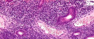
The diagnostic signs of the pathological condition of the cervical tissue are clearly expressed, since the pathological cells are significantly increased in size, they have a variety of shapes, some cellular structures merge, while others disintegrate. Pathological cells developing on the cervix are characterized by cell degeneration, as well as their vacuolization.
Precancer of the uterus - what to do?
If the underlying conditions can be managed without surgery, then the obligate variant requires serious surgical interventions. Precancer of the uterus is a precarious balance over the edge of an abyss : balancing between recovery and the appearance of a malignant tumor, it is important to accurately and correctly follow the doctor’s recommendations and prescriptions. There are many chances to prevent the development of carcinoma, but we remember that the enemy is insidious and implacable. Refusal to surgically remove the uterus can cost a woman her life (by leaving a source of cancer cells, conditions are created for cancer progression). Therefore, in case of hyperplasia, the doctor will treat with tablets, and in case of any adenomatous changes in the cells, one must agree to surgery.
The article was published in Yandex.Zen Onkos
Diseases with atypia
The most common causes of atypia are:
- Postmenopausal squamous atypia of the cervix.
- Dysplasia.
- Leukoplakia.
Postmenopausal squamous atypia
This disease occurs in women over 50 years of age. This is characterized by two or more nuclear cells. In these cells, the nucleus doubles in size.
Dysplasia
This disease is classified as a precancerous condition. Since normal cells of the vaginal part of the uterine cervix turn into atypical ones. Violations of all indicators occur: structural changes, rapid growth, periods of maturation and destruction are disrupted.
Dysplasia develops in 3 stages. In mild cases, a small area of the cervix is affected, namely the lower stratified epithelium. The moderate stage is characterized by the development of atypical cells at the level of the lower layer of the epithelium. A severe form of dysplasia is the presence of atypical cells in all its layers. This stage is considered non-invasive cancer.
Leukoplakia
This disorder is characterized by an atypical change in the squamous epithelium, namely the process of keratinization of individual areas of the cervix. As a result, white spots or plaques visually appear on it.
This disease can often be diagnosed only during examination by a gynecologist, since the symptoms are not pronounced.
Precancerous diseases depending on location
Female reproductive system
Precancerous diseases of the female reproductive system include diseases of the genitals and mammary glands. Predisposing factors for their development are hormonal disorders, human papillomavirus, oral contraceptives, heredity, early and late menarche, multiple births, lack of childbirth and a history of breastfeeding. The group of precancerous diseases of the female reproductive system includes the following:
- Cervical dysplasia.
- Kraurosis of the vulva and vagina.
- Some types of ovarian tumors, such as serous or mucinous.
- Endometrial hyperplasia.
- Leukoplakia of the cervix and vagina.
Precancerous diseases of the mammary gland include some types of mastopathy, hormonal hyperplasia, ductal hyperplasia, etc.
Treatment of precancerous diseases of the female reproductive system depends on the specific pathology. In some cases, anti-inflammatory therapy and hormonal treatment are required, in others - surgical intervention. Regular examination by a gynecologist, ultrasound of the pelvic organs and mammary glands, as well as mammography from a certain age (after 40 years) will help to detect the problem in a timely manner. In most cases, preventive measures come down to timely treatment of hormonal disorders, proper contraception and prevention of abortions. Some diseases cannot be prevented.
Separately, it is worth dwelling on cervical dysplasia. This disease is asymptomatic, so a woman may not be aware of its presence. However, it is easily detected using routine cancer screening, which is offered by any antenatal clinic. It is based on a cytological examination of a scraping of the cervix; some programs also offer the determination of the human papillomavirus.
Treatment of dysplasia involves different options for removing the pathological focus. For dysplasia of the first or second degree, gentle treatment is indicated, for example, radio wave surgery, laser vaporization, cryodestruction and many other options. These methods do not leave rough scars and do not subsequently affect the patient’s reproductive function.
When treating severe dysplasia, a more radical intervention is necessary - conization of the cervix or amputation of the cervix. Such operations in the future require careful pregnancy planning and special approaches to its management.
After treatment, the patient should undergo regular examinations with oncocytology tests in order to detect a possible relapse in time. First, for the first year they are done once every three months, the next year once every 6 months, and then annually. Prevention of precancerous diseases of the cervix is vaccination against the human papillomavirus and the use of barrier methods of contraception.
Precancerous skin diseases
Precancerous skin diseases include the following pathologies:
- Bowen's disease. Can affect any area of the skin. At the initial stage, it looks like a red spot with uneven contours, which gradually transforms into a copper-red plaque with a velvety surface. On its surface there may be areas of pigmentation change and scales. If left untreated, it progresses to squamous cell skin cancer.
- Keir's erythroplasia is a type of Bowen's disease, only localized on the penis.
- Paget's disease. It is most often localized on the skin of the chest, but can be on any part of the body. At the initial stage, it resembles eczema - it looks like a red spot that itches and hurts, there may be tingling and burning. Then the neoplasm grows, scales and ulcers appear on it. If left untreated, the tumor begins to infiltrate the subcutaneous tissue and transforms into cancer.
- Xeroderma pigmentosum is a rare hereditary skin disease. It occurs in three stages. The first occurs at the age of 2-3 years and is manifested by red inflammatory spots that appear after exposure to the sun. Later, areas with disturbed pigmentation remain in these places. The second stage develops after a few years. Atrophic changes and telangiectasia form in the affected area. The skin becomes mottled, various growths appear on it in the form of warts and crusts. There may be sores. At the same time, eye symptoms are noted - conjunctivitis, photophobia, corneal clouding. The third stage develops in adolescence and is characterized by various skin neoplasms of both benign and malignant nature, from angioma and fibroma to squamous cell carcinoma and melanoma.
Precancerous diseases of the gastrointestinal tract
Precancerous diseases of the gastrointestinal tract include various pathologies:
- Peptic ulcer disease. The likelihood of malignancy depends on the location of the ulcer and its size. The larger the defect, the higher the likelihood of its malignant transformation. Ulcers of the curvature of the stomach become malignant in 100% of cases.
- Atrophic gastritis is accompanied by a decrease in the acid-producing function of the stomach and atrophic changes in its mucosa. The probability of malignancy is about 13%.
- Adenomatous polyps of the stomach and colon. The larger the polyp and the longer it exists, the greater the likelihood of it degenerating into cancer.
- Crohn's disease and ulcerative colitis are chronic inflammatory diseases of the large intestine.
- Barrett's esophagus is characterized by gastric metaplasia of the epithelium in the lower third of the esophagus. Develops due to chronic reflux.
- Colon polyposis syndrome. Characterized by multiple intestinal lesions. In some patients, the number of polyps exceeds a hundred, and they always degenerate into cancer.
Patients with such pathologies require regular medical monitoring, including esophagogastroscopy and colonoscopy. If there is a high risk of malignant transformation, surgical treatment is performed, for example, polyps or part of the colon are removed, ablation of dysplasia foci is performed, or other minimally invasive methods are used.
Lungs
Lung cancer usually develops over a long period of time against the background of the following precancerous changes:
- Pneumosclerosis is a thickening of lung tissue.
- Pneumoconiosis is an occupational disease caused by prolonged inhalation of industrial dust. Its variety is anthracosis, an occupational disease that develops due to the deposition of coal dust in the lungs.
- Chronic nonspecific lung diseases, for example, chronic obstructive pulmonary disease, chronic bronchitis.
- Scar changes after tuberculosis.
Patients with precancerous lung diseases are subject to clinical observation.
Genitourinary system
The most common precancerous disease of the urinary system is leukoplakia of the bladder - the transformation of the urothelium that lines the bladder into stratified squamous epithelium. It manifests itself as an increased urge to urinate and chronic pelvic pain. Subject to surgical excision using minimally invasive methods.
Precancerous diseases of the thyroid gland
A precancerous disease of the thyroid gland is an adenoma - a conditionally benign tumor that grows from the glandular tissue of the organ and has its own hormonal activity, which in some cases leads to symptoms of hyperthyroidism. These include rapid heartbeat, anxiety, irritability, weight loss, arrhythmias, etc. Treatment involves surgical removal.
As we see, in most cases, precancerous diseases are subject to either dynamic observation or surgical excision. In rare cases, the problem can be eliminated with medication. However, timely surgery helps to avoid transformation into cancer and, as a result, difficult treatment with extensive interventions, chemotherapy and radiation therapy. In many cases, it even helps save lives.
At the European Clinic, experienced oncologists monitor patients with precancerous diseases.
The modern equipment of the clinic allows you to quickly provide adequate treatment in accordance with European standards. Book a consultation around the clock +7+7+78
Diagnostics
Atypical changes in the cervix should be diagnosed on time, since otherwise the altered cells can transform into malignant ones. The first diagnostic method is an examination by a gynecologist in a chair. During the examination, the doctor takes a smear from the vagina for a cytological examination. Often the examination is carried out immediately with a colposcope. That is, with the help of an optical apparatus that allows you to examine the cervix with all the subtleties and diagnose even small areas of atypia.
The smear and scraping are examined by performing a Papanicolaou cytogram. After staining the biological material, it will be clear what type of pathological process is inflammation or malignant.
The following studies are also required:
- General and biochemical blood tests;
- Blood and urine test for hormones;
- Biopsy.
Cervical cancer
In cancer, proliferating leukoplakia is noted. It is located in the area of the ectocervix. White, lumpy lesions with clearly defined boundaries rise above the epithelium.
A clear sign that the process has taken a malignant course is the polymorphism of vascular and epithelial structures. They can be of different shapes, differ in size, shade (white, yellow, transparent). It is impossible to determine the vascular pattern. The Schiller test result is negative.
Fields of atypical epithelium are areas of epithelium surrounded by wriggling pinkish and red stripes, having clear boundaries and characterized by polymorphism. Their relief is concave. They are usually located in the vaginal area of the cervix.
Papillary zone of atypical epithelium - polymorphic foci are located in the area of the external os of the cervical canal. Colposcopy reveals atypical epithelium, which looks like thick, overgrown yellowish layers.
The atypical transformation zone is characterized by the presence of “rims” of epithelium that surround the ductal openings. The rims are polymorphic in nature. Branching vessels remain visible even after treatment with acid, that is, their adaptive hypertrophy takes place.
Atypical vascularization - proliferation of atypical vessels occurs. They are characterized by uneven expansion and do not respond to the action of drugs. The boundaries of such a pathological zone can only be seen using the Schiller test. After it, the area of the epithelium with altered vessels remains unstained.
Cancer in situ
At this stage of the malignant process, epithelial cells undergo malignancy, but the carcinoma still lacks the ability to infiltrate and metastasize. The typical localization of the formation is at the border between columnar and squamous epithelium. In young patients, the external pharynx is more often affected; in mature women, the cervical canal is usually affected.
There are two types of carcinoma in situ:
undifferentiated and differentiated. Cancer is called differentiated if its cells retain the ability to mature. With undifferentiated neoplasms, the epithelial layer loses its layering. Patients with this diagnosis complain of pain in the lower abdomen, bloody discharge and leucorrhoea.
Microinvasive cancer
Such tumors are considered intermediate between carcinoma in situ and the invasive form. They are relatively compensated and not too aggressive. Since such carcinoma belongs to the preclinical stage, no specific manifestations are observed with it.
Invasive cervical cancer
At this stage, the disease manifests itself with bloody discharge, leucorrhoea, and severe pain. Pain sensations are mainly concentrated in the abdomen, rectum, and lower back. As the disease progresses, the tissue of the pelvic lymph nodes is involved in the malignant process. And then the pain begins to radiate to the thigh area.
Since the vessels in the tumors are injured, bleeding often occurs. Leucorrhoea may be serous or mixed with blood. The discharge often smells unpleasant. Leucorrhoea appears due to the fact that as a result of the disintegration of the tumor, the lymph nodes are opened.
If the tumor invades the bladder, then the patient’s urge to empty it becomes more frequent. The ureter is compressed, so pyo- and hydronephrosis develops, and over time, uremia. When the tumor spreads to the rectum, the patient suffers from constipation, and blood and mucus are present in her stool, and fistulas form.
Publication date 12/05/2016
Treatment
The treatment method is selected depending on how deeply the epithelium is affected. Therefore, conservative or surgical treatment is used.
Conservative therapy
Drug therapy is effective if atypia is diagnosed at an early stage. Complex treatment is aimed at relieving the inflammatory process, stopping abnormal processes in cells, and it is also necessary to restore normal microflora in the vagina. It is very important for a woman to take medications to strengthen her immune system.
If there is a history of additional infectious diseases, then it is necessary to prescribe antibacterial, antifungal, and antiviral drugs.
For cervical atypia, topical medications are the most effective. That is, suppositories, and physiotherapy in the form of douching with various solutions can also be prescribed.
Surgical treatment
Various methods are used for this. The modern and most common is laser. A special beam affects atypical cells, dehydrating them. This method is contactless and gentle.
The radio wave method involves exposure to radio waves. This procedure can be carried out in all clinics. But after this a relapse may occur.
Dysplasia is often treated by treating the cervix with liquid nitrogen. After this, the disease loses its activity and is rejected.
Cauterization of atypical areas is also carried out using electric current. Electrical waves are sent to a special loop that is in direct contact with the affected areas. This method is the most traumatic and painful. Also, such cauterization leaves scars and, as a result, deformation of the cervix occurs.
The surgical method involves performing operations with a scalpel. That is, during the operation, atypical areas are excised. Sometimes complete removal of the cervix or resection of the uterus is required. The latter operation is required for a malignant process.
Precancer of the uterus - background and obligate conditions
Every woman needs to monitor her periods. Disturbances in rhythm and abundance can be one of the first signs indicating uterine precancer. There are 2 variants of diseases:
- Background conditions (endometrial hyperplasia, uterine polyp);
- Obligate precancer of the uterus (endometrial adenomatosis, adenomatous polyp).
Initial changes in the form of hyperplasia extremely rarely lead to cancer (the risk is no more than 1%), but they are obligate - almost every second woman is pushed to the edge of the abyss (40%). It is important to consider risk factors that provoke precancerous changes:
- age (over 35 years);
- metabolic and endocrine diseases (the combination of obesity, diabetes and high blood pressure is especially dangerous);
- hormonal disorders (many estrogen hormones, few gestagens);
- gynecological diseases (fibroids, endometriosis, ovarian cyst);
- chronic inflammation in the reproductive organs (endometritis, cervicitis, adnexitis);
- prolonged wearing of a foreign body in the uterus (spiral);
- frequent abortions (2 or more).
Cancer is insidious, patient and unforgiving - having received its chance, the tumor will begin to grow slowly and gradually, going through all the stages of pre-tumor growth.