Endometriosis is a systemic and immune-dependent disease. Removal of the uterus is not a radical operation, but only a symptomatic treatment that is carried out under circumstances.
The main indications for surgical interventions are as follows:
- combination of adenomyosis and uterine fibroids;
- heavy bleeding, which leads to a serious decrease in hemoglobin levels;
- combination of adenomyosis and endometrial pathology (polyps, hyperplasia);
- ovarian cysts (including endometrioid) and adenomyosis in menopausal women.
Removal of the uterus should be approached in a differentiated manner, because without this organ a woman loses her reproductive function.
Ovarian endometriosis usually occurs in young girls. Treatment of pathology is often carried out in preparation for pregnancy or as a stage of IVF. Removal of the uterus for ovarian endometriosis is rarely performed, for example, in the following cases:
- If during surgery to remove cysts their malignant nature cannot be ruled out. Endometriosis is a benign pathology. But in 5-7% of cases, malignant cells may appear in the lesions, most often when the ovaries are affected.
- In women in menopause or after 45 years of age when combined with uterine fibroids and endometrial pathology.
Removal of the ovaries and uterus leads to castration - a condition identical to menopause occurs abruptly. The level of estrogen drops, which leads to regression of other foci of endometriosis.
This is the most radical way to get rid of the disease , but along with it other problems arise: hot flashes, sweating, a tendency to gain weight, mood changes, destruction of bone tissue and other problems. If only one ovary is removed (with or without the body of the uterus), this condition develops gradually. Sometimes the remaining appendage takes on the function of both, which leads to its hypertrophy (enlargement) and often cystic changes.
The following factors speak in favor of surgical intervention:
- severe endometriosis that is not amenable to conservative treatment. Removal of the uterus in this case leads to an improvement in well-being;
- if for a number of reasons taking pills or giving injections is impossible to suppress endometriosis, for example, with multiple drug allergies;
- combination with other diseases of the genital organs: large fibroids or multiple pathologies of the endometrium, ovarian cysts;
- heavy menstruation with a constant decrease in hemoglobin levels and lack of effect from other treatment.
In such situations, removal of the uterus significantly improves the woman's quality of life. Blood counts normalize and manifestations of endometriosis go away. There is no need to remove the uterus:
- if a woman is planning a pregnancy or is under 35 years of age;
- in cases where conservative treatment has not yet been applied;
- with mild endometriosis and the absence of other indications for surgery.
Possible consequences after removal of the uterus for endometriosis:
- a woman will never be able to bear a child on her own;
- even if the ovaries are preserved, menopause occurs faster than with the preserved organ;
- adhesive disease may develop, especially against the background of previously established endometriosis, and this can cause pain and discomfort in the lower abdomen;
- operations for endometriosis with removal of the uterus often require highly qualified and experienced surgeons and are accompanied by a high risk of postoperative complications;
- After removal of the uterus, a woman may notice changes in sensations in the intimate area, problems with bowel movements and urination.
Surgical options:
- amputation: removal of only the body while preserving the neck;
- extirpation: if the cervix is removed in addition to the body, the vagina is subsequently completed blindly.
The issue of appendages is decided individually in each case . Resection of part of one or both, complete removal of the right or left, or all of them may be performed.
The operation can be performed laparoscopically or laparotomically (classic version) . At the same time, other endometriotic ectopia in the peritoneum, intestines, bladder and other places are removed. Preference should be given to laparoscopic surgery , since the degree of trauma is less, recovery is much faster, and wounds on the skin of the anterior abdominal wall after laparoscopy are almost invisible over time.
a) Laparotomy and b) laparoscopic method
Recovery after surgery takes at least 2 months, and in some cases longer . In the case of severe endometriosis, when this particular pathology has become the main indication for intervention, taking hormonal drugs for replacement purposes (to prevent menopause symptoms and improve the woman’s general well-being) is not recommended. The doses of estrogen contained there can provoke the progression of the disease.
If endometriosis was a concomitant indication, and many foci of other localization were not identified, then such drugs can be used. Depending on the woman’s age, these can be regular oral contraceptives (preference should be given to “Klaira”, “Janine”, “Bonade”) or adapted ones (“Femoston” and the like).
After the rehabilitation period, a woman can lead her usual lifestyle, including intimate relationships. Even after removal of the uterus, you should regularly visit your doctor, especially if amputation was performed, to monitor the condition of the cervix.
Read more in our article about hysterectomy for endometriosis.
Classification and degrees of development
Experts highlight:
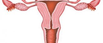
- genital endometriosis, which can be internal (if pathological foci are detected in the body of the uterus or cervical canal) and external (if, for example, the ovaries, fallopian tubes, vaginal part of the cervix, pelvic peritoneum, vagina are affected);
- extragenital endometriosis (in this case, lesions can be found outside the reproductive system: in the gastrointestinal tract, lungs and other organs);
- combined form.
The degrees of endometriosis are determined based on the number, location, size of heterotopias and the depth of penetration of the lesions into the tissue. 1st degree – a small number of superficial lesions; 2nd degree - heterotopias become larger and begin to penetrate deep into the tissues (for example, with adenomyosis at this stage the middle layers of the muscular layer are affected); 3rd degree – deep layers of tissue are affected; 4th degree - heterotopias are present in large numbers, growing deep into the tissue and affecting other organs located nearby. A common adhesive process is diagnosed.
Rules for the operation
Surgery for endometriosis is performed three days before the onset of menstruation.
A common form of endometriosis, in which the ovaries and pelvic peritoneum are affected, conglomerate tumors and chocolate cysts are diagnosed, requires immediate removal. In this case, doctors can completely remove the uterus and ovaries. Conservative surgery, during which the unaffected ovary is preserved, is indicated for girls who want to give birth to a child. In this case, complete removal of all endometrioid tumors is indicated.
During a comprehensive diagnosis and examination, the doctor checks the uterus and peritoneum for the presence of lesions. If patients are found to have widespread pathological formations, then surgery for endometriosis has its own difficulties. With this course of the disease, tissue that is very close to the bladder, ureter and rectum is affected. Due to the high likelihood of injury, doctors limit themselves to removing not all endometrioid heterotopias that are located inside the abdominal cavity. Even with such an operation, the development of pathological processes in the future stops. Surgical treatment in women of menopausal age is carried out with radical removal of the uterus and appendages.
Removal of foci of adenomyosis in menopausal women is carried out with ablation - excision of the basal layer to prevent further growth of the endometrium. Young women undergo less invasive surgery. Curettage, cauterization of lesions and hormonal treatment are carried out, introducing a medicated menopause for 6-9 months.
Symptoms of endometriosis in women
At the initial stage, there may be no symptoms of endometriosis, and it is often detected during preventive examinations or diagnostic measures associated with other diseases. In later stages, endometriosis usually has the following symptoms:
- dysmenorrhea (painful periods). Up to 60% of women with endometriosis experience this symptom;
- pain in the pelvic area, which is felt by approximately every 5 patients;
- dyspareunia (pain during intimate contact);
- menorrhagia. Menstruation, during which greater than usual blood loss occurs, is characteristic primarily of adenomyosis. This symptom is less common (approximately 2-16% of patients);
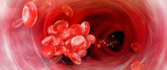
- anemia developing as a result of menorrhagia, which is caused by constant blood loss exceeding the norm;
- pain during urination or defecation;
- spotting intermenstrual discharge from the genital tract (usually in the second half of the cycle), which is characteristic of vaginal endometriosis. With adenomyosis, brownish spotting usually occurs immediately before and after menstruation;
- infertility, which is a fairly common symptom of endometriosis. According to statistics, up to 40% of women with this pathology experience difficulties conceiving.
How is endometrial polyp removed?
An endometrial polyp is a growth of excess tissue inside the uterine cavity. A polyp is formed due to excessive growth of endometrial cells. There may be several or one such growth, and the size of the polyps can be a couple of millimeters or several centimeters.
Neoplasms on the uterine mucosa do not respond to standard treatment, and surgical intervention is usually used to remove the polyp. Modern medicine allows such operations to be performed with high efficiency. Using hysteroscopy as a diagnostic method, you can visualize the uterine cavity and find out the exact location of the polyp during surgery.
Removal of an endometrial polyp is a mini-operation to eliminate neoplasms (polyps) on the walls of the uterus or its cervix.
An endometrial polyp is diagnosed accidentally, during a routine examination, ultrasound, or in the process of identifying the cause of infertility. The disease is practically asymptomatic or manifestations may indicate other diseases. An endometrial polyp can cause the following symptoms:
- slight spotting before or after menstruation;
- bleeding between periods;
- increased discharge during menstruation;
- discharge with blood clots before or after menopause;
- infertility and failure to conceive through IVF.
Weakness, apathetic mood, and fainting may also develop. When a large polyp forms, it can put pressure on neighboring organs, thus causing discomfort and spasms. Painful sensations in the lower abdomen systematically appear, daily discharge (leucorrhoea) acquires a thicker consistency and darker color. A polyp during pregnancy can negatively affect the development of the fetus. And women of childbearing age risk remaining infertile if endometrial polyps from the uterus and cervix are not removed in time. Such neoplasms are dangerous because the indicated symptoms are characteristic of other diseases, and women do not always consult a doctor in time to restore their health.
Diagnostics
Differential diagnosis plays an important role, since the signs of endometriosis are not specific and can be characteristic of various diseases of the pelvic organs. To identify the disease and determine its stage, the doctor may prescribe tests such as:
- computer (CT) and magnetic resonance (MRI) imaging, with the help of which the doctor can determine the nature of the pathology and the presence of lesions in various organs. These methods are more often used for severe forms of endometriosis, for example, retrocervical, when there is a suspicion of involvement of the rectal wall or ureters in the endometrioid infiltrate;
- Ultrasound examination of the pelvic and abdominal organs is a nonspecific method, i.e. it will not allow you to determine the localization of the lesions, but there is a certain group of narrow specialists - ultrasound diagnostic doctors who specialize in endometriosis and can describe a certain anatomical picture. But ultrasound is excellent for identifying adenomyosis;
- hysteroscopy, which is performed if adenomyosis is suspected, makes it possible to study the uterine cavity in detail for the presence of endometrioid foci. But this method is also non-specific;
- Laparoscopy is a minimally invasive surgical intervention that allows you to identify pathological foci even at an early stage and eliminate them in a timely manner. It is the “gold standard” in the diagnosis of endometriosis;
- and the simplest and most informative method for diagnosing endometriosis and identifying endometrioid infiltrates is a routine bimanual examination by a doctor on a chair, i.e. The doctor can feel painful lumps in the vagina with his hands.
Recovery after laparoscopy
Laparoscopy for endometriosis requires some recovery time. Typically, the patient goes home after the surgery is completed, especially if it is just a diagnostic procedure.
However, there are some cases where you may need to spend an overnight stay in the hospital, for example if the endometriosis was extensive and the surgeon took a long time to remove all the damage.
Your doctor may prescribe painkillers for your surgery. Rest and avoiding excess stress on the abdomen may also help.
Avoid heavy lifting for the first few weeks after surgery to reduce the chances of a hernia.
Recovery time after laparoscopy varies, but if there are no complications, most patients can return to their normal activities within a week.
The first time after undergoing a laparoscopy procedure may be more difficult and painful than usual. If necessary, take time to rest and prepare additional painkillers and hygiene products in advance.
Recovery Tips
There are many ways to help you recover faster after laparoscopy. These include:
- Comfortable delivery home and care in the first days after laparoscopy.
- Placing a pillow or sweater between the seat belt and the abdomen to reduce pressure on the wounds during the ride home.
- During transport, the patient may vomit, so it is better to have a container with you in this case.
- Drink peppermint tea to relieve stomach pain due to the presence of gas. The gas can cause pain in the abdomen and shoulder and can take up to several weeks to leave the cavity.
- Taking quiet walks in the first days after the procedure will help get rid of gas in the abdominal cavity faster.
- Sanitary pads can help manage vaginal bleeding caused by laparoscopy. You should not use tampons or insert anything into your vagina during the recovery period.
- Recover as long as possible, do not go to work or study, trying to do this at home.
After laparoscopy, a woman may be emotional, cry, or feel weak.
It is important to take steps slowly after laparoscopy for endometriosis to ensure a complete recovery.
Our advantages
All operations are performed by surgeons with extensive experience
We do not prescribe tests, studies or operations unless absolutely necessary
We use the best latest generation equipment
Conservative therapy
Treatment involves taking hormonal medications, which are necessary to reduce the level of sex hormones. As a first-line drug, the doctor can use gonadotropin-releasing hormone analogues and gestagens. For adenomyosis - installation of a levonorgestrel intrauterine system (Mirena). As a second line, the doctor may recommend taking COCs (but this group of drugs is not specific for the treatment of endometriosis). Danazol (a synthetic androgen) has proven itself in the treatment of endometriosis, allowing not only to sharply reduce estrogen production, but also to directly affect heterotopias, promoting their regression. In addition, as a result of its use, pain is reduced. Non-steroidal anti-inflammatory drugs (ibuprofen, naproxen) can be prescribed as symptomatic treatment for pain. You also need to remember that drug therapy is temporary and does not relieve the patient of the disease! These drugs are used to obtain certain clinical bonuses that the doctor needs to achieve the desired clinical effect, for example, preparing a patient for an IVF protocol who cannot undergo surgical treatment.
Surgical treatment of endometriosis
Surgery is an effective treatment method - the “gold standard”. The operation means laparoscopic access, and the scope of the operation is the removal of all detected foci and infiltrates of endometriosis, including endometrioid (“chocolate”) ovarian cysts.
In order to prevent relapses for patients who do not plan a future pregnancy, so-called suppressive therapy may be prescribed after surgery - drugs from the group of gonadoliberin agonists (diferelin, buserelin, zoladex, etc.) or gestagens that suppress the hormonal function of the ovaries. The result of treatment is a condition called artificial menopause, during which relapse is impossible, but after discontinuation the chances of relapse increase again. If a woman is planning a pregnancy, treatment for endometriosis should be discussed with both a reproductive specialist and an operating gynecologist with experience in endometriosis surgery. Before laparoscopy, an assessment of ovarian reserve is necessary, after which further strategies for achieving pregnancy are discussed. This is especially true for patients of late reproductive age (over 35 years old). Young women (under 35 years of age) after combined treatment of endometriosis are recommended to plan spontaneous pregnancy over the next 12 months (depending on the stage of endometriosis and the age of the patient). If pregnancy does not occur, it is necessary to decide on IVF without waiting for the depletion of the ovarian reserve or relapse of endometriosis.
Indications for surgical treatment
The main goal of any treatment for endometriosis should be complete removal of pathological foci . Only surgery can fully cope with this task, and subsequent hormone therapy can be aimed at preventing relapses of the disease. However, there are situations when surgical intervention cannot be avoided. So the indications for the operation are:
- retrocervical localization of endometriosis;
- the presence of an endometrioid ovarian cyst;
- endometriosis of the uterus (adenomyosis), occurring together with fibroids, complicated by uterine bleeding;
- ineffectiveness of drug therapy even for uncomplicated forms of endometriosis.
Methods for preventing endometriosis
The earlier the disease is detected, the greater the chance of completely eliminating foci of endometriosis without resorting to surgery. The main preventive measures are:
- regular scheduled examinations with a gynecologist;
- examination for the presence of endometriosis in patients with dysmenorrhea;
- careful monitoring of the condition of women who have undergone any surgical intervention on the uterus;
- timely treatment of inflammatory diseases of the pelvic organs;
- choosing COCs as a means of contraception.
Sign up for an appointment with the doctor
Types of endometrial polyp
- A fibrous polyp is formed primarily from fibrous connective tissue cells. It may contain collagen fibers and only single glands lined with non-functioning epithelial cells.
- Glandular fibrous polyp is rare and predominates in women with a stable menstrual cycle. Consists of glands of irregular shape and varying lengths. The phenomena of inflammation and circulatory disorders in formations of the glandular-fibrous type are more common than in others.
- Adenomatous endometrial polyp is rare in its pure form. Tumors with focal adenomatosis are more common. Polyps of this type can transform into malignant tumors, especially after menopause, against the background of metabolic and endocrine disorders.
Surgical technique
Stages of uterine amputation:
- The doctor creates access to the pelvic organs: makes one large incision or several small ones,
- Crosses the ligaments that hold the uterus in the pelvic cavity,
- Performs hemostasis - stops bleeding in the vessels supplying the uterus,
- Mobilizes the uterine appendages, crosses and ties the fallopian tubes,
- Cuts off the body of the uterus from the cervix,
- Removes the body of the uterus from the pelvic cavity,
- Places sutures on the cervical stump.
During extirpation, the doctor completely separates the uterus from the pelvic ligaments and vaginal walls, performs hemostasis and sutures the wound.