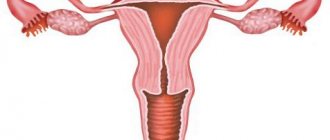
Hysteroresectoscopy is a modern surgical method for the treatment of submucosal nodes, an operation to remove the submucosal node of the uterus - by hysteroresectoscopy, intrauterine synechiae, polyps (hysteroresectoscopy of uterine polyp) and hyperplastic processes of the endometrium using a hysteroscope and the electrical equipment attached to it by grinding, excision or cauterization of the pathological focus. You can treat not only the uterine cavity, but also the cervical canal. This is called hysteroresectoscopy of the cervical canal polyp.
Services table
| Service name | Price |
| Initial consultation with a gynecologist | 2,300 rub. |
| Ultrasound gynecological expert | RUB 3,080 |
| Insertion of an intrauterine contraceptive device | 4,500 rub. |
| Hysteroscopy | RUB 22,550 |
| Repeated consultation with a gynecologist | 1,900 rub. |
| Taking a smear (scraping) for cytological examination | 500 rub. |
| Laparoscopy (difficulty category 1) | 61,000 rub. |
| Program "Women's Health after 40" | RUB 31,770 |
| Treatment of the cervix (medication) in 1 procedure | 800 rub. |
| Diagnostic curettage | 12,000 rub. |
In this case, the operation is considered low-traumatic and does not require laparotomy access (through the abdominal cavity) since it is performed by introducing equipment through the natural opening - the vagina and the cervical canal. In this case, the diagnosis and removal of pathological foci of all structures occurs: hysteroresectoscopy of the cervical canal, uterine cavity, orifice of the fallopian tubes.
What is hysteroresectoscopy
Hysteroresectoscopy is considered the best method for diagnosing and treating many gynecological diseases:
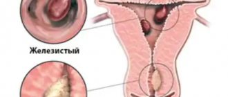
- polyps on the mucous membrane of the uterus (endometrium) and the membranes of the cervical canal;
- uterine hyperplasia;
- uterine fibroids located in the submucosal and muscular layer of the organ (submucosal fibroids);
- adhesions inside the uterus;
- intrauterine septa.
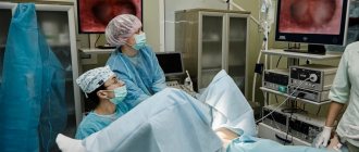
Another name for hysteroresectoscopy is surgical hysteroscopy. The essence of the operation is the delicate removal of large-scale pathological lesions that cannot be eliminated with medications or other minimally invasive methods. Gynecologists claim that hysteroresectoscopy is as serious an intervention as a full-fledged abdominal operation, but less traumatic, since a miniature instrument is used. The hysteroscope for hysteroresectoscopy is equipped with a set of surgical instruments: loops, forceps, coagulators, laser and high-frequency “scalpels”.
Compared to classical laparoscopy, hysteroresectoscopy differs in the way it accesses the walls of the organ. No incisions are made on the abdominal wall, since instruments for visualizing the surgical field and resection of pathological foci are inserted through the woman’s genital tract: the vagina and cervical canal. This reduces the likelihood of complications and shortens the recovery period. However, anesthesia is mandatory during hysteroresectoscopy.
Karl Storz is a leading manufacturer of medical equipment:
Equipment for minimally invasive surgery by Karl Storz (Germany) is used today all over the world. It has proven itself to be extremely reliable; the most modern technologies are used in its development. Operating endoscopes from the German company Karl Storz have been associated with tradition, high technology and quality for more than 70 years.
Hysteroresectoscopy with the help of reliable equipment from the German company Karl Storz allows you to save a woman from many unpleasant diseases in the most careful way.
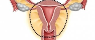
Hysteroresectoscopy of the uterus
The use of hysteroresectoscopy of the uterus is justified in the presence of submucous myomas with a diameter of no more than 5 cm, small, medium and large polyps without signs of malignancy. Various tools are used for this:
- To excise a flat polyp in the uterus, loop and spherical electrodes are used. The neoplasm is removed in a loop layer by layer, and then the remaining wound is coagulated with a spherical electrode.
- To excise a pedunculated polyp, a loop electrode, hook or electric knife (high-frequency electric scalpel) is used. The tumor bed is coagulated with a laser or a cylindrical electrode.
- Fibroids, the bodies of which protrude into the uterus, are removed using a loop. The doctor cuts off the tissue layer by layer, after which he coagulates the resulting wound with a ball electrode or laser.
In addition, uterine resectoscopy for polyps or fibroids can be performed using a laser. In this case, the doctor “evaporates” the tumors using a contact or non-contact method. Most often, this method is used if there is no need to collect biological tissues for histology. If such a study has not been carried out previously, it is recommended to use the methods described above.
Manipulations usually take no more than half an hour. During the operation, the woman is under anesthesia. After the tumors in the uterus are removed, she is transferred to the ward. The patient can leave the clinic on the same day if she has no signs of complications.
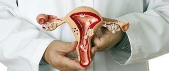
Hysteroresectoscopy at the ABIA clinic, St. Petersburg:
Our clinic performs all examinations necessary for an accurate diagnosis: video colposcopy, ultrasound and ultrasonic hysterosalpingoscopy, laboratory tests. When solving various gynecological problems, the clinic’s gynecologists use modern diagnostic and treatment equipment and equipment from leading manufacturers of medical equipment.
When polyps and myomatous nodes are identified, our gynecologists will select the optimal and effective therapy, which may include hormonal therapy or surgical treatment. If necessary and if indicated, sick leaves are issued for the period of treatment.
Hysteroresectoscopy of synechia
Synechiae or intrauterine septa made of connective tissue are effectively treated using hysteroresectoscopy. These neoplasms are thin film-like or dense, leathery membrane-like cords that connect the walls of the uterus to each other. Synechiae is considered the most common cause of infertility in women.
To remove the synechia fusion, various endoscopic instruments are used:
- thin films of synechiae are torn with the body of the hysteroscope, their remains are excised using endoscopic scissors;
- synechiae of medium density and thickness are cut with endoscopic scissors;
- dense synechiae, consisting of fibrous tissue, are dissected with an electric scalpel.
For multiple synechia, several instruments are used. The manipulations are monitored both through the hysteroresectoscope camera and using ultrasound. If there is a risk of damage to the uterine wall, laparoscopy is additionally used.
Bleeding usually does not occur with this type of intervention, so the woman can go home immediately after recovering from anesthesia.
Anesthesia
When a hysteroresectoscopy operation is performed, a variety of anesthesia can be used, depending on the patient’s concomitant diseases, her current condition, as well as the duration and scope of the operation and even the patient’s personal wishes. It can be intravenous, general (rarely used), endotracheal anesthesia (in case of a long operation), spinal anesthesia (if the operation lasts no more than thirty to forty minutes). Therefore, how long hysteroresectoscopy lasts and how anesthesia is administered directly depends. Usually, this issue is first discussed between a gynecologist and an anesthesiologist, after which the patient can be given a choice; in any case, no manipulation will be performed without her written permission. And she will be offered the most optimal anesthesia option in this particular case.
Using the method for endometrial ablation
Ablation (sometimes called ablation) is used in hysteroresectoscopy to remove hyperplastic tumors of the endometrium of the uterus. With this pathology, an atypical growth of areas of tissue lining the uterus occurs. To eliminate such tumors, endometrial ablation is used - deep destruction of the mucous membrane inside the uterus. To do this, various techniques are used, which are implemented using hysteroresectoscopy for endometrial hyperplasia:
- Combination of laser and loop electrode. First, the doctor, under the control of a hysteroscope, destroys the upper layer of the endometrium with a laser, leaving longitudinal grooves on it in the direction from the mouths of the fallopian tubes to the cervical canal. Next, the strips of remaining endometrium between the grooves are removed using a loop electrode. Tissues that accumulate in the cervical canal area are removed periodically so that they do not limit the view. Some of them are sent for histology.
- Spherical or cylindrical electrode. With them, the doctor “shades” the entire surface of the endometrium of the uterus: first the front wall, then the sides, and lastly the back.
Both methods lead to complete removal of the endometrium. Later, connective tissue forms in its place.
Important! After ablation, a woman cannot conceive and carry a pregnancy due to the absence of the endometrium, so the procedure is performed if the woman does not plan to have children.
Ablation is practiced not only for hyperplasia. It is used to eliminate menorrhagia and untreatable uterine bleeding, as well as adenomyosis. As in the previous case, the woman loses the ability to conceive and bear children. Very often, the endometrium is completely replaced by connective tissue, as a result of which the patient stops menstruation, although the ovaries function normally.
Preparation
During the preparatory period, the patient should do a number of studies:
- Standard gynecological examination using a speculum, as well as palpation of the uterus and its appendages.
- Vaginal smear. By collecting biomaterial from the urethra, cervical canal and vagina, the state of the flora can be determined.
- Clinical blood test, determination of group and Rh factor, blood test for RW, hepatitis and HIV. Determine blood clotting (coagulogram).
- Macroscopic and microscopic examination of urine, which can detect renal failure.
- Ultrasound of the pelvic organs (through the anterior abdominal wall or transvaginally).
- Electrocardiogram and fluorogram.
Before the planned hysteroscopy, the patient will be required to consult with related specialists: therapist, cardiologist, anesthesiologist. In addition, she should inform her doctor about the presence of any drug allergic reaction, suspicion of pregnancy, and medications taken on an ongoing basis.
Before undergoing hysteroscopy, a woman should adhere to the following recommendations: 2 days before the study, exclude sexual contact, a week before the scheduled procedure, do not douche and do not use store-bought gels and foams for washing.
A week before hysteroscopy, do not use medicated vaginal suppositories (with the exception of those prescribed by the gynecologist); in case of persistent constipation, the day before the examination, clean the intestines with an enema. 2 days before the procedure, start taking sedatives if prescribed by a doctor, 5 days before hysteroscopy start taking antibiotics if prescribed by a gynecologist.
On the morning of the procedure, you should refrain from eating and drinking. The patient must perform hygiene procedures, shave the pubic and groin area, and empty the bladder immediately before entering the examination room. All unnecessary items (jewelry, mobile phone) remain in the room. To the hospital, the patient must take with her slippers, socks, a change of underwear, a robe, as well as sanitary pads, which will be needed after the procedure due to heavy vaginal discharge.
In order for the uterine cavity to be better visualized, it is expanded using some medium
What is the difference between hysteroscopy and hysteroresectoscopy?
At their core, the methods of hysteroscopy and hysteroresectoscopy are similar: they are performed using the same instrument to visualize the inner surface of the uterus. However, there are several differences between them:
- Hysteroscopy is an exclusively diagnostic procedure, while hysteroresectoscopy, in addition to diagnosis, includes the treatment of gynecological diseases using minimally invasive and surgical methods.
- To perform hysteroscopy, you do not need such serious preparation as when planning hysteroresectoscopy. These two methods differ in the duration of the intervention: hysteroscopy lasts about 10 minutes, while hysteroresectoscopy lasts from 15 to 45 minutes.
- Hysteroscopy is performed on an outpatient basis with local anesthesia, while hysteroresectoscopy is always performed under general anesthesia.
In most cases, hysteroscopy is used for primary diagnosis and monitoring of uterine recovery after interventions. Hysteroresectoscopy combines a diagnostic and therapeutic procedure, so it is more self-sufficient.
Office hysteroscopy with RDV
It is possible to perform hysteroscopy with RDV - separate diagnostic curettage, which can be carried out for both research and therapeutic purposes. Curettage allows not only to determine the presence of a pathological process, but also its degree. An examination with curettage is indicated if there is suspicion of:
- tumor processes;
- hypoplasia;
- polyps;
- fibroids;
- endometriosis.
In addition, curettage examination is widely used in the treatment of prolonged and heavy menstrual flow, inflammatory processes after childbirth or abortion, as a method of excision of adhesions in the uterine cavity.
Treatment after resectoscopy
In addition to directly eliminating pathologies inside the uterus, it is important to pay attention to rehabilitation and treatment after hysteroresectoscopy. The further functioning of the female reproductive system will depend on how correctly hormonal drugs and therapeutic procedures for recovery are selected.
The basis of restorative treatment after hysteroresectoscopy are drugs containing hormones:
- Oral contraceptives, which compensate for the lack of female hormones and promote the normal formation of the endometrium, and also prevent untimely pregnancy - “Yarina”, “Regulon” and others. They must be taken for 3 to 6 months.
- In addition to the listed medications, the hormonal drug Premarin is prescribed. Recommendations regarding its use apply only to cases where uterine fibroids were removed during hysteroresectoscopy. Therapeutic therapy using this drug helps endometrial tissue to regenerate faster. You should take the drug for no more than 4 months.
- Antigonadotropins "Buserelin", "Danazol" or "Zoladex", which are necessary to control the growth of the endometrium. These drugs are used after curettage and ablation of the endometrium of the uterus.
Before the patient is discharged from the hospital, she is prescribed antibiotics from the cephalosporin group for a week. They are needed to prevent infections. To eliminate unpleasant symptoms after hysteroresectoscopy, the use of non-steroidal anti-inflammatory drugs (Diclofenac, Nimesulide or Ibuprofen in tablets or injections) is indicated. Since penetration into the uterine cavity was carried out through the vagina, therefore, there are also damages in it, patients are prescribed vaginal NSAIDs “Terzhinan” or “Betadine”.
Full recovery after hysteroresectoscopy occurs on average after 4-6 weeks. During this time, the woman is advised to abstain from sexual contact and be especially careful to maintain the cleanliness of the perineum. Pregnancy and childbirth can be planned at least 6 months after surgery.
Hysteroscopy: description and indications for use
The term hysteroscopy consists of two words: hystero - uterus, scopia - examination. From this it becomes clear that hysteroscopy is a procedure whose purpose is to examine the internal genital organs.
This manipulation is performed using a special instrument with an optical device called a hysteroscope. In appearance, it resembles a long tube with a camera attached to the end. With its help, it is possible to examine in detail the condition of the uterus, cervix, and determine the presence of certain neoplasms. The examination takes place in a gynecological chair and lasts on average from 5 to 30 minutes.
Hysteroscopy is divided into 2 main types:
- Diagnostic hysteroscopy
- Surgical (or therapeutic) hysteroscopy.
In the first case, hysteroscopy is one of the diagnostic methods. It is indicated in the following situations:
- Suspicion of neoplasms in the organs of the reproductive system (fibroids, polyps, cysts, etc.);
- Suspicion of cancer;
- Pathologies of development and structure of organs;
- Infertility;
- Disruptions of the menstrual cycle;
- Control examinations during drug treatment, after operations, childbirth.