What is uterine inflexion?
Normally, the uterus is fixed on movable ligaments in the middle of the pelvis, in the space between the bladder and rectum. The body of the organ is slightly tilted forward compared to the cervix and vagina. This anatomical feature is optimal for sperm to penetrate the uterus and cause pregnancy. If the location of the organ differs from normal, this is a sign of a bent uterus. It is incorrect to call such a condition a disease; rather, in this case we are talking about pathology.
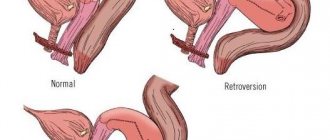
The following types of atypical location of the uterus are distinguished:
- Retroflexion (bending the uterus backward) is one of the most common types of unusual position of the uterus. The organ is located in such a way that its axis is deviated towards the spine. If the angle of inclination is slight, the atypical position often goes unnoticed. However, with a significant deviation back, prolapse and even prolapse of the uterus is possible.
- Anteflexion (anterior bending of the uterus) implies an inclination of the organ forward, towards the bladder, while the cervix remains in its normal position. This condition can be represented by anteversion - bending of the cervix.
- Lateroflexion is the bending of the uterus to the left/right, towards one of the ovaries.
Uterus during early pregnancy
Let's start with the fact that from the first days of conceiving a baby, a woman's body experiences revolutionary changes. However, the uterus itself in the early stages of pregnancy remains without any special modifications, which cannot be said about its cervix. As a rule, it is in this place that significant restructuring occurs first.
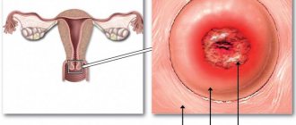
So, immediately after fertilization, the uterine cervix changes its color. If before pregnancy it had a predominantly light pink color, then after the birth of a new life, the cervix becomes darker, acquiring a lilac color. This is explained by the fact that after conception, blood flow in the main reproductive organ of the female body increases, small vessels dilate, changing the color of the cervix.
Another sign of early pregnancy is softening of the cervical epithelium. Already from the first days after conception, the cervical canal becomes more elastic, since after nine months (during childbirth) it will have to increase hundreds of times.
Normally, the cervix is slightly elevated and the cervical canal is open during ovulation. After conception, her position changes, she goes down a little.
An interesting point: by examining the shape of the uterine canal, a gynecologist can easily determine not only the current, but also previous pregnancies. In women who have previously experienced childbirth, the cervix has the shape of a cone, while in primigravidas it is wider and shaped like a cylinder.
In the early stages of pregnancy, when even ultrasound diagnostics do not show pregnancy, the doctor can conclude that conception has occurred based on the external characteristics of the uterus and its location.
Causes of uterine bending back, forward, to the side
There are congenital and acquired forms of this pathology. With the congenital variety, deviation of the uterus compared to the normal position develops in utero. In the second case, the cause of the bend is diseases of the pelvic organs. First of all, this concerns infectious and inflammatory processes that caused the development of adhesions. Also, the cause of a bent uterus can be heavy lifting, lack of movement caused by a long-term illness, weakening of the pelvic floor muscles, and the appearance of neoplasms.
The uterus during pregnancy to the touch
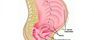
After three months of pregnancy, the uterus can not only be examined using ultrasound diagnostics, but also palpated through the abdomen. At this stage of gestation, the gestational age can be determined by how high the uterine fundus is located. Its level is measured using an ordinary measuring tape, the extreme end of which is applied to the pubis. Let's take a closer look at how the uterus rises over the weeks of pregnancy:
- At the sixteenth week of pregnancy, the uterine fundus is located 6 cm above the pubis. At this time, the upper part of the organ is felt approximately in the middle between the navel and the pubic bone.
- By the twentieth week, the uterus has enlarged so much that its bottom is 12 cm above the pubis.
- By the twenty-fourth week of pregnancy, the uterine fundus is located 20 cm above the level of the pubic bone, and the lower part is located approximately near the navel.
- At twenty-eight weeks, the reproductive organ can already be felt quite high, since its bottom is located at a level of 24 cm above the pubis.
- By the end of the thirty-second week, the fundus of the uterus is located 30 cm above the level of the pubic bone, that is, the upper part of the organ is located in the middle between the navel and the sternum.
- At the thirty-sixth week, the uterine fundus is 34-36 cm from the level of the pubic bone.
- Towards the end of pregnancy, at the fortieth week of pregnancy, the fundus of the uterus drops again; it can be felt at a level of 30 cm above the pubis.
It should be emphasized that the norms for the size of the uterus during pregnancy are given approximately, since these parameters directly depend on each individual organism.
The uterus and its normal location
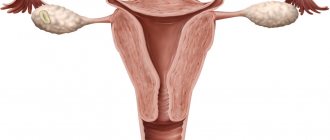
A hollow organ consisting of muscle tissue is called the uterus. Normally, it is located between the bladder and intestines in the pelvic space; the fallopian tubes extend from it to the right and left, through which the uterus is connected to the ovaries.
A healthy organ is located deviated from the symphysis pubis; an angle of 70-100 degrees is formed between the body and the neck. The position of the uterus is ensured by a complex of muscles and ligaments that are highly elastic.
Due to them, it takes a deflected position forward, backward, right or left - when filling the bladder and intestines.
How does the uterus rise during pregnancy?
During pregnancy, the uterus stretches and continuously increases in accordance with the growth of the fetus in it.
In addition, it is gradually shifting. If for approximately the first three months the organ is located in the abdominal cavity, then by the fourth month its bottom drops and is located between the navel and pubis. By the fifth month, the uterine fundus is at the level of the navel, and in the sixth, seventh, eighth, ninth and tenth months it reaches the lower edge of the chest. Towards the end of pregnancy, the uterus is so high that it puts pressure on the diaphragm, making breathing difficult. It also puts pressure on organs in the abdominal area: it compresses the bladder, intestines and stomach. That is why, when a rounded tummy appears, frequent urination and some digestive disorders in expectant mothers are observed.
Causes
The source of displacement of the uterus to the right or left can be congenital anomalies, age-related changes, or diseases of the reproductive department. Other reasons for the rejected state are presented:
- injuries in the sacrococcygeal spine;
- surgical interventions;
- decreased elasticity of ligaments, constant lifting of heavy objects;
- muscle weakness in the pelvis;
- malignant and benign tumors;
- diseases of nearby organs;
- hypodynamia, adhesive processes.
Symptoms of position change
A deviated position to the right or left when the uterus is displaced often has no manifestations and is determined when attempts at conception are unsuccessful. In other variants the problem is expressed:
- pain during menstruation or cycle disorders;
- intestinal dysfunction, inflammatory processes in the pelvic organs;
- discomfort in the lumbar region, pain during sexual intercourse;
- dryness of the vaginal mucosa, loss of sexual desire.
Consequences
The deviated position of the uterus to the right or left when displaced causes:
- difficulty conceiving;
- heavy menstruation or lack thereof;
- infertility, organ loss.
Pathology during pregnancy
If the uterus remains mobile, then the deviated position does not become a problem with conception. During gestation, the organ straightens as the fetus grows; in some patients, a significant displacement to the right or left can cause serious complications.
The pronounced pressure of the organ on the bladder and intestines becomes a source of development of congestion and problems with their release. Prolonged compression provokes inflammation of the kidneys, septic peritonitis, gangrene of the bladder, and spontaneous abortion.
Displacement of the uterus, consequences
Congenital features of the placement of the uterus, in the absence of concomitant diseases, do not in any way affect the woman’s health and her reproductive capabilities.
Another thing is displacement of the uterus, which occurs as a result of pathological phenomena occurring in the pelvic area. Often such processes occur without any symptoms, and displacement of the uterus is the only warning signal indicating the presence of problems in the pelvis.
Acquired displacement of the uterus is not, according to popular opinion, the root cause of pain during sexual intercourse, infertility and heavy menstruation - it is just one of the symptoms, the presence of which is explained by various pathologies of adhesive or inflammatory nature.
Diagnosis and treatment
Confirmation of uterine displacement to the right or left is carried out:
- gynecological examination;
- colposcopy;
- ultrasound examination;
- taking a smear for infection.
X-ray helps to identify the deviated position of the organ.
Surgical intervention
The operation is recommended for prolapse or displacement of other pelvic organs. Plastic surgeries help restore the ligamentous apparatus and return it to its original position. Complete prolapse requires excision of the uterus and pelvic floor plastic surgery.
Exercises
At the initial stage, with minimal pain, the patient is prescribed to perform Kegel and Yunusov exercises. These techniques allow you to return from a deviated state to its original position.
Description
The uterus is a hollow muscular organ that is pear-shaped. Its length for a nulliparous woman is 7–8 centimeters, and for a woman who has given birth is 8–9.5 centimeters. The uterus is located in the central part of the small pelvis between the rectum (back) and the bladder (front). The part of the uterus that is much wider is called the body, and the narrower part is called the cervix. The uterus communicates with the external environment through a canal (about a centimeter in length), which passes through the cervix and opens into the vagina. In the normal normal state, an angle of at least 70 degrees is formed between the cervix and the body of the uterus.
The uterus is a very mobile organ. Without difficulty, the uterus, suspended by ligaments, can move to the back - to the sacrum, or to the sides - to the side walls of the pelvis, as well as up and down. Such mobility is the norm for the activities of adjacent organs. For example, when the bladder fills, the uterus, rising up, does not put pressure on it. The position of the uterine appendages and the uterus itself depend on the state of the pelvic floor and the muscles of the peritoneum, because they are its basis, as well as on the ligaments that secure and fix the suspended uterus.
The location of the female genital organs changes with age. For example, the uterus is located slightly higher in childhood, and by the period of maturation it begins to descend slightly. With the advent of old age, the genitals atrophy, and the uterus deviates back, becoming deeply planted in the pelvic cavity.
Prevention
Preventing drift to the right or left begins with the following recommendations:
- prohibition on lifting and carrying heavy objects weighing more than 10 kg;
- daily hygiene procedures - to avoid the development of inflammation;
- timely treatment of diseases of the reproductive department;
- giving up a sedentary lifestyle;
- exclusion of hypothermia, frequent change of sexual partners;
- regular check-ups with a gynecologist.
A shift to the right or left can be caused by both physiological and pathological sources.
The uterus is an unpaired muscular organ that is necessary for bearing a fetus. Normally, the uterus is located in the pelvis, between the bladder and intestines, and on both sides of the uterus there are its appendages: the ovaries and fallopian tubes. Uterine displacement is a situation in which an organ changes its location. As a result, the normal distance between the pelvic organs changes.
It is worth noting that normally the uterus constantly moves left and right, forward and backward, for example, when the bladder fills. Also, the uterus changes location as a woman ages, which is also a physiological norm. In a girl, the uterus is located higher than in a woman of reproductive age. But in some cases the displacement is too strong, which is a pathology.
How does the uterus enlarge during pregnancy?
Its size certainly changes during the period of gestation. First, the walls of the organ grow - they become thicker, build up the mucous membrane, to which the fertilized egg that has left the fallopian tube will subsequently be attached. In addition to the mucous layer, the muscle tissue of the organ also increases in size, since it needs to accommodate the growing child. By the end of pregnancy, the uterus enlarges approximately 500 times its original size. That is why an experienced gynecologist can easily determine the approximate period of a woman’s “interesting position” by examining the size of the reproductive organ.
It should be noted that during pregnancy, changes also occur in the vagina - the woman’s labia minora become somewhat darker than they were before. By the end of the fifth week, the shape of the uterus changes: from pear-shaped to round. And already in the eighth week of pregnancy, the organ can be compared in size to a woman’s fist. In the twelfth week it grows even more. At this time, the uterine fundus is located at the level of the pubis.
Causes
The uterus is tilted to the left, what does this mean, patients are interested. Such a violation means that the uterus has shifted and is not located physiologically. Pathology can occur for the following reasons:
- gynecological diseases, such as inflammation or ovarian cyst;
- oncological diseases, large tumors;
- adhesions in the pelvis;
- heavy physical activity;
- weak muscles and ligaments of the pelvis;
- passive lifestyle;
- consequences of surgery;
- intestinal pathologies, constipation;
- connective tissue dysplasia;
- congenital developmental anomalies.
Women often experience displacement of the uterus after difficult labor, as well as those patients who have given birth many times. The likelihood of uterine displacement also increases if there is a large amount of abdominal fat.
Symptoms
Uterine displacement can be accompanied by various symptoms. In some cases, the pathology is asymptomatic and is discovered when a woman is examined for infertility. The fact is that if the uterus is displaced, then the chances of pregnancy are very low.
Common symptoms of uterine displacement are:
- menstrual irregularities, pain during menstruation;
- vaginal dryness, decreased sexual desire, lack of orgasm;
- frequent exacerbations of inflammatory pathologies of the uterus and appendages;
- pain in the lower abdomen and lumbar region;
- infertility.
If your uterus hurts during pregnancy
Pain in the reproductive organ can bother a woman at any stage of pregnancy. They are associated with the growth and contraction of the uterine muscles. So, in the early stages of pregnancy, a woman may feel a slight tingling sensation when the embryo settles on the wall of the uterus. The sensations in this case cause minor discomfort, so do not worry if you feel a slight pain in the lower abdomen, which soon disappears.
During the growth of the uterus, the expectant mother may feel a nagging pain on both sides of the abdomen. The female reproductive organ has good elasticity and the ability to expand. When the uterus increases in size, mild and quickly passing pain may occur. Moreover, such processes often continue until childbirth.
Relaxation of the uterus occurs due to the hormone progesterone, which prevents its contraction. With a lack of this hormone, uterine tone may occur during pregnancy - a pathology caused by uterine contractions. This condition can cause spontaneous miscarriage. Hypertonicity causes severe aching pain. In such a situation, you should immediately seek help from a doctor.
Note that uterine contractions are also possible when a woman has a full bladder. A heating pad, which should not be too warm, will help relieve severe pain due to uterine tone. Also, the expectant mother must control her digestion and avoid constipation, since they can also negatively affect the condition. Remember that any pain in the uterus during pregnancy may be mild and should not bother you. If they begin to worsen, be sure to consult your doctor.
Treatment
Usually, displacement of the uterus is a consequence of some disturbance in the reproductive system. Pathology does not occur on its own, so treatment is based on eliminating the root cause.
To identify the cause of uterine displacement, a woman undergoes a pelvic ultrasound, laboratory tests of urine and blood, and smears. If necessary, laparoscopy and other diagnostic methods are performed. Based on the results of the examination, a diagnosis is established and treatment is prescribed.
If an inflammatory process is detected, anti-inflammatory therapy is indicated. Treatment of weakened muscles and ligaments of the pelvic floor requires exercise to strengthen the muscles. In this case, the woman should perform Kegel exercises.
For tumors, adhesions, and cysts, surgery may be required. As a rule, a laparoscopy is performed, with the help of which the doctor can remove all existing abnormalities. Gynecological massage can also help return the uterus to its place.
Uterine displacement does not always require treatment. Sometimes such a disorder does not have any effect on reproductive function and the woman successfully becomes pregnant. If no concomitant pathologies are found, you need to think about treatment if infertility is observed.
Prevention
Prevention of uterine displacement involves preventing various gynecological pathologies. To do this, you must follow the following recommendations:
- lead an active lifestyle, play sports and do exercises;
- eat right, avoid obesity and exhaustion;
- do not drink alcohol or smoke;
- have sex regularly, use proper protection, avoid promiscuity;
- do not lift heavy objects;
- avoid overwork and stress;
- dress according to the weather. do not overcool;
- undergo regular examinations with a gynecologist.
A healthy lifestyle and regular preventive examinations will help prevent the development of pathology, and if it occurs, quickly identify and treat it.