Ovarian adhesions are a disease in which ovarian tissue fuses with neighboring organs. This creates scars.
Fusion occurs due to the formation of films and fibers. They glue organs together.
Most often, adhesions between the ovary and the uterus are observed, and less often, adhesions between the ovary and intestines are observed.
As a result of the process, blood circulation is disrupted, which prevents pregnancy.
Ovary in adhesions: what does it mean?
Pathology affects the organ in the following cases:
surgical intervention.
The operation violates the integrity of the ovaries and leads to disruptions in the processes of blood clotting and cell renewal. The body is trying to restore tissue structure. Failures in this process lead to the formation of connective tissue that glues organs together. Moreover, the pathology spreads to neighboring organs. At risk are women after an abortion or cesarean section. If there are other ovarian diseases: cyst, endometritis, hypoplasia, the likelihood of adhesive disease increases;- chronic or acute inflammatory diseases of the ovaries. These processes are accompanied by fluctuations in the number of leukocytes in the blood. The affected cells injure the ovarian stroma - the dense membranes that connect the tissues. The stroma contains blood vessels that supply the follicles with the necessary substances for functioning. Damage to the membranes creates favorable conditions for tissue growth and division of “infected” cells. As a result, tissue sticks together and scars form. The situation is aggravated by increased synthesis of fibrin, a protein that forms fibers, the clots of which form a blood clot.
There are frequent cases when adhesions form after laparoscopy of an ovarian cyst.
Factors that contribute to the formation of adhesions include:
- internal bleeding. When the ovary is damaged, blood or intercellular fluid is released. This material forms films that glue organs together;
- incorrect insertion of the intrauterine device;
- sexually transmitted infections;
- endometriosis is a gynecological disease characterized by the growth of endometrial tissue beyond the organ;
- ectopic pregnancy;
- treatment with antibiotics, antimicrobial agents;
- ruptures during childbirth;
- hypothermia;
- hysteroscopy – examination of the uterine cavity with a special device for diagnostic or treatment purposes.
Why is the ovary soldered or located behind the uterus. Displacement of the uterus - left, right
The main cause of displacement of the appendage is adhesions in the pelvis. The occurrence of an adhesive process in which the right ovary (or left) is affected is influenced by the following factors:
- Gynecological operations (abortion, cesarean section), when the integrity of the appendage is disrupted, which provokes deviations in the processes of blood clotting and cell restoration. Instead of regeneration, connective tissue is formed, gluing organs to each other.
- Concomitant pathologies of the reproductive sphere (cyst, endometritis, etc.). Due to the affected cells, the stroma of the appendages suffers, and the processes of local blood supply are disrupted. Abnormal cells begin to divide, pathological tissues grow, which leads to the appearance of scars.
- The ovary is pulled towards the uterus under the influence of the following factors:
- violation of the rules for inserting an intrauterine device;
- venereal diseases;
- endometriosis, in which the tissue of the uterine lining extends beyond its boundaries;
- ectopic pregnancy;
- use of antibacterial agents;
- ruptures during labor;
- hypothermia;
- performing hysteroscopy.
Symptoms
Often the pathology passes without obvious symptoms. The first symptoms may appear several years after the start of the process.
Adhesive disease manifests itself:
- nagging pain in the lower abdomen, radiating to the lower back;
- menstrual irregularities;
- painful periods;
- unpleasant sensations during physical activity, sexual intercourse;
- intestinal disorders;
- temperature rise.
The pain syndrome during the disease is mild. It manifests itself as a dull pain covering one side. A change in the nature of discomfort indicates complications of the adhesion between the uterus and ovary.
In this case, the pain is severe, because the patency of the fallopian tubes is impaired. As a result, the menstrual cycle is disrupted. The duration of the delay is 2-3 months.
The adhesive process is characterized by a yellowish-green discharge. A bloody “spot” may appear.
Often, symptoms of adhesive disease occur after hypothermia or a course of physiotherapeutic procedures.
Features of dietary nutrition
With this pathology, you do not need to adhere to special diets. Proper and balanced nutrition is necessary, which will help the body restore its functionality, increase the level of the immune system to prevent the ingestion of dangerous viruses that can aggravate the condition.
DETAILS: Streptoderma - symptoms and treatment of the disease
You need to eat as many fruits and vegetables as possible, preferably fresh or steamed. Drink plenty of liquids and freshly squeezed juices.
It is not recommended to consume in large quantities:
- Fatty and fried foods;
- Spicy dishes;
- Alcohol.
To prevent displacement, consume as many vitamins and microelements as possible. A huge amount of which is found in sea fish, seaweed, and cereals.
Don't neglect dairy products. They saturate the body with necessary substances that increase immunity. It is healthy and important to eat a lot of nuts and dried fruits, especially prunes. It cleanses the body of toxins and prevents frequent constipation.
Diagnostics
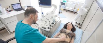
After a gynecological examination, the patient is sent for research:
- Ultrasound of the pelvic organs . This study is not enough, because It is difficult to identify adhesions;
- laparoscopy. During diagnosis, small punctures are made in the abdominal cavity to examine organ tissue;
- MRI . Using this method, you can see the smallest changes in the organ;
- microflora analysis;
- Hysterosalpingography is an X-ray diagnostic method based on the injection of a special substance into the uterus and fallopian tubes. The procedure is carried out from 5 to 11 days of the menstrual cycle.
Help from folk remedies
Traditional methods help with displacement of the uterus, but before carrying out the procedures you should consult with a specialist so as not to aggravate the condition of the disease.
Douching with medicinal infusions is effective for this pathology:
- To prepare, mix sweet clover, marshmallow leaves and chamomile in equal quantities. Pour one spoon of the resulting mixture into a glass of boiling water and leave covered for half an hour. Strain, syringe twice a day, a quarter cup.
- Douching with an infusion of St. John's wort and cinquefoil effectively helps with displacement. To prepare, pour 2 liters of water into a container, put it on the fire, when it boils, add 4 tablespoons of chopped St. John's wort, cook for half an hour. Then add a spoonful of bloodroot and boil for another 5 minutes. Strain and use twice a day.
- Pour a glass of water into a container, add a spoonful of crushed oak bark. Boil over low heat for 5-10 minutes. After straining, dilute a little with cold water. Use half a glass every day for 2 weeks.
- Mix 2 servings of St. John's wort, one each of horsetail and chamomile. Pour a spoonful of the mixture into a glass of water and place on low heat. After 15-20 minutes of boiling, turn off and let sit for half an hour. Strain, take a quarter glass three times a day.
- Dilute a teaspoon of tannin in 200 ml of warm boiled water and take half a glass twice a day.
Often, displacement of the uterus is associated with inflammatory processes.
To get rid of unpleasant symptoms you can use the following recipes:
- Mix chamomile, peppermint, and valerian root one spoon at a time. Pour 200 ml of boiling water over one spoon of the resulting mixture and let it brew for 1-2 hours. Drink before meals three times a day.
- Pour 4 tablespoons of chamomile into a glass of boiling water, let it brew for half an hour, strain, drink half a glass twice a day. You can wash yourself with this infusion, it gives a good result.
- Make an infusion of plantain leaves, it will help relieve inflammation and pain. pour a spoonful of crushed, dried herbs into two glasses of boiling water, leave for 20-30 minutes, strain, drink a teaspoon three times a day.
- If spasms occur in the pelvic area, an infusion of prickly plum flowers will help. Pour 2 tablespoons into a glass of boiled cold water and leave overnight. Afterwards, strain, take half a glass three times a day.
- A decoction of acacia flowers effectively relieves pain. Pour boiling water over a spoonful of flowers, leave for 30 minutes and drink before eating.
With the help of such herbal preparations you can douche or take a bath. They effectively fight inflammation and help relieve pain.
Is infertility at risk?
If there are adhesions on the ovaries, is it possible to get pregnant? One of the main causes of infertility is adhesive disease. Most often, with this pathology, a woman cannot become pregnant. Adhesions displace organs. This disrupts the anatomical connection between them.
Schematic representation of adhesions
The fallopian tube has special outgrowths that normally help the fertilized egg move into the womb. With adhesive disease, these outgrowths stick together. Because of this, the egg dies when it enters the abdominal cavity. With the development of pathology, the outgrowths disappear. A scar appears in their place.
It is possible to get pregnant with an ovarian adhesion. Even if the disease is in advanced form.
Thanks to new methods of reproductive medicine, pregnancy is possible in the absence of both fallopian tubes. Of course, the chances are decreasing, but they are there. If after treatment a woman does not become pregnant, she is offered an IVF procedure.
Adhesions are dangerous with a high risk of ectopic pregnancy, so if there is a problem, it is important to eliminate it.
Chances of pregnancy
As mentioned earlier, the bending of the ovary behind the uterus (left or right) is often a manifestation of the adhesive process. Difficulties in getting pregnant are caused by a violation of the anatomically correct location of the reproductive organs.
A woman who finds out that her ovary has gone behind the uterus, of course, doubts the possibility of conception. To normalize the condition of the reproductive organs, the help of a qualified gynecologist is required.
To get pregnant, you need to undergo treatment. If it is not effective, then IVF is performed. Since adhesions increase the risk of attachment of the fertilized egg outside the reproductive organ, it is necessary to direct all efforts to eliminate it.
Treatment of the disease
At the initial stage of a disease such as ovarian adhesions, treatment involves the use of the following drugs:
- antibiotics;
- anti-inflammatory drugs;
- thrombolytics;
- enzymes;
- vitamins.
Electrophoresis with the introduction of calcium, magnesium and zinc through the skin has a positive effect. Thanks to physical therapy, the adhesions become thin and stretch easily.
Having detected adhesions on the ovaries, treatment can be carried out in a sanatorium setting. If possible, the patient is treated with natural mineral water. The woman is advised to move more to prevent the appearance of new adhesions.
In advanced cases of the disease, laparoscopy is used in therapy. The operation is painless and performed under general anesthesia. It helps eliminate and separate fabric constrictions.
Laparoscopy
Then a layer of polymer film is applied to the ovaries and a barrier liquid is injected. This measure prevents the re-formation of adhesive disease. But there is no 100% guarantee. Adhesions after ovarian laparoscopy may return.
After the operation, the patient is prescribed treatment with antibiotics and drugs that prevent the formation of blood clots. Physical activity is prohibited for two weeks. Next, the doctor evaluates the effectiveness of the operation and subsequent treatment. In some cases, the patient is additionally prescribed a course of physical therapy.
In addition to laparoscopy, there are other methods of surgical intervention in the adhesive process:
- laser therapy. The method is based on the action of light flux;
- electrosurgery. Pathogenic tissues are destroyed when heated under the influence of high frequency current;
- aquadissection - dissection of adhesions using water.
Sometimes patients are afraid of surgical adhesions on the ovaries. Treatment with folk remedies is the solution to this problem. However, it is far from the most recommended.
Operations are performed in extreme cases when medications, vitamins and manipulations do not help.
The ovaries are pressed against the rib of the uterus. The ovaries are pressed against the rib of the uterus. The left ovary is adjacent to the rib of the uterus.
One of these types is subserous myomatous tumor.
What is subserous uterine fibroid?
Typically, such formations have a fairly wide base, connected to the uterine tissues through a thin stalk. This leg is a kind of channel through which nutrition for the tumor passes. The nodule can be single in nature, although multiple small formations are also found.
Causes
The main factor contributing to the development of benign subserous tumors in women is changes in the hormonal status of women.
According to doctors, a tumor is not able to form in a healthy body, so its development requires specific prerequisites such as:
- Surgical manipulations in the female genitourinary organs. This category includes various diagnostic curettages, abortions, laparoscopic examinations, etc. These interventions injure the uterine muscle tissue, which provokes the formation of a tumor;
- Genetically determined predisposition to this pathology;
- A history of surgery to remove fibroids;
- Various types of inflammation or infection in the genitourinary area;
- Long-term contraception with hormonal contraceptives;
- Tumors in the appendages or mammary tissues;
- Functional problems in the thyroid and adrenal structures.
Typically, the causes of the disease are a group of factors that, to varying degrees, influence the formation of the myomatous process in the uterine body.
Clinical picture
Externally, a subserous tumor looks like a node. This type of myomatous formation is considered the safest, since it is distinguished by its external uterine localization and grows towards the retroperitoneal space. Such a tumor does not provoke an increase in the uterine body and cannot affect the menstrual cycle.
Such a formation does not harm pregnancy, which occurs without difficulties with such a disease; a subserous formation cannot harm delivery, however, it is quite capable of causing a spontaneous miscarriage.
Sometimes reproductive difficulties occur if the formation is located in close proximity to the tubes and compresses them.
Since the formation grows into the abdominal space, it develops mainly without symptoms, at least during the period while the node is of insignificant size. When the formation grows, it begins to compress the surrounding tissues and organic systems, which leads to their functional disorders.
In general, subserous myomatous formation can be characterized by the following symptoms:
- Frequent constipation;
- Hemorrhoidal inflammation;
- Difficulty and frequent urination, causing pain;
- Hyperthermia
- Hypersweating, dizziness;
- Discomfortable sensations in the abdomen and lower back when sitting or standing;
- Painful symptoms of an aching nature, localized in the area above the pubis, in the lower back, and lower half of the abdominal wall.
Painful symptoms tend to occur during prolonged periods of standing, long walking, or heavy physical activity. Often the pain becomes cramping in nature, which indicates the development of an exacerbation or active growth of the tumor.
The severity of pain symptoms is usually determined by the parameters, location and development of the myomatous node. If the tumor fuses with the abdominal wall, the patient will constantly suffer from pain.
Varieties
Subserous myomatous nodes can be multiple or single.
Single ones are distinguished by the presence of a shell like a capsule.
Multiple formations are found somewhat less frequently, but they are accompanied by more severe painful symptoms.
If multiple myomatosis becomes large in size, then adjacent structures are compressed, which disrupts their activity. In addition, they are divided into interstitial or intramural uterine fibroids.
Intramural
Intramural subserous fibroids are localized on the outer uterine layer. This formation is considered the “safest”, since it does not affect the size of the uterine body and the menstrual cycle, and does not interfere with reproductive abilities.
Such a tumor is formed from smooth muscle fibers, as well as connective tissue structures. It is believed that such fibroids occur in a kind of lighter version.
Interstitial subserosal
The subserous-interstitial myomatous node is formed in the thickness of the wall of the uterine body, but grows towards the small pelvic cavity. Such a formation belongs to the category of mixed tumors and is somewhat different from a traditional subserous tumor.
It develops in the muscle layer, and therefore leads to a slight increase in the body of the uterus. An interstitial subserous myomatous node can negatively affect surrounding tissues, but its size rarely exceeds 10 cm.
Stages
Experts stage the development of such myomatous formations into several stages:
- The first stage – active growth is observed, the tumor is characterized by the full course of metabolic processes and increased vascular permeability;
- The second stage is characterized by rapid progression, but it is still impossible to detect a formation without microscopic examination;
- At the third stage, myomatous formation is easily detected during a medical examination.
Typically, such tumors are of a multiple nature, when several formations develop simultaneously.
How dangerous is this disease?
The danger of subserous fibroids lies in the high probability of torsion of the pedicle, through which the formation receives the necessary nutrition. This complication is usually caused by atrophic changes in tissue.
Tissue death is usually accompanied by hyperthermic symptoms, intense pain, severe tachycardic symptoms, increased sweating, dizziness and other intoxication symptoms.
Diagnosis and treatment
When diagnosing subserous myomatous formations, the following diagnostic measures are most often used:
- Echography;
- Magnetic resonance imaging, which allows you to obtain a layer-by-layer image of the tumor;
- Diagnostic curettage in this case is carried out to determine changes in the mucous uterine tissues;
- Hydrosonographic diagnostics - allows you to detect even the smallest tumors and determine the degree of their development;
- Laparoscopic examination is used if it is impossible to differentiate the pathology.
Most often, treatment of the subserous form of uterine fibroids is surgical, especially in the presence of large formations, heavy blood loss, torsion of the pedicle, rapid growth and progression of the tumor.
In the photo, removed subserous uterine fibroids
To carry out surgical procedures, several common techniques are used, such as laparoscopic myomectomy, which involves removing the tumor through several punctures through enucleation.
In addition, hysteroscopic myomectomy is used, when the node is removed through the vagina. In difficult cases, the formation is removed along with the appendages and uterus, i.e., a total hysterectomy is performed.
In recent years, FUS ablation has become widely used, which involves removing a tumor using ultrasound.
Treatment with folk remedies
Among the traditional methods of treating subserous uterine fibroids, healing ointments, extracts, medicinal tinctures or vaginal suppositories are especially popular among patients. Usually, for the preparation of such medicines, raw materials are used in the form of poisonous plants that have antitumor activity.
Therefore, such medications should be taken with extreme caution. Experts do not rule out the presence of a therapeutic effect from such healing remedies of traditional medicine, however, they recommend their use as additional therapy and in the absence of contraindications.
Prognosis and prevention
Subserous forms of myomatous formations are characterized by a high rate of benignity, and therefore do not have a tendency to malignancy. The prognosis is generally positive, however, if there is rapid growth and progression of the pathology, surgical intervention may be necessary.
Even benign tumors can significantly ruin a woman’s life and deprive her of opportunities for motherhood. Therefore, it is necessary to contact a specialist when the first suspicion of pathology appears.
In addition, to prevent myomatous formations, it is necessary to avoid abortions, give birth and breastfeed a child until the age of 30, promptly treat gynecological problems, monitor hormonal status, and avoid long-term hormonal contraception.
This is the only way to protect yourself from the occurrence of serous uterine myomatous formations, and, therefore, to avoid possible complications such as infertility, purulent-inflammatory processes in the uterus and total hysterectomy, which deprives the patient of a chance for successful motherhood.
The video shows laparoscopic surgery for subserous uterine fibroids:
Source: https://yanaorgo.ru/yaichniki-prizhaty-k-rebru-matki-yaichniki-prizhaty-k-rebru-matki.html
Possible complications
Infertility is the main consequence of adhesive disease; if not treated in time, other complications arise:
- the growth of adhesions into neighboring organs, which causes their displacement;
- contact between the ovaries, uterus and fallopian tubes is disrupted;
- the level of contractions and patency of the fallopian tubes decreases;
- threat of ectopic pregnancy;
- disruptions in the ovulation process. Nerve endings and blood vessels in the genitals are pinched. It is very difficult for the egg to penetrate the uterine cavity.
At the first signs of the disease, it is not recommended to delay examination by a gynecologist. Timely diagnosis of adhesive disease will simplify treatment.
Prevention of adhesions
- avoid hypothermia and the appearance of inflammatory processes in the body;
- maintaining local immunity;
- preventing the occurrence of infections;
- regular sex with a regular partner;
- using a condom during sexual intercourse;
- avoiding termination of pregnancy;
- timely diagnosis of cystic formations of female ovaries;
- avoidance of promiscuity with different partners;
- regular visits not only to the gynecologist, but to the gastroenterologist, since with pathologies in the gastrointestinal tract, adhesions can form between the ovary and the intestines;
- control over the menstrual cycle;
- moderate exercise is necessary;
- adherence to a diet regimen (overeating and fasting are not allowed).
- use of high-quality threads for suturing wounds;
- Powdered gloves are not allowed;
- instruments must be sterile;
- normalization of blood circulation during surgery;
- insertion of drainage for the outflow of blood and inflammatory fluid;
- it is necessary to avoid electrical or laser exposure to the peritoneum;
- do not allow the peritoneum to dry out.