General information about the cervix and its examination
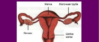
The cervix is a part of the reproductive organ, which is a hollow body measuring 2.5 x 3 cm. It can be called the connecting link between the vagina and the uterus.
A woman can independently feel this area at home by completely immersing her middle finger into the genital tract. As soon as the fingertip touches the bulge, there is no need to move further - the desired element has been found.
By doing such diagnostics every day for several cycles, a woman will learn to determine the position of the cervix before menstruation, calculate ovulation and select days favorable for conception. Or vice versa, he will understand on which days it is necessary to strengthen contraceptive measures.
The doctor examines the patient's genital tract on a chair using a special mirror. At home, it will be convenient for a woman to examine the cervix in several positions:
- Sitting on the toilet.
- Squatting.
- Standing, but one leg placed high.
The study is not carried out during menstruation. On “clean” days, the organ is felt once a day before bed at the same time. If a gynecological disease of an infectious-inflammatory nature is suspected or with the onset of bleeding, the diagnosis is canceled.
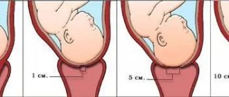
The speed of its detection helps determine whether the cervix is high or low on a particular day. If the location is high, the area is difficult to palpate; if it is low, it can be detected immediately. The amount of openness is judged by the recess. If a small gap is felt, the uterus is closed. If the hole is round and deep, then the organ is open.
Despite the simplicity of this method, doctors do not recommend examining internal organs yourself. The ban is justified for the following reasons:
- When the hole is open, there is a risk of damage to the uterus by pathogenic bacteria. Because of them, the ovaries become inflamed and obstruction of the fallopian tubes develops. In advanced cases, the disease can result in infertility.
- Since the cervix drops slightly before menstruation, inept palpation can injure it and provoke the development of erosion. The pathological focus is susceptible to infection and is dangerous in terms of oncological degeneration.
- The woman does not see the true state of the uterus. The gynecologist evaluates the real picture visually using a mirror.
The shape of the cervix before menstruation should resemble a pupil. Deviation indicates estrogen deficiency and improper functioning of the corpus luteum.
Video:
Self-examination techniques
How to examine your own cervix? To begin with, you can familiarize yourself with the anatomy of the female organs using photos, and only then proceed to action.
Before diagnosis, you should carefully prepare your own hands for examination and do not forget to wear sterile gloves. The examination is carried out using two fingers, which are slowly inserted into the vaginal opening.
Body position during the procedure: squatting or on the toilet, or standing on one leg, lift the other and place it on a chair or bathtub.
Simply put, the border after the vagina is the cavity of the cervix, a kind of bridge between the uterus and the vagina. When penetrating the vagina, you need to move slowly along the cervix. Any lady can independently feel the female organ. When the fingers come into contact with a small bulge or tubercle, we can assume that the uterus has been found.
With regular self-examination, over time, a girl will be able to accurately determine in which phase of the cycle her reproductive system is. This procedure will become invaluable during the period of planning future offspring.
Position of the cervix in different phases of the cycle
The entrance to the uterine cavity changes in different periods of the cycle. The cyclic work of the ovaries goes through the ovulatory, follicular and luteal phases. Uterine functions change from secretory to menstrual and proliferative.
The composition of the fluid in the cervical canal also changes. It can be studied in detail in laboratory conditions. At home, a woman can analyze the consistency.
The cervix feels like this:
- Soft.
- Elastic.
- Dense.
- Loose.
- Solid.
Its position is either high or low.
Secretory stage
The tone of the uterus in the 2nd half of the cycle decreases and after ovulation, closer to menstruation, the cervix becomes soft. The external pharynx opens to such an extent that the tip of the finger penetrates inside.
The high position of the cervix helps sperm reach the fallopian tube faster. The glandular part of the cervical canal intensively produces transparent whitish mucus. The alkaline index of the secretion reaches 8 units. The mucus doesn't stretch.
In the absence of a fertilized egg, the body begins to prepare for endometrial rejection. A plug forms in the canal, the neck hardens and falls. The pharynx closes, the cervical canal narrows. In case of pregnancy, the cervix does not change its position. It will be difficult to feel the pharynx manually at home.
Experts know exactly what the cervix looks like on the eve of menstruation, and characterize it as follows:
- Located below.
- It has a loose soft surface.
- The external pharynx is open, the canal is expanded.
- Little cervical mucus is produced.
- The secretion has a sticky, thick consistency.
- pH < 6.5 units.
Thus, before the start of menstruation, the cervix becomes completely ready to reject bloody discharge. Along with internal changes, symptoms of PMS appear - nagging pain in the lower abdomen and deterioration in general well-being.
Proliferative stage
At the proliferation stage, the endometrium is restored in the uterus. It forms its own connective substances and blood network. The proliferative stage lasts until ovulation occurs. It begins in the 2nd half of the follicular phase.
After menstruation, the cervix undergoes changes again:
- Descends into the vagina.
- The external pharynx narrows greatly.
- The endocervix becomes denser.
- The amount of cervical fluid increases.
- The pH level gradually increases to 7.3 units.
The endocervix is the mucous part of the canal. Its epithelium produces cervical fluid. Its role is to protect the uterine cavity from microbial invasion. When the egg is released, the mucus thins and makes it easier for the male seed to penetrate the uterus.
Menstrual stage
Critical days are a difficult stage for the body. You are not feeling well and your genitals are at risk of infection. For this reason, it is not recommended to examine the uterus manually, and it is not recommended to constantly use hygienic tampons.
During the menstrual period, the cervix has other characteristics:
- She's hanging low.
- He is in low tone.
- The acidity level is approaching 7 units.
- The vaginal epithelium is slightly flaky.
- Cervical fluid is secreted in moderate volumes.
- The external pharynx is slightly open, the opening is directed towards the vaginal vault.
Menstruation occurs during the first segment of the follicular phase of the ovaries. Next, the reproductive system begins to prepare for the next ovulation.
The value of cervical palpation as a diagnostic method
Daily palpation of the cervix, including before menstruation, helps to track cyclic changes. In order not to injure or infect a delicate organ, it is necessary to cut off the nail on the desired finger and polish the edge of the plate.
For greater safety, it is better to perform the procedure with sterile gloves or a fingertip. It is recommended to record all data collected before the onset of bleeding in a notebook.
The purpose of diagnosis is to assess the position of the cervix before menstruation and during other phases of the cycle, as well as during pregnancy, if it has occurred. It is also important to know the density of the organ at the proliferative and secretory stages of MC.
The information collected over several months will help the woman with family planning and will signal the need to see a doctor if there are any abnormalities.
Is it necessary to carry out diagnostics?
The cervical self-diagnosis procedure may seem strange to many, but believe me, there are many positive things in it:
- Accurately determine the favorable period for pregnancy;
- Know the period when the risk of getting pregnant is minimal;
- The uterus changes before menstruation, which means the woman will know for sure that her period is coming soon;
- In addition, with the help of palpation, a neoplasm on the organ can be detected in the early stages, which increases the chance of quickly getting rid of the problem.
However, you should still be careful when self-diagnosing:
- Remember that before menstruation the cervical canal is slightly open, so the risk of infection increases, which can lead to an inflammatory process in the female body;
- Rapid and careless movements during an examination can cause injury, which in the future can develop into erosion. Of course, it doesn't always happen this way, but the risk is there;
- You should not worry if during the examination a strange lump or something else is discovered. Only a doctor can accurately determine the level of complication, if any.
In any case, whether or not the cervix is diagnosed before menstruation or during other phases of the menstrual cycle, it is worth visiting a gynecologist twice a year. This is the key to women's health.
Organ sizes
Some girls may wonder how much a woman's uterus weighs. Experts say that in a completely healthy girl who has not yet given birth, the mass of the organ does not exceed 50 grams. If there has already been a pregnancy, then this figure reaches 100 grams.
The length of the organ ranges from 7 to 8 centimeters, and its width is no more than 5 centimeters. When the layers of the uterus, in particular the muscular structure, hypertrophy, which occurs during the intrauterine development of the child, these indicators definitely increase. Inside, the cavity is no more than 5-6 cm in size, since its walls are quite thick.
In the absence of congenital or acquired anomalies of structure and development, the reproductive organ is localized in the middle part of the small pelvis, parallel to the bone structures. Since the level of physiological mobility of the uterus is quite high, it can move freely to the areas of the most closely located structures. In this case, temporary bends occur.
If the bladder is not filled with biological fluid, then the fundus of the uterus is directed forward towards the peritoneum. When it stretches, when it is filled with urine, the organ temporarily bends backward and approaches the intestine.
Essential elements
You can see what the uterus looks like in pictures, of which there are many on the Internet. But such a review cannot provide a clear understanding of the anatomical features of the organ. The following photo shows in close-up what a woman’s uterus looks like.
Parts of the reproductive organ. Source: ru.wikipedia.org
It can be studied in more detail through an ultrasound examination. The first part is the fundus of the uterus. It is located at the top and has a convex shape. In the middle part there is an expanded cavity - this is the body of the organ, the neck is in the lower part, and it is narrowed.
Walls
The walls of the uterus also consist of several layers. The first is the serous membrane, called the perimetrium. The tissues are called external because they face the cavity and are closely connected with the integument of the intestines and bladder. The main components here are connective fibers.