Ligamentous apparatus of the uterus
This is pathology. Often, along with the ligaments, part of the peritoneum extends into the groin area. This is where cysts form. Another name for this disease is Nucca cysts. They are circular in shape and filled with liquid. In some cases, they can reach the size of dropsy.
Roughly speaking, a cyst is a blockage of the uterine glands by epithelial cells. Because of this, the outflow of secretory fluid stops and a sac of fluid forms. As a result, the uterine canal narrows, and this leads to infertility.
Sometimes such cysts are located on the vaginal part of the cervix. They are called Nabothian cysts. They are blue in color and filled with bloody fluid. What causes these cysts?
Causes of the disease
The risk group of patients with round ligament cysts is over 30 years of age. Why? Because it was at this age that almost all women gave birth or were pregnant.
And childbirth, as is known, is the main reason for the development of this type of pathology. However, over the past few years, this diagnosis has also been made to nulliparous women under 30 years of age. Mainly due to the formation of inflammatory phenomena.
Below are the main reasons for the development of round ligament cysts.
- Endometrial injuries. Mechanical damage can occur during surgical manipulations in the uterus, or when the intrauterine device is worn incorrectly.
- Injuries to the genital tract. These are abortions or, one of the causes of such injuries, violence or harsh sexual intercourse.
- Termination of pregnancy artificially. In addition to abortion by curettage, late-term abortions can be used. This also harms the health of the uterus and ligaments.
- Colpitis. This is a viral inflammation of the vagina. Caused by either an individual bacterium or a whole group of microorganisms.
- Cervit. This pathology arises and develops in the cervix. Cause: streptococci.
- Endometritis. Inflammation of the inner lining of the uterus.
- Polyps in the uterus contribute to the development of round ligament cysts.
- Erosion in the cervix.
- Adenomyosis. This is the growth of the endometrium of the uterus into other layers of the organ.
All of the above diseases and mechanical damage can directly or indirectly affect the appearance and development of a round ligament cyst. Can a woman suspect that she has begun to develop this pathology?
Symptoms
Often this disease does not manifest itself in any way. It can provoke the development of other pathologies or inflammations, and they, in turn, will make themselves felt with all sorts of symptoms. This cyst has a blurry picture. Of course, if the pathology of the round ligaments is in a woman’s body for a long time, some symptoms begin to appear. Which?
- Pain. For gynecological diseases, pain is the main reason why a woman decides to visit a doctor. With a round ligament cyst, the pain can be either nagging or sharp when changing body position.
- Failure of the menstrual cycle. He may be delayed, or, conversely, come too often.
- The volume of menstrual flow changes. A woman notices that her periods have become scanty or too heavy.
- Sometimes there is a change in body weight due to hormonal imbalance.
- In an advanced version of the pathology, the woman experiences an elevated temperature.
But what if the woman doesn't experience any of this? In this case, if the development of formations in the round ligament is suspected, doctors conduct a thorough diagnosis.
Diagnostics
This type of pathology can be diagnosed at the first visit to the gynecologist. During the examination, the doctor, using palpation and a “mirror,” can confidently diagnose “cystic growths on the round ligament of the uterus.” However, to be sure, or if the diagnosis requires additional reassurance, some more steps are taken.
- Colposcopy. Examination of female internal organs using a special colposcope apparatus. During this procedure, the vagina, cervix and adjacent muscles are clearly visible. This inspection can be simple or extended. During an extended examination, medicinal solutions are used. For example, acetic acid, Lugol. In the case of a cyst, this procedure additionally determines whether the pathology is a consequence of a viral infection.
- Ultrasound. This procedure makes it possible to diagnose a cyst with a probability of up to 92%. Using this study, doctors observe echogenic changes. During an ultrasound, the exact location and size of the tumor is determined.
- Bacteriological analysis. This test is recommended for those who have complaints of pain, itching and unpleasant discharge. The following symptoms are often observed with formations on the round ligament. During the procedure, a sample is taken from the vagina, cervix and urethral canal. The biomaterial is then examined in the laboratory for the presence of STDs.
- Cytological diagnosis. This study is carried out when a woman experiences discomfort in the genitals. The main task is to study cells for the presence of cancer. The study is carried out in laboratories using a microscope. The material is collected in a gynecological chair. Samples are taken from the cervix and urethra.
After diagnosis, the doctor prescribes treatment.
Treatment options
As a rule, all treatment for a cyst consists of its elimination. Drug treatment is carried out after removal of the formation in the round ligaments. Many people try to avoid abdominal surgery. Fortunately, modern medicine makes it possible to eliminate cysts with minimal loss of time and health.
- Treatment with radio waves. A procedure with minimal complications. Lasts a few minutes. Tool: radio wave knife. With its help, an incision is made on the cyst and the contents are sucked out using a vacuum. The walls of the formation swell and collapse. After the procedure, dark discharge is observed - this is normal.
- Cryodestruction. This operation occurs using liquid nitrogen. I influence the cyst with low temperatures and, as a result, it is destroyed. It can be performed on an outpatient basis immediately after the end of menstruation.
- Laser destruction. It is highly recommended for patients who have problems with blood clotting or have diabetes. The operation is performed using a laser knife. What's the advantage? The incision is immediately followed by sealing of the vessels. Blood loss is minimal.
As you can see, treating this pathology is not so difficult. It is very important to seek help in time so that a rupture does not occur and the contents do not spill into the peritoneum.
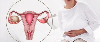
Definition
A cystic formation on the round ligament of the genital organ is a benign pathology. This ligament begins in the area of the fallopian tubes, extends along the sides of the pelvis through the inguinal canal, and ends in the area of the labia.
Cysts are round in shape, usually filled with fluid and in some cases reach the size of dropsy.
The secretion that accumulates inside the benign formation is the secretion of the nabothian glands. That is, a cystic tumor causes blockage of the ducts. Because of this, the glands significantly increase in size, and squamous epithelium accumulates in them.
How is a cyst different from a fibroid or tumor? The term tumor usually describes a tissue change. It may be benign or malignant. Malignant tissue changes are called cancer. This diagnosis causes concern for both patients and doctors.
Source: https://yazdorov.win/venericheskie-bolezni/svyazochnyj-apparata-matki.html
How does the intervention work?
The bougienage procedure is carried out in a hospital setting under general anesthesia. Local anesthesia is used less frequently because it is considered ineffective. The type of anesthesia is determined by the doctor after a complete examination of the woman. The surgical technology involves the introduction of special instruments into the cervical canal.
Attention! Without anesthesia, the patient will feel pain, and the risk of injury to the mucous membranes due to sudden movements will increase.
The diameter of the bougie increases gradually; if the technology is not followed, cervical rupture is likely. In case of accidental injury, the risk of re-occlusion of the cervical canal increases. Correctly performed dilation guarantees the absence of complications. Bougienage is prohibited if the fusion occurs against the background of a malignant process. In this case, radical operations are used. Bougienage is not performed if a woman has had false amenorrhea for 6 months. Hormonal testing can help detect this condition. In this case, plastic canalization of the cervix is performed.
After the operation, the woman can go home, but sometimes the doctor recommends staying in the hospital. This condition reduces the likelihood of bleeding. Outpatient treatment is acceptable in the absence of pathologies of the heart and blood vessels. Local anesthesia is used if general anesthesia cannot be administered.
Ovaries and fallopian tubes. Reproductive system. human anatomy
This internal muscular organ of the reproductive system has a pear-shaped shape that is flattened in front and behind. In the upper part of the uterus on the sides there are branches - fallopian tubes, which pass into the ovaries. The rectum is located behind, and the bladder is located in front. The anatomy of the uterus is as follows. The muscular organ consists of several parts:
- The fundus is the upper part, which has a convex shape and is located above the line of origin of the fallopian tubes.
- A body into which the bottom smoothly passes. It has a cone-shaped appearance. It narrows downwards and forms an isthmus. This is the cavity leading to the cervix.
- Cervix - consists of the isthmus, cervical canal and vaginal part.
The size and weight of the uterus vary from person to person. The average weight for girls and nulliparous women reaches 40–50 g.
The anatomy of the cervix, which is a barrier between the internal cavity and the external environment, is designed so that it protrudes into the anterior part of the vaginal vault. At the same time, its posterior arch remains deep, and the anterior arch, on the contrary.
Features of the ligaments of the internal genital organs of women
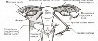
The ligaments connecting the uterus, tubes, ovaries with the pelvic walls, as well as among themselves, form their suspending and fixing apparatus, providing the internal organs with a normal physiological position.
The uterine suspensory apparatus includes the following ligaments:
The broad ligament is a pair, consisting of two layers of peritoneum adjacent to the surface of the uterus in front and behind in the frontal plane. On the sides it extends to the walls of the pelvis, at the bottom its leaves pass onto the parietal peritoneum of the pelvic diaphragm. Its structure consists of 3 parts - the mesentery of the uterus, fallopian tube, and ovary.
Features of the development of cervical diseases, symptoms
The ovaries are located on both sides of the uterus and are defined respectively as right and left.
The size of the gland is 3-4 cm in length, 1-1.5 cm in width, but these parameters may vary depending on the woman’s age, condition, for example, pregnancy, menopause.
The ovaries are connected to the uterus using a special ligament; their location is asymmetrical; often the gland located on the right is larger than the left.
The maintenance of the gonads in the pelvic area is ensured by the ligamentous apparatus, which relates directly to the ovaries. It allows them to move slightly relative to other organs, which is necessary during pregnancy, when the uterus significantly increases in size.
If we consider the anatomical structure of a woman’s ovaries, the gland consists of various functional layers. Initially there is a junction of the germinal epithelium, followed by a dense protein shell, consisting, among other things, of connective tissue and elastic fibers. It is this layer that forms a kind of stroma or base that helps maintain the shape of the organ.
- Internal or cerebral. It consists mainly of special loose tissue and many vessels.
- Outer layer or cortex. It is more dense in structure, contains connective fibers and maturing follicles.
Mature follicles surround the ovary, their approximate size is 1-2 cm in diameter. An interesting feature of the development of follicles is the dominance of one of them. If it has reached a significant size, larger than the others, then all resources are directed to the maturation of the egg, and the growth of other follicles is suspended.
Pathological changes often occur on the vaginal surface of the cervix, in the “transition zone” of one epithelium to another. When the localization of this zone changes, background diseases arise: ectopia (pseudo-erosion), ectropion, leukoplakia, erythroplakia, polyps and, as a consequence, malignant neoplasms of the cervix.
Ectopic cervix
Ectopia (pseudo-erosion) is a disease of the cervix, characterized by the emergence of columnar epithelium on the vaginal surface of the cervix. Women with irregular menstrual cycles are predisposed to developing this disease.
The clinical picture is somewhat blurred. Such women may complain of the release of blood clots after intercourse and the pain of the process of intercourse itself. The disease can be recognized during a medical examination of the cervix in the mirror: the surface of the organ will not be pale pink, but a bright red hue.
Ectropion is an eversion of the mucous membrane of the cervical canal into the vagina, with the formation of scars and ectopia on the cervix. This disease occurs as a result of unsuccessful diagnostic or surgical intervention on the uterus, as well as after labor or abortion.
Leukoplakia is a disease associated with the appearance of keratinized areas on non-keratinized epithelium. Leukoplakia can be simple or proliferative. The proliferative form is a precancerous condition and when biological material is taken from this area (biopsy), atypical cells are found.
With erythroplakia, the cervix is affected locally, closer to the external os. The pathogenesis of the disease is the thinning of the stratified squamous epithelium and, as a result, tissue atrophy. The etiology of erythroplakia has not yet been studied. The photo clearly shows red spots in the ectocervix area.
Cervical polyps
Polyps are growths of various sizes on the mucous membrane of the cervical canal. According to their structure, polyps are epidermal and glandular. When examined in a speculum, the polyps are oblong and leaf-shaped. It is possible to diagnose and differentiate such polyps only with an ultrasound examination of the pelvic area.
Cervical endometriosis is a disease in which tissue similar in structure to the endometrium is located on the vaginal surface of the cervix. Normally, such tissue should be located in the uterine cavity.
The main reasons for the development of this disease:
- cervical injuries due to violence, childbirth or abortion, as well as medical interventions;
- genetic predisposition;
- hormonal disbalance.
Patients with cervical endometriosis usually do not present characteristic complaints. There is discomfort during sexual intercourse, bleeding after intercourse, as well as before and after menstruation.
Source: https://cher-crb.ru/organy-malogo-taza/slizistyj-sloj-matki.html
How to prevent overgrowth
Compliance with the following rules helps prevent the development of the problem:
- timely treatment of various inflammatory processes affecting the central canal and cervix;
- regular visits to the gynecologist to identify inflammatory processes and malignant neoplasms;
- prevention of unwanted pregnancy, reducing the frequency of abortions;
- refusal of self-medication;
- compliance with the rules of a healthy lifestyle;
- the use of barrier methods of contraception to prevent bacterial infection.
Ligaments of the uterus: round, broad and cardinal
One of the main organs of the female reproductive system is the uterus. It is located in the small pelvis, in its middle part.
This organ is characterized by mobility and can occupy different positions relative to other organs. This may be influenced by the size of other organs and their condition, as well as the individual characteristics of each female representative.
However, there are certain rules according to which the norm and pathology in the location and functioning of the uterus are determined. Typically, its longitudinal axis should run along the pelvic axis, and the bottom of the organ is inherently tilted forward.
The concept of the ligamentous apparatus of the uterus
To maintain the body of the uterus in a normal position, the body has a ligamentous apparatus, which includes ligaments and fascia. Each element is characterized by certain functions, thanks to which the organ maintains its position while remaining sufficiently mobile.
The ligamentous apparatus is a complex system consisting of different types of ligaments. There are three main components of this system. This:
- Suspensory. Its main function is to connect the uterus to the walls of the pelvis to hold it in the required position. It is formed by the following ligaments: broad ligaments of the uterus;
- round ligaments of the uterus;
- ovarian ligaments;
- suspensory ligaments of the ovary.
- main uterine;
sacrouterine;
During pregnancy, the uterus increases in size, which is necessary for the full development of the fetus. At this time, the ligaments surrounding it are stretched so as not to hamper its growth.
In addition, during pregnancy, the load on the ligamentous apparatus increases, since its main function is to support the uterus in a certain position. To achieve this, the elements of the ligamentous apparatus thicken in order to cope with their functions. Such changes occur under the influence of hormones.
These changes that occur during pregnancy can often cause unpleasant and even painful sensations in the expectant mother. This phenomenon is normal, and the pain can be both acute and mild, short or long-lasting.
They can occur especially often in the second trimester of pregnancy, when a woman suddenly changes position, or during prolonged physical activity.
During the second pregnancy (and subsequent ones), pain due to sprains is characterized by greater severity. However, you should not think that any pain during this period is caused by changes in the ligamentous apparatus. If pain occurs too often, and along with it occurs:
- bleeding;
- elevated temperature;
- pain in the lumbar region;
- chills;
- difficulty urinating, etc.,
you should consult a doctor. Such symptoms during pregnancy may indicate the presence of abnormalities in pregnancy or an infection in the genitourinary tract. The doctor will conduct the necessary tests to make the correct diagnosis.
Also, the occurrence of pain in the pubic area during pregnancy may indicate the presence of disturbances in the functioning of the pelvic ligaments. Therefore, if you discover this symptom, it is advisable to consult a doctor.
In general, the ligamentous apparatus adapts to pregnancy relatively easily, however, excessive and frequent pain should be a cause for concern.
Main types
Among the elements that make up the ligamentous apparatus, not only those relating to the uterus are distinguished, but also the ligaments of the ovaries, as well as the muscles of the perineum. Directly related to the uterus itself are:
- wide,
- round,
- cardinal,
- sacrouterine.
Wide
The broad ligament of the uterus is double, parts of which are located on the right and left. They are located in the small pelvis, in its frontal plane. This element continues the serous layer of the integument of the body of the organ in front and behind.
It extends from its edges and is attached to the walls of the pelvis from the inside. Each part is split into two leaves, between which in the upper part there are fallopian tubes. At their base, between the sheets, there is fiber, through the bottom of which the uterine artery runs.
These parts of the ligamentous apparatus have almost no effect on the location of the organ, since they lie freely and allow the uterus to maintain mobility. Smoothness is not typical for this type of ligaments, since they cover:
- ovaries;
- fallopian tubes;
- ovarian ligaments;
- round uterine ligaments.
Because of this, mesenteries are formed in their thickness to accommodate each element.
Round
The round ligament of the uterus is also double and runs on both sides. Their total length reaches 15 cm. They consist of smooth muscle and connective tissue. The round ligaments lie within the broad ligaments.
They begin in the area of the lateral parts of the uterine body, slightly lower than the fallopian tubes, and pass to the side walls of the pelvis. Then they pass through the inguinal canal and exit into the tissue of the labia.
Thanks to the presence of this part of the ligamentous apparatus, the uterus does not tip back.
Cysts, fibroids and fibromyomas sometimes form in this element. They, as well as malignant tumors developing in this area, often do not manifest themselves at all. Only when they grow under the influence of hormones can pain occur in the groin and lower abdomen. All formations that arise in this area can only be treated surgically.
Features of cardinal ligaments
The cardinal ligaments are the basis for the broad ligaments. They are located in the cervix area in the form of rounded dense cords that secure the organ on both sides.
In essence, it is a thickened lower part of the broad ligaments, in which, as development progressed, the amount of connective and smooth muscle tissue increased.
The function of this element is to prevent forward and backward displacement of the uterus. In their thickness lie the uterine vessels, as well as the ureters. Between the individual sections of the ligaments, a parametrium formed from fiber is formed.
Sacrouterine
This is the last element of the ligamentous apparatus of the uterus. Such ligaments also consist of connective and smooth muscle tissue. They originate on the back of the cervix and are fixed in the muscle tissue of the rectum.
Some of their fibers continue further - to the sacral vertebrae. Their role is to create a counterweight to the round uterine ligaments. Thanks to their presence, the uterus does not deviate forward, maintaining its position.
In addition to those listed, the location of the uterus in the pelvic cavity is also influenced by other ligaments, for example, the own ovarian ligaments. They pass from the uterus to the ovaries. But their presence has an effect mainly on the ovaries; for the uterus they are not so significant.
Let us know about it - rate it Loading...
Source: https://ginekologii.ru/ginekologiya/matka/kruglaya-svyazka-matki.html
Organ height in expectant mothers
An increase in uterine size, a change in its density and shape is a characteristic sign of bearing a child.
In the early stages of pregnancy, asymmetry of the muscular organ is often recorded, which frightens inexperienced expectant mothers. However, there is no cause for concern. Protrusion of one of the uterine sides occurs due to the development of the fertilized egg. Gradually, during the growth process, the fertilized egg is located in the uterine cavity, and this asymmetry is eliminated.
At 12 weeks, the body of the uterus becomes four times larger, while the fundus stands high and is located in the area of the upper border of the symphysis pubis.
Gynecologists begin measuring and recording the height of the uterine fundus from about 14 weeks.
Due to the anatomical characteristics of each woman, a deviation from the norm of 1-2 centimeters is allowed. But in order to prevent the development of pathologies, the doctor must take into account all the criteria:
- physiological characteristics of a woman before conception (height, weight);
- uterine parameters;
- fruit size;
- pelvic circumference.
Simultaneously with the height of the mother's womb, obstetricians identify the volume of amniotic fluid, which affects the fetus. This value also depends on the physiological characteristics of the female body and on the presence of subcutaneous fat.
Ligamentous apparatus of the uterus
Ligaments forming the apparatus Connects the uterus to the walls of the pelvis Paired wide uterine suspensory ligaments of the ovary Own ligaments of the ovary Round ligaments of the uterus Fixes the position of the organ, during pregnancy it is stretched, providing the necessary mobility ligament of the uterus Forms the pelvic floor, which is a support for the internal organs of the genitourinary system Muscles and fascia of the perineum (outer, middle, inner layer) Anatomy The uterus and appendages, as well as other organs of the female reproductive system, consists of developed muscle tissue and fascia, which play a significant role in the normal functioning of the entire reproductive system.
Characteristics of the hanging apparatus
The suspensory apparatus consists of paired ligaments of the uterus, thanks to which it is “attached” at a certain distance to the walls of the pelvis. The broad uterine ligament is a transverse fold of the peritoneum. It covers the body of the uterus and the fallopian tubes on both sides. For the latter, the structure of the ligament is an integral part of the serous covering and mesentery. At the lateral walls of the pelvis it passes into the parietal peritoneum. The suspensory ligament arises from each ovary and has a wide shape. Characterized by durability. The uterine artery passes through it. The own ligaments of each of the ovaries originate from the uterine fundus on the back side below the branch of the fallopian tubes and reach the ovaries. The uterine arteries and veins pass inside them, so the structures are quite dense and strong. One of the longest suspensory elements is the round ligament of the uterus. Its anatomy is as follows: the ligament looks like a cord up to 12 cm long. It originates in one of the corners of the uterus and passes under the anterior sheet of the broad ligament to the internal opening of the groin. After which the ligaments branch into numerous structures in the tissue of the pubis and labia majora, forming a spindle. It is thanks to the round ligaments of the uterus that it has a physiological inclination anteriorly.
Structure and location of fixing ligaments
The anatomy of the uterus should have suggested its natural purpose - bearing and giving birth to offspring. This process is inevitably accompanied by active contraction, growth and movement of the reproductive organ. In this connection, it is necessary not only to fix the correct position of the uterus in the abdominal cavity, but also to provide it with the necessary mobility. It is for such purposes that fixing structures have arisen. The uterine ligament consists of plexuses of smooth muscle fibers and connective tissue, radially located towards each other. The plexus surrounds the cervix in the area of the internal os. The ligament gradually passes into the pelvic fascia, thereby fixing the organ to the position of the pelvic floor. The vesicouterine and pubic ligamentous structures originate from the lower anterior part of the uterus and are attached to the bladder and pubis, respectively. The uterosacral ligament is formed by fibrous fibers and smooth muscle. It extends from the back of the cervix, envelops the rectum on the sides and connects to the fascia of the pelvis on the sacrum. In a standing position, they have a vertical direction and support the cervix.
Supporting apparatus: muscles and fascia
The anatomy of the uterus implies the concept of “pelvic floor”. This is a set of muscles and fascia of the perineum that make it up and perform a function that supports the woman’s internal genital organs. The pelvic floor consists of an outer, middle and inner layer. The composition and characteristics of the elements included in each of them are given in the table: Fixing apparatus of the internal genital organs of a woman
consists of suspending, securing and supporting devices that ensure the physiological position of the uterus, tubes and ovaries.
The suspensory apparatus
is a complex of ligaments that connect the uterus, tubes and ovaries to the walls of the pelvis and to each other.
This group includes the round, broad ligaments of the uterus, as well as the suspensory and proper ligaments of the ovary. The round ligaments of the uterus
(lig. teres uteri, dextrum et sinistrum) are a paired cord 10-15 cm long, 3-5 mm thick, consisting of connective tissue and smooth muscle fibers, which are a continuation of the outer muscular layer of the uterus.
Starting from the lateral edges of the uterus, slightly lower and anterior to the beginning of the fallopian tubes on each side, the round ligaments pass between the leaves of the broad uterine ligament (intraperitoneal) and are directed first outward, almost horizontally, and then anteriorly and downward, to the side wall of the pelvis, retroperitoneally. On their way, the round ligaments
cross the obturator vessels and nerve, the middle umbilical ligament with the obliterated umbilical artery passing through it, the external iliac vessels with the lower epigastric vessels extending from them, and then enter the internal opening of the inguinal canal.
Their distal third is located in the canal, then the ligaments exit through the external opening of the inguinal canal and branch in the subcutaneous tissue of the labia. The broad ligaments of the uterus
(lig. latum uteri, dextrum et sinistrum) are frontally located duplications of the peritoneum, which are a continuation of the serous cover of the anterior and posterior surfaces of the uterus away from its “ribs” and split into sheets of parietal peritoneum of the side walls of the small pelvis - from the outside.
At the top, the wide ligament of the uterus is closed by the fallopian tube, located between its two layers; below, the ligament splits, passing into the parietal peritoneum of the pelvic floor. The broad ligament of the uterus
distinguishes the following parts: mesentery of the fallopian tube (mesosalpinx);
mesentery of the ovary (mesovarium); mesentery of the uterus (mesometrium), which includes the rest (large) part of the broad ligament of the uterus, located below the ligament proper and the mesentery of the ovary. Between the leaves of the broad ligament
(mainly at their base) lies fiber (parametrium), in the lower part of which with one and on the other side is the uterine artery.
The broad ligaments of the uterus
lie freely (without tension), follow the movement of the uterus and cannot, naturally, play a significant role in maintaining the uterus in a physiological position.
Speaking about the broad ligament of the uterus, it is impossible not to mention that with intraligamentary tumors of the ovaries located between the leaves of the broad ligament, the usual topography of the pelvic organs is disrupted to one degree or another. The suspensory ligaments of the ovary
(lig. suspensorium ovarii, dextrum et sinistrum) go from the upper (tubal) end of the ovary and fallopian tube to the peritoneum of the lateral wall of the pelvis.
These relatively strong ligaments, thanks to the vessels (a. et v. ovaricae) and nerves passing through them, keep the ovaries suspended. The proper ligaments of the ovary
(lig. ovarii proprium, dextrum et sinistrum) are a very strong short fibrous-smooth muscle cord connecting the lower (uterine) end of the ovary with the uterus, and pass through the thickness of the broad ligament of the uterus. The fixing, or
actually fixing, apparatus
(retinaculum uteri) is a system of “compression zones” that form the basis (skeleton) of ligaments that are in close connection with the parietal and visceral fascia of the pelvis.
The compaction zones consist of powerful connective tissue cords, elastic and smooth muscle fibers. In the fixing apparatus, the following parts are distinguished: the anterior part
(pars anterior retinaculi), which includes the pubovesical or pubovesical ligaments (ligg. pubovesicalia), which continue further in the form of the vesicouterine (vesico-cervical) ligaments (ligg. vesicouterina s. ve-sicocervicalia);
the middle part
(pars media retinaculi), which is the most powerful in the fastening apparatus system;
it mainly includes the system of cardinal ligaments (ligg. cardmalia); the posterior part
(pars posterior retinaculi), which is represented by the uterosacral ligaments (ligg. sacrouterine). Some of the listed
ligaments
should be discussed in more detail.
The vesico-uterine
, or vesico-cervical, ligaments are fibromuscular plates that cover the bladder on both sides, fixing it in a certain position, and keeping the cervix from moving posteriorly. The main, or
main (cardinal) ligaments of the uterus
are a cluster intertwined dense fascial and smooth muscle fibers with a large number of vessels and nerves of the uterus, located at the base of the wide uterine ligaments in the frontal plane.
The uterosacral ligaments
consist of muscular-fibrous bundles (m. rectouterinus) and extend from the posterior surface of the cervix, arcuately covering the rectum from the sides (weaving into its side wall), and are fixed to the parietal layer of the pelvic fascia on the anterior surface of the sacrum.
Raising the peritoneum covering the top, the sacrouterine ligaments form rectouterine folds (plicae rectouterinae). The supporting (supporting) apparatus
is united by a group of muscles and fascia that form the floor of the pelvis, above which the internal genital organs are located, described in detail in our article.
Source: https://zodiacc.ru/info/svjazochnyj-apparat-matki/
Ligaments of the uterus
Today we’ll talk about the uterine ligaments. This is a good topic both before Module 5 (“About relationships”, we will still talk about a woman, and therefore about the uterus), and before Module 3 (“Internal organs”), and just to understand what this organ is connected to and how affects the musculoskeletal system and other organs.
The ligaments of the uterus are divided into two groups according to their main functions:
- Hanging apparatus.
- Fastening apparatus.
Round ligaments
They depart from the corners of the uterus (slightly anteriorly and below the origin of the tubes), go inside the broad ligament to the internal openings of the inguinal canals. Having passed the inguinal canal, the round ligaments fan out into the tissue of the pubis and labia majora.
The round ligaments pull the fundus of the uterus anteriorly. What are the conclusions? If during pregnancy, during menstruation, the labia majora pull, it means there is tension on the part of the uterus.
Will the condition of these ligaments affect symphysitis, discrepancy of the pubic symphysis? Yes, sure!
Broad ligaments of the uterus
- double layers of peritoneum, continuing the membrane covering the anterior and posterior surfaces of the uterus, extending to the side walls of the pelvis.
These ligaments do not particularly secure the uterus from the sides, they lie freely, follow the uterus when it moves, and serve more for packaging! The pipes lie on top of them, the fiber (parametrium) lies at the base, in the lower part of which there are vessels, nerves and the ureter.
Suspensory ligaments of the ovary
- These are continuations of the broad ligaments of the uterus from the ampulla of the tube to the pelvic wall. These ligaments hold the ampullary end of the tube and ovary quite well in a suspended state.
Thus, the uterus is still attached to the sides of the pelvic bones by the tubes (+ remember that our ovary is fixed to the pelvic wall). The ovarian artery and vein pass through the thickness of the ligaments.
What does this mean? If the ligaments stick together and have low mobility, the trophism (nutrition) of the ovary will be disrupted.
Anchoring apparatus of the uterus
Galina thinks that it would be possible not to write everything written above, but to leave only about these ligaments, and you would already have a picture that the uterus affects EVERYTHING!
See:
The anchoring apparatus consists of connective tissue cords with an admixture of smooth muscle fibers (i.e., having a tendency to contract uncontrollably under the influence of stress with the subsequent formation of fibrosis), which are directly connected to the muscles of the lower part of the uterus.
Ligaments run from the lower part of the uterus to the posterior, lateral and anterior walls of the pelvis, thoroughly fixing the position of the uterus in the pelvis. So what does that mean?
If we have a pelvic distortion, scoliosis, had hip dysplasia in childhood, or have a spasm in the cecum or sigmoid colon, then the ligaments will pull the uterus to the side, at the same time creating tension in the opposite tube and ovary. That’s why everything there is so poorly corrected with medication, but so excellent with your hands!
Uterovesical ligaments
They go from the lower part of the uterus to the bladder, cover it and continue to the pubic symphysis, changing the name to vesico-pubic. These ligaments secure the bladder and cervix anteriorly. And we draw a conclusion about the mutual influence of these two organs on each other. And this sheds light on the cause of attacks of cystitis after sexual intercourse. Is it clear why? Adjustable by hand.
Cardinal (main) ligaments of the uterus
They depart from the body of the uterus at the level of the internal uterine os (where the cervix meets the body of the uterus). They are dense connective tissue with a small inclusion of muscle fibers. They are located at the base of the broad ligaments and go to the lateral walls of the pelvis. What is the connection? With hip joints!
Innervation of the uterus and other female reproductive organs
The internal genital organs are innervated by the sympathetic and parasympathetic autonomic nervous system. The nerves going to the uterus are usually sympathetic. On their way, spinal fibers and structures of the sacral nerve plexus are attached. Contractions of the uterine body are regulated by the nerves of the superior hypogastric plexus. The uterus itself is innervated by branches of the uterovaginal plexus. The cervix usually receives impulses from the parasympathetic nerves. The ovaries, fallopian tubes, and adnexa are innervated by both the uterovaginal and ovarian plexuses of nerves.