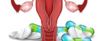
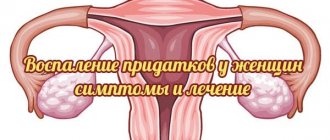
Frequent diseases of the reproductive system include inflammation of the appendages in women (uterus, fallopian tubes, ovaries). The disease is also called adnexitis or salpingoophoritis and occurs when the organs of the reproductive system are affected by infection (genital and non-genital).
Once in a woman’s body, the infection affects both the fallopian tubes and the ovaries. This may happen:
- ascending (mainly) from the vagina and uterus;
- descending route (hematogenous, from the cecum and sigmoid colon in the peritoneum);
- through the lymphatic tract or blood vessels (in rare cases).
Most often, inflammation of the uterine appendages is accompanied by the formation of adhesions, which aggravates the disease itself and complicates treatment. Therefore, it is highly advisable not to delay your visit to the doctor in order to eliminate the infection and prevent undesirable consequences.
Causes
The appendages become inflamed under the pathological influence of cocci (streptococci, staphylococci, gonococci), Escherichia coli, Koch bacillus (mycobacterium tuberculosis), actinomycetes (radiant fungi). Koch's bacillus, a pale spirochete, enters the tubes and ovaries from infected organs through the bloodstream. This route of infection is called hematogenous. The remaining bacteria penetrate the appendages along the ascending path.
The vagina is inhabited by opportunistic microorganisms. When certain factors appear, they become pathogenic, causing adnexitis. Pathagents penetrating from outside displace beneficial microflora from the vaginal area. Bacteria spread along an ascending path, reaching the tubes and gonads, leading to their inflammation and corresponding symptoms. Pathogens can also be carried through the lymphatic system.
Provoking factors:
- promiscuity in intimate partners, their frequent change;
- sex without a barrier contraceptive. A regular partner may not be sick himself, but may be a carrier of the infection. If a woman has a weak immune system and/or menstruation, she will most likely become infected;
- STD;
- stagnation of blood in the pelvis caused by irregular sexual intimacy, physical inactivity, obesity, constipation;
- tuberculous damage to any organ;
- chronic infectious process in the absence of treatment during relapse (tonsillitis, caries, sinusitis, bronchitis, cystitis, endometritis, vulvitis, vaginitis, others);
- low immune status, susceptibility to frequent colds;
- long-term antibiotic therapy;
- having sex during menstruation. Through the open cervical canal with regulation, microorganisms easily penetrate into the appendages, causing inflammation;
- stress negatively affects the production of hormones;
- severe illnesses that weaken the body;
- lack of treatment for urogenital infections;
- hormonal imbalance;
- unsuccessfully installed IUD. The intrauterine contraceptive itself is not the cause of salpingoophoritis, but it aggravates the course of the pathological process. Therefore, before treatment, the IUD is removed;
- regular physical fatigue;
- constant diets, fasting, as a result - vitamin deficiency, deficiency of microelements;
- ignoring hygiene procedures;
- using other people's towels, napkins, linen for hygiene;
- improper hygiene when the pathogen is introduced from the intestines;
- swimming in open water, especially during menstruation;
- intimacy after surgical treatment, when the gynecologist’s recommendations are not followed;
- surgical interventions on the reproductive organs;
- childbirth, especially complicated ones;
- invasive diagnostic procedures.
Pregnant women are less at risk of inflammation of the appendages. This is due to changes in the composition and quality of cervical mucus. During pregnancy, it thickens, performing a barrier function, trapping bacteria. If a woman has chronic adnexitis, then a relapse is possible. This is explained by intense hormonal changes during the gestational period.
Where are they located?
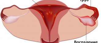
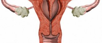
The epididymis is located between the ovary and the end of the fallopian tube. It is possible to determine their location even more precisely: between the peritoneum of the uterus (its widest part) and the ovary, the end of the tube. This wide part of the uterus with its adjacent organs is susceptible to various diseases. Most often this is inflammation of the appendages in women. We will consider the symptoms and treatment of this disease below. The appendage tubes are on average 10-12 cm, physiologically the right one is longer than the left one. These organs are also divided into specific departments, each of which performs its own functions.
Classification
Inflammation of the appendages is classified as follows:
| By localization | With the flow | By pathogen type |
| Left-handed | Acute – abrupt onset, severe symptoms. Recovery in a few days | Bacterial – caused by different groups of bacteria |
| Right-handed | Subacute – lasts several days or months | Fungal – the cause is a fungal infection |
| Two-way | Chronic – occurs if acute inflammation is not treated. Characterized by an undulating course, unexpressed symptoms |
Diagnosis
Diagnostic measures begin with collecting anamnesis - determining the frequency and regularity of the menstrual cycle. At the same time, the doctor performs an intrauterine intervention. A physical examination allows you to determine such abnormalities as an enlarged uterus, its compaction, and so on.
Laboratory diagnostic methods are represented by blood tests and smears. Acute endometritis is characterized by an increase in erythrocyte sedimentation rate and leukocyte count. In parallel with this, C-reactive protein appears, indicating the presence of an inflammatory process in the body. When diagnosing the disease, microscopy of a vaginal smear plays an important role. Instrumental diagnostic methods are represented by ultrasound, biopsy and hysteroscopy.
Signs of inflammation of the appendages
In 2/3 of patients there is an acute onset. In 1/3, the disease begins “sluggishly”, with “erased” symptoms, characteristic of many genitourinary infections.
The acute stage is characterized by:
- cramping or nagging severe pain in the lower abdomen or only on the affected side. Pain increases during coitus, lifting weights, and playing sports. Irradiation to the lumbar region and anus is noted. This is the main symptom of the pathology;
- fever 38 degrees or higher;
- joint, headache, muscle pain;
- sweating, weakness;
- cardiopalmus;
- dyspeptic disorders due to intoxication;
- severe vaginal itching, which intensifies during urination, after sex, and hygiene procedures with soap. A burning sensation in the vagina is another characteristic manifestation of inflammation;
- frequent urination;
- problems with the menstrual cycle. Regulations may increase or, on the contrary, become scarce. The regularity of the cycle is disrupted;
- bleeding outside the cycle;
- when the process is started, an unpleasant odor from the genitals and purulent-serous discharge appear.
With chronic inflammation of the appendages, an asymptomatic course is often observed.
May occur:
- unexpressed periodic aching pain;
- painful sex and bowel movements;
- non-cyclic bleeding;
- mucous vaginal discharge;
- unpleasant odor that increases during menstruation;
- Lack of treatment leads to cycle disruption. With purulent inflammation, amenorrhea sometimes occurs;
- low-grade fever during relapse.
Pain syndrome worsens the quality of intimate life. Libido decreases, irritability and emotional tension appear. A woman may refuse intimacy without knowing the reasons for her condition. You just need to see a doctor, undergo an examination and a therapeutic course.
If there is pain in the area of the right appendage, the cause is acute abdominal syndrome
The term "acute abdomen" is used to refer to symptoms that occur with inflammation of the pancreas (pancreatitis) and peritonitis. Acute abdomen syndrome means that the patient has:
1 sharp or nagging pain in the area of the right appendage;
2 high body temperature;
3 vomiting;
4 weakness;
5 muscle tension in the anterior abdominal wall.
When the diagnosis is confirmed, the patient is urgently hospitalized in the surgical department, where the necessary care is provided.
Diagnostics
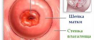
With inflammation of the female organs, consult a gynecologist. At the appointment, the patient is asked what is bothering her, her medical history is ascertained, and a gynecological examination is performed. Using a mirror, the doctor detects enlargement, thickening, swelling of the appendages, redness of the mucous membrane, and uneven surface. There is a change in tissue structure, pathological discharge, unpleasant odor, and pain on palpation.
It turns out whether abortions, curettage, or operations were performed. Was there pregnancy and childbirth, method of delivery. How does menstruation happen and how long does it last? Questions are asked about intimate life.
After examination and conversation, a diagnostic complex is prescribed:
- blood biochemistry;
- general urine test;
- PCR testing to exclude STDs;
- a smear from the infected area for bacterial culture to determine the type of microflora so that an effective antibiotic can be selected;
- transvaginal ultrasonography. For pregnant women, ultrasound is performed transabdominally;
- pregnancy test. In some cases, ectopic pregnancy may have symptoms similar to inflammation of the appendages.
If a tuberculous form of adnexitis is suspected, menstrual blood is taken for bacterial culture and a Pirquet or Mantoux test is performed. For differential diagnosis of inflammation of the appendages or in case of an unclear diagnosis, the following can be performed:
- hysterosalpingography - X-ray contrast examination of the fallopian tubes and uterus;
- colposcopy – a detailed examination of the uterine cavity using a colposcope (binocular with a lighting device);
- hysteroscopy – the uterus and appendages are examined using a hysteroscope. As with colposcopy, biopsy material can be taken;
- Laparoscopy – allows you to simultaneously examine organs and perform surgical procedures.
A biopsy is needed to exclude atypical cell degeneration and assess the risk of neoplasms. MRI and computed tomography with contrast can be performed.
Female uterus
Before we begin to describe the disease, let us briefly consider the anatomical structure of a woman, more precisely, the uterus.
So, the female uterus refers to a muscular, hollow organ that is located between the bladder and the rectum. The uterus acts as a receiving organ that receives all the hormones produced by the ovaries. The surface of the organ is covered with a special layer, which, at the moment of activation of reproductive function, experiences significant changes. These transformations are caused by preparation for the process of fertilization.
Treatment
In severe cases, hospitalization is required, in others it can be treated on an outpatient basis. The main way to eliminate inflammation of the appendages is antibacterial therapy. For effective treatment, broad-spectrum drugs are needed. The duration of the course, medications, dosage, and frequency of administration are selected individually. For mild salpingoophoritis, the patient takes the tablets for 5-7 days. In severe cases, antibiotics are administered intravenously or intramuscularly.
To relieve symptoms, painkillers, antihistamines, antifever, and anti-inflammatory drugs are prescribed. Multivitamin complexes are used to strengthen the immune system. Local treatment must be carried out in combination with systemic treatment. Hygienic baths with antiseptic solutions are made, vaginal suppositories or tablets with an antibacterial effect are prescribed, and the uterine cavity is sanitized.
After the acute phase of inflammation is removed, physiotherapy is used.
| Highly effective physiotherapeutic methods | Action |
| Medicinal electrophoresis | Relieves pain, swelling |
| Laser therapy | Improves microcirculation, shortens recovery period, normalizes metabolism |
| Ultrasound | Helps soften adhesions and normalize hormonal levels |
| Ural Federal District | Kills bacteria |
| Paraffin therapy | Stimulates ovarian hormone production |
| Magnetotherapy | Accelerates the regeneration of damaged tissues |
During the rehabilitation period, gynecological massage is useful. It improves muscle tone and blood circulation in the pelvic organs. Massage helps to resolve adhesions, normalize menstrual function, hormonal balance, and increase libido. Physiotherapy has a calming effect and has a beneficial effect on the emotional background. For chronic inflammation of the appendages, sanatorium treatment helps to prolong remission. Balneotherapy and mud tampons have a good effect.
In case of a complicated course, the patient is hospitalized for surgical intervention. In modern gynecological surgery, organ-preserving techniques are used. This is especially true for women of reproductive age planning pregnancy.
Laparoscopy is most often used. Under systemic anesthesia, the patient is given 3-4 small punctures in the abdominal wall. Gas is injected into the peritoneum to expand the surgical space. A laparoscope and microinstruments are inserted into the incisions. The surgeon uses manipulators to eliminate the problem. It can dissect adhesions, remove damaged tissue, polyps, cysts, myomatous nodes, and small tumors. A biopsy sample is taken for morphological examination. The entire procedure is displayed on the screen. The doctor visualizes the organs under significant magnification. Therefore, with a proven technique, errors are practically eliminated.
If inflammation of the appendages is advanced, there is a risk of pipe rupture, a purulent abscess has developed, or there is a danger to the woman’s life, then the appendages are removed in older patients.
To quickly restore the menstrual cycle, oral contraceptives are prescribed in minimal doses.
After surgery it is recommended:
- temporary abstinence from sexual intercourse. In each case, a specific period is determined;
- do not eat hot, spicy, pickled, sour foods. Do not drink alcohol or hot drinks;
- do not take hot baths, do not visit baths, saunas, do not swim in open reservoirs and public pools;
- you cannot lift weights;
- do not engage in active sports;
- don't get too cold.
Minor physical activity is allowed so that there is no stagnation in the pelvic organs. You need to drink plenty of fluids: non-hot tea, non-acidic fruit drinks, compotes, juices, clean water. Ultrasonography is prescribed to monitor the effectiveness of treatment.
Routes of infection
Due to the etiological factors described below, the mucous membrane of the organ can become inflammatory in nature. The development of the process may be associated with E. coli, chlamydia, or other pathogens.
In addition, pathogens are represented by viruses and protozoan microorganisms. From the above it follows that endometritis occurs against the background of infection entering the organ. Below we will consider ways of infection penetration.
So, infection occurs in the following ways:
- The first path is upward.
- The second is lymphogenous.
- Hematogenous.
Often the infection enters the organ through the external genitalia. After the infection process starts, the inflammatory process is activated. Due to the fact that the mucous membrane adheres tightly to the muscle layer, the process starts in the muscle tissue. If the inflammation is characterized by a chronic course, the disease is identified as metroendometritis. The process is of a short-term nature, which over time involves the uterine appendages or pelvic peritoneum.
Complications
Inflammation of the appendages develops rapidly. If not treated in time, it can become complicated by the following pathological conditions:
- various forms of menstrual cycle disorders. Oligomenorrhea occurs in 3% of women who have recovered from the disease;
- pain syndrome;
- hydrosalpinx - sacs with fluid;
- pyosalpinx - purulent exudate (in the tube), pyovar (ovary);
- cicatricial adhesive process;
- tubo-ovarian tumor - the tube enlarges due to the accumulation of pus and becomes similar to a tumor. It has nothing to do with neoplasm;
- hypofunction, ovarian depletion;
- sexual dysfunction;
- hormonal imbalance;
- damage to the pelvic tissue.
- ectopic pregnancy (up to half of cases).
Infertility is the most severe consequence of inflammation for women of childbearing age. The most dangerous thing is an abscess. It can lead to rupture of the fallopian tube. If purulent contents enter the abdominal cavity, peritonitis will develop. This is a life-threatening condition that requires immediate surgery.
Pain in the lower abdomen on the right and genitourinary diseases
Pain in the area of the right appendage may occur due to:
1 infection with sexually transmitted diseases (associated symptoms - copious vaginal discharge of various colors and consistency, burning and itching in the genital area);
2 pedicle torsions or cyst ruptures;
3 inflammation or apoplexy of the ovary;
4 adhesions in the fallopian tube.
Puncture of follicles as part of the IVF protocol and ectopic pregnancy can also provoke acute pain in the area of the right appendage: in the first case, the problem is solved by observing bed rest, in the second - through surgery.
Prevention
Inflammation of the appendages in its clinical picture and symptoms is similar to many diseases of the genitourinary system, especially of infectious etiology. Therefore, there is no specific prevention; you need to follow some rules:
- treat inflammation without allowing the process to become chronic or develop complications;
- refuse to have sex during menstruation;
- be selective in sexual relationships;
- It is advisable to use a condom during sexual intercourse. This will not only protect you from infection, but also from unwanted pregnancy. Curettage of the uterus is a risk factor. A barrier contraceptive will not prevent infection;
- do not overcool;
- treat colds in a timely manner;
- visit the dentist - microbes in the oral cavity from caries, unhealthy teeth or gums can be a source of infection that enters the genital tract through the bloodstream;
- be physically active to prevent stagnation in the pelvis;
- maintain hygiene regularly;
- do not use other people’s towels or hygiene products;
- do not swim in unfamiliar bodies of water, especially during menstruation;
- strengthen the immune system: harden yourself, make sure that your diet contains enough useful microelements and that your food is fortified. It’s good to take vitamin courses every six months or a year. Only a doctor can prescribe medications. Hypervitaminosis is as dangerous as vitamin deficiency.
If alarming symptoms appear, it is better to immediately get checked by a doctor so as not to start the inflammatory process. Medical examinations cannot be ignored. Once a year you need to undergo a gynecological examination.
Possible diseases
Women often complain of increased muscle tone in the lower abdomen. This may also indicate diseases that affect the ovaries and epididymis. Only a doctor can determine the cause of the discomfort.
Doctors identify the following main pathologies:
- Failure in the hormonal background of the female body.
- Neoplasms, ovarian cyst.
- Development of inflammatory processes.
Diseases of the pelvic organs are fraught with infertility and other health-threatening complications for women. You should not delay a visit to the gynecologist or self-medicate.
Ultrasound for endometrium
Ultrasound examination is associated with some difficulties due to the characteristics of the anatomical structure of the upper female genital organs. Despite some difficulties, this non-invasive research method is often used in diagnosing pathologies not only of the uterus, but also of the appendages.
Ultrasound detects symptoms only in 35 percent of cases. Ultrasound often reveals indirect symptoms of inflammation of the uterus, however, they are all nonspecific and may indicate another disease.
The following changes in the acute course are clearly visible on ultrasound:
- Quenching the endometrium.
- Purulent clots.
The chronic form of the disease is characterized by pathological changes that are difficult to detect on ultrasound. However, this disease is accompanied by changes in the structure of the uterine mucosa, which are detected by ultrasound.
The following echo signs are characteristic of chronic endometrium:
- Reduction and increase in the thickness of the uterine mucosa.
- Abnormal position of the uterus.
- Adhesions in the uterus.
Since inflammation can also affect adjacent female genital organs, in particular, the ovaries and fallopian tubes, changes can be noted on ultrasound.
Despite this, ultrasound is an indicative diagnostic method, which, in combination with other methods, makes it possible to establish an accurate diagnosis. Ultrasound examination allows us to exclude a number of diseases that have identical symptoms. A complete and detailed examination includes ultrasound, biopsy, and hysteroscopy.
Due to the fact that the diagnostic process includes an integrated approach, the data obtained during ultrasound gives the right to make a diagnosis such as endometritis.
Therapeutic measures
Treatment of endometritis begins immediately after an accurate diagnosis is made. If the pathology was detected in a timely manner, treatment takes place on an outpatient basis, but under the mandatory supervision of the attending physician. Otherwise, the patient is sent to the hospital. As for the treatment regimen, in most cases it is the same and consists of the following points:
- Antibacterial therapy.
- Mechanical cleaning of the uterine cavity.
- Detoxification.