Ligamentous apparatus of the uterus
Ligaments forming the apparatus Connects the uterus to the walls of the pelvis Paired wide uterine suspensory ligaments of the ovary Own ligaments of the ovary Round ligaments of the uterus Fixes the position of the organ, during pregnancy it is stretched, providing the necessary mobility ligament of the uterus Forms the pelvic floor, which is a support for the internal organs of the genitourinary system Muscles and fascia of the perineum (outer, middle, inner layer) Anatomy The uterus and appendages, as well as other organs of the female reproductive system, consists of developed muscle tissue and fascia, which play a significant role in the normal functioning of the entire reproductive system.
Characteristics of the hanging apparatus
The suspensory apparatus consists of paired ligaments of the uterus, thanks to which it is “attached” at a certain distance to the walls of the pelvis. The broad uterine ligament is a transverse fold of the peritoneum. It covers the body of the uterus and the fallopian tubes on both sides. For the latter, the structure of the ligament is an integral part of the serous covering and mesentery. At the lateral walls of the pelvis it passes into the parietal peritoneum. The suspensory ligament arises from each ovary and has a wide shape. Characterized by durability. The uterine artery passes through it. The own ligaments of each of the ovaries originate from the uterine fundus on the back side below the branch of the fallopian tubes and reach the ovaries. The uterine arteries and veins pass inside them, so the structures are quite dense and strong. One of the longest suspensory elements is the round ligament of the uterus. Its anatomy is as follows: the ligament looks like a cord up to 12 cm long. It originates in one of the corners of the uterus and passes under the anterior sheet of the broad ligament to the internal opening of the groin. After which the ligaments branch into numerous structures in the tissue of the pubis and labia majora, forming a spindle. It is thanks to the round ligaments of the uterus that it has a physiological inclination anteriorly.
Structure and location of fixing ligaments
The anatomy of the uterus should have suggested its natural purpose - bearing and giving birth to offspring. This process is inevitably accompanied by active contraction, growth and movement of the reproductive organ. In this connection, it is necessary not only to fix the correct position of the uterus in the abdominal cavity, but also to provide it with the necessary mobility. It is for such purposes that fixing structures have arisen. The uterine ligament consists of plexuses of smooth muscle fibers and connective tissue, radially located towards each other. The plexus surrounds the cervix in the area of the internal os. The ligament gradually passes into the pelvic fascia, thereby fixing the organ to the position of the pelvic floor. The vesicouterine and pubic ligamentous structures originate from the lower anterior part of the uterus and are attached to the bladder and pubis, respectively. The uterosacral ligament is formed by fibrous fibers and smooth muscle. It extends from the back of the cervix, envelops the rectum on the sides and connects to the fascia of the pelvis on the sacrum. In a standing position, they have a vertical direction and support the cervix.
Supporting apparatus: muscles and fascia
The anatomy of the uterus implies the concept of “pelvic floor”. This is a set of muscles and fascia of the perineum that make it up and perform a function that supports the woman’s internal genital organs. The pelvic floor consists of an outer, middle and inner layer. The composition and characteristics of the elements included in each of them are given in the table: Fixing apparatus of the internal genital organs of a woman
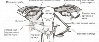
consists of suspending, securing and supporting devices that ensure the physiological position of the uterus, tubes and ovaries.
The suspensory apparatus
is a complex of ligaments that connect the uterus, tubes and ovaries to the walls of the pelvis and to each other.
This group includes the round, broad ligaments of the uterus, as well as the suspensory and proper ligaments of the ovary. The round ligaments of the uterus
(lig. teres uteri, dextrum et sinistrum) are a paired cord 10-15 cm long, 3-5 mm thick, consisting of connective tissue and smooth muscle fibers, which are a continuation of the outer muscular layer of the uterus.
Starting from the lateral edges of the uterus, slightly lower and anterior to the beginning of the fallopian tubes on each side, the round ligaments pass between the leaves of the broad uterine ligament (intraperitoneal) and are directed first outward, almost horizontally, and then anteriorly and downward, to the side wall of the pelvis, retroperitoneally. On their way, the round ligaments
cross the obturator vessels and nerve, the middle umbilical ligament with the obliterated umbilical artery passing through it, the external iliac vessels with the lower epigastric vessels extending from them, and then enter the internal opening of the inguinal canal.
Their distal third is located in the canal, then the ligaments exit through the external opening of the inguinal canal and branch in the subcutaneous tissue of the labia. The broad ligaments of the uterus
(lig. latum uteri, dextrum et sinistrum) are frontally located duplications of the peritoneum, which are a continuation of the serous cover of the anterior and posterior surfaces of the uterus away from its “ribs” and split into sheets of parietal peritoneum of the side walls of the small pelvis - from the outside.
At the top, the wide ligament of the uterus is closed by the fallopian tube, located between its two layers; below, the ligament splits, passing into the parietal peritoneum of the pelvic floor. The broad ligament of the uterus
distinguishes the following parts: mesentery of the fallopian tube (mesosalpinx);
mesentery of the ovary (mesovarium); mesentery of the uterus (mesometrium), which includes the rest (large) part of the broad ligament of the uterus, located below the ligament proper and the mesentery of the ovary. Between the leaves of the broad ligament
(mainly at their base) lies fiber (parametrium), in the lower part of which with one and on the other side is the uterine artery.
The broad ligaments of the uterus
lie freely (without tension), follow the movement of the uterus and cannot, naturally, play a significant role in maintaining the uterus in a physiological position.
Speaking about the broad ligament of the uterus, it is impossible not to mention that with intraligamentary tumors of the ovaries located between the leaves of the broad ligament, the usual topography of the pelvic organs is disrupted to one degree or another. The suspensory ligaments of the ovary
(lig. suspensorium ovarii, dextrum et sinistrum) go from the upper (tubal) end of the ovary and fallopian tube to the peritoneum of the lateral wall of the pelvis.
These relatively strong ligaments, thanks to the vessels (a. et v. ovaricae) and nerves passing through them, keep the ovaries suspended. The proper ligaments of the ovary
(lig. ovarii proprium, dextrum et sinistrum) are a very strong short fibrous-smooth muscle cord connecting the lower (uterine) end of the ovary with the uterus, and pass through the thickness of the broad ligament of the uterus. The fixing, or
actually fixing, apparatus
(retinaculum uteri) is a system of “compression zones” that form the basis (skeleton) of ligaments that are in close connection with the parietal and visceral fascia of the pelvis.
The compaction zones consist of powerful connective tissue cords, elastic and smooth muscle fibers. In the fixing apparatus, the following parts are distinguished: the anterior part
(pars anterior retinaculi), which includes the pubovesical or pubovesical ligaments (ligg. pubovesicalia), which continue further in the form of the vesicouterine (vesico-cervical) ligaments (ligg. vesicouterina s. ve-sicocervicalia);
the middle part
(pars media retinaculi), which is the most powerful in the fastening apparatus system;
it mainly includes the system of cardinal ligaments (ligg. cardmalia); the posterior part
(pars posterior retinaculi), which is represented by the uterosacral ligaments (ligg. sacrouterine). Some of the listed
ligaments
should be discussed in more detail.
The vesico-uterine
, or vesico-cervical, ligaments are fibromuscular plates that cover the bladder on both sides, fixing it in a certain position, and keeping the cervix from moving posteriorly. The main, or
main (cardinal) ligaments of the uterus
are a cluster intertwined dense fascial and smooth muscle fibers with a large number of vessels and nerves of the uterus, located at the base of the wide uterine ligaments in the frontal plane.
The uterosacral ligaments
consist of muscular-fibrous bundles (m. rectouterinus) and extend from the posterior surface of the cervix, arcuately covering the rectum from the sides (weaving into its side wall), and are fixed to the parietal layer of the pelvic fascia on the anterior surface of the sacrum.
Raising the peritoneum covering the top, the sacrouterine ligaments form rectouterine folds (plicae rectouterinae). The supporting (supporting) apparatus
is united by a group of muscles and fascia that form the floor of the pelvis, above which the internal genital organs are located, described in detail in our article.
Source: https://zodiacc.ru/info/svjazochnyj-apparat-matki/
Ligamentous apparatus of the uterus
The uterus is one of the most important organs of the female body, which is located in the pelvic area. Its main characteristic is mobility. This means that the uterus can take different places in relation to other nearby organs. These properties of the uterus are influenced by the size and condition of the organs, as well as the structural features of each woman’s body.
Despite everything, special standards have been developed by which doctors determine the normal and pathological functioning of the uterus. It is believed that the norm is the location of the longitudinal axis of the uterus along the pelvic axis, and its bottom is slightly tilted forward.
Ligamentous apparatus
To support the body of the uterus, every female body has a ligamentous apparatus, which includes fascia and ligaments. Each component has certain functions that help maintain the uterus in a certain position, but at the same time, without reducing its mobility.
The ligamentous apparatus is a very complex system, which includes ligaments of different types. It is customary to distinguish 3 main components:
- Suspensory. Its main task is to hold the uterus in the desired position by attaching it to the pelvic wall. Main ligaments: broad, round, ligaments that support the ovaries.
- Fixing. They help secure the uterus in the correct position, have the ability to stretch during gestation, and the organ can move, although not actively. Includes: main uterine, vesicouterine and main.
- Supportive. From the ligaments that are included in this component, the pelvic floor is formed, which in turn provides support for other internal organs. The basis of this type of ligamentous apparatus is the muscles and fascia that are located in the perineal area.
During gestation, the uterus significantly increases its size, which is very important for the full development and formation of the fetus. This is facilitated by the ligaments that are located around the uterus. They stretch, but do not hinder the growth of the organ.
Figure taken from ppt-online.org
Doctors also note that during pregnancy the load on the entire ligamentous apparatus actively increases. The uterus increases in size, its weight also increases. To hold it, the ligaments become thicker so that they can perform their functions. All these changes occur under the influence of hormones.
There are situations when these changes provoke the appearance of unpleasant or painful sensations. It is believed that this may be normal, and the nature of the pain can vary from mild to prolonged.
Most often, pain worries a woman in the second and third trimesters when changing body position or excessive physical activity. It is noted that with each subsequent pregnancy the pain becomes stronger and more acute.
It is important to understand that not all unpleasant changes in well-being are provoked by changes that occur in the ligamentous apparatus. It is important to consult your doctor for help if the following symptoms appear along with pain:
These symptoms are especially dangerous in pregnant women, since they indicate deviations in the process of pregnancy or the development of an infectious process in the genitourinary system. Only an experienced specialist can competently conduct an examination, evaluate the test results and prescribe the necessary therapy.
Pain that occurs in the pubic area may indicate problems with the pelvic ligaments. A woman’s body is designed in such a way that the ligamentous apparatus can adapt to bearing a fetus without any problems, but frequent pain and its severity should signal the woman to urgently consult a doctor.
Types of ligaments
The ligamentous apparatus includes not only the ligaments of the uterus, but also the ligaments that touch the ovaries and the muscles of the perineum. The following ligaments can be attributed to the uterus:
- wide;
- round;
- cardinal;
- sacrouterine.
Wide
The broad ligament is a double ligament and is located in the pelvic area, but has no influence on the location of the organ itself. These ligaments are not smooth as they enclose the fallopian tubes and ovaries.
Round
The round ligament also has a double structure and reaches a length of up to 15 cm. This type of ligament is found in the thickness of the broad ligament. Due to the fact that the body has such structures, the uterus is held vertically and does not tip back.
There are situations when cysts, fibroids or fibroids can form in the ligaments, which may not manifest themselves in any way. Only their active growth and the influence of hormones provoke the appearance of severe pain in the groin and lower abdomen. Treatment for this condition is exclusively surgical intervention.
Cardinal ligaments
The basis of the broad ligaments are the cardinal ligaments. They are located near the cervix and are round, dense cords. Their main task is to fix the uterus on both sides and prevent it from moving forward or backward. In the thickness of these ligaments pass the vessels that lead to the uterus, as well as the ureters.
Sacrouterine
These ligaments are the last elements of the uterine ligamentous apparatus. The basis of the ligament is connective and smooth muscle tissue. The ligament begins in the posterior part of the cervix and ends at the rectum.
A certain amount of fibers reach right up to the sacral vertebrae. Their task is to act as a counterweight to the round uterine ligaments, which holds the uterus and does not allow it to deviate forward.
Ligaments are a very important part in a woman’s body. Thanks to their proper functioning, she is able to carry and give birth to a baby and does not allow the uterus to descend. Every woman should monitor her health and consult a doctor if there are any deviations.
Source: https://uterus2.ru/other/kruglaya-svyazka-matki.html
Necessary diagnostics
A specialist can detect the incorrect location of the uterus during a classic examination, during which fingers are inserted into the patient’s vagina, and at the same time the surface of the lower abdomen is palpated. The woman is asked to push and the doctor determines the degree of prolapse if it is suspected.
For some deviations, a specialist may try to return the uterus to its place manually. If it is mobile, this will happen without much discomfort for the woman and the location of the organ will change.
To confirm the diagnosis and determine the degree of deviation of the uterus from the correct location, it is necessary to additionally perform an ultrasound examination of the pelvic organs or computed tomography. After this, the gynecologist will select the necessary therapy.
Stretching of the round ligament of the uterus during pregnancy: causes and pain relief
Pain when the uterine ligaments are sprained is very common and it is almost impossible to avoid them. During pregnancy, ligaments are greatly stretched, especially the round ligaments.
The large round ligament holds the uterus on both sides, keeping it in place. When carrying a baby, these ligaments become more elastic, which increases pain in the side and groin during various movements. Pain in the ligaments is not constant and appears only with tension.
The major ligament is one of several that hold the uterus in the abdominal cavity. As the uterus increases in size and weight, the ligament becomes stretched during pregnancy. It becomes tense, long and thick.
Painful sensations can be of a different nature, but they are all fleeting:
Painful symptoms caused by sprains appear in the groin area and move to the outer side of the thigh. Pain may appear involuntarily on both sides. If such symptoms occur, you should consult a gynecologist:
- bleeding;
- severe pain;
- pain that occurs even after sleep;
- painful sensations during bowel movements;
- obsessive dull pain.
These symptoms, together with sprains during pregnancy, are very dangerous.
Correct location of the ovaries
To the right and left of the body of the uterus and slightly behind it, below the funnels of the fallopian tubes, are the female reproductive glands.
In newborn girls, the organs are located quite high above the pelvic inlet and are very mobile, moving forward and backward (from the front to the back wall of the abdominal cavity) and right and left.
By the age of five, the ovaries in girls gradually descend into the pelvic cavity and take their rightful place.
The ovaries are not located symmetrically relative to each other. The right one, being somewhat heavier than the left one, is located lower. However, this difference in mass and location does not affect their structure and functions.
The female genital organ is designed in such a way that it is able to change its location, while always remaining behind the bladder.
The ovary is surrounded from above by the fallopian tube and is attached to the broad ligament of the uterus through the mesentery. The tubular end is strengthened by an additional suspensory ligament with blood and lymphatic vessels.
It also has its own ligament, which connects its uterine extremity with the lateral surface of the uterus.
Thus, everyone is held in a physiological position thanks to three ligaments. The ovaries of a nulliparous woman are located vertically, while those of a woman who has given birth are slightly displaced relative to the axis of the spinal column.
Prevention and treatment
To prevent sprain of the round ligament during pregnancy, you must adhere to preventive measures:
- do not wear high-heeled shoes;
- avoid physical activity that will lead to injury;
- When moving on uneven surfaces, keep the position of your feet under control;
- do not carry heavy bags;
- don't jump;
- engage in special exercises that will strengthen tendons and muscles.
There are some ways to reduce pain in the ligaments of the uterus:
- wearing a special belt or bandage;
- it is important to avoid sudden movements;
- if severe pain occurs, take a horizontal position;
- A warm bath will help reduce tension.
Treatment for sprained ligament teres and pregnancy pain includes:
- Taking analgesics. It is possible to take paracetamol, but after prior consultation with a specialist.
- Exercises. Yoga and stretching are beneficial for expectant mothers. Exercise helps maintain muscle function. Before gymnastics, be sure to consult a doctor.
- Move smoothly. Avoid sudden movements, change positions strictly smoothly.
- Lie down or sit down if experiencing severe pain.
- Warm compress. It will help cope with severe tension, the muscles relax, and the pregnant woman begins to feel much better. Be sure to get your doctor's approval before using heat.
If the condition worsens, bleeding and fever appear, be sure to consult a specialist or call an ambulance. Pain can be caused by various reasons, which can lead to complications in the future.
Ligaments of the uterus: round, broad and cardinal
One of the main organs of the female reproductive system is the uterus. It is located in the small pelvis, in its middle part.
This organ is characterized by mobility and can occupy different positions relative to other organs. This may be influenced by the size of other organs and their condition, as well as the individual characteristics of each female representative.
However, there are certain rules according to which the norm and pathology in the location and functioning of the uterus are determined. Typically, its longitudinal axis should run along the pelvic axis, and the bottom of the organ is inherently tilted forward.
The concept of the ligamentous apparatus of the uterus
To maintain the body of the uterus in a normal position, the body has a ligamentous apparatus, which includes ligaments and fascia. Each element is characterized by certain functions, thanks to which the organ maintains its position while remaining sufficiently mobile.
The ligamentous apparatus is a complex system consisting of different types of ligaments. There are three main components of this system. This:
- Suspensory. Its main function is to connect the uterus to the walls of the pelvis to hold it in the required position. It is formed by the following ligaments:
- broad ligaments of the uterus;
- ovarian ligaments;
- suspensory ligaments of the ovary.
round ligaments of the uterus;
- main uterine;
sacrouterine;
During pregnancy, the uterus increases in size, which is necessary for the full development of the fetus. At this time, the ligaments surrounding it are stretched so as not to hamper its growth.
In addition, during pregnancy, the load on the ligamentous apparatus increases, since its main function is to support the uterus in a certain position. To achieve this, the elements of the ligamentous apparatus thicken in order to cope with their functions. Such changes occur under the influence of hormones.
These changes that occur during pregnancy can often cause unpleasant and even painful sensations in the expectant mother. This phenomenon is normal, and the pain can be both acute and mild, short or long-lasting.
They can occur especially often in the second trimester of pregnancy, when a woman suddenly changes position, or during prolonged physical activity.
During the second pregnancy (and subsequent ones), pain due to sprains is characterized by greater severity. However, you should not think that any pain during this period is caused by changes in the ligamentous apparatus. If pain occurs too often, and along with it occurs:
- bleeding;
- elevated temperature;
- pain in the lumbar region;
- chills;
- difficulty urinating, etc.,
you should consult a doctor. Such symptoms during pregnancy may indicate the presence of abnormalities in pregnancy or an infection in the genitourinary tract. The doctor will conduct the necessary tests to make the correct diagnosis.
Also, the occurrence of pain in the pubic area during pregnancy may indicate the presence of disturbances in the functioning of the pelvic ligaments. Therefore, if you discover this symptom, it is advisable to consult a doctor.
In general, the ligamentous apparatus adapts to pregnancy relatively easily, however, excessive and frequent pain should be a cause for concern.
Main types
Among the elements that make up the ligamentous apparatus, not only those relating to the uterus are distinguished, but also the ligaments of the ovaries, as well as the muscles of the perineum. Directly related to the uterus itself are:
- wide,
- round,
- cardinal,
- sacrouterine.
Features of cardinal ligaments
The cardinal ligaments are the basis for the broad ligaments. They are located in the cervix area in the form of rounded dense cords that secure the organ on both sides.
In essence, it is a thickened lower part of the broad ligaments, in which, as development progressed, the amount of connective and smooth muscle tissue increased.
The function of this element is to prevent forward and backward displacement of the uterus. In their thickness lie the uterine vessels, as well as the ureters. Between the individual sections of the ligaments, a parametrium formed from fiber is formed.
Forms
Displacement of the uterus can occur along a vertical plane (up and down), around the longitudinal axis and along a horizontal plane.
Displacement of the uterus along the vertical plane includes elevation of the uterus, prolapse, prolapse and inversion of the uterus. When lifted, the uterus moves upward, its bottom is located above the plane of the entrance to the pelvis, and the vaginal part of the cervix is above the spinal plane. Pathological elevation of the uterus occurs when menstrual blood accumulates in the vagina due to atresia of the hymen or lower part of the vagina, with voluminous tumors of the vagina and rectum, with encysted inflammatory effusions in the pouch of Douglas. Lifting (elevation) of the uterus can also occur when it is fused with the anterior abdominal wall after laparotomy (caesarean section, ventrofixation).