In patients over 50 years of age who consult a gynecologist about polycystic ovary syndrome, in 75% of cases the presence of stringy endometrial hyperplasia is detected.
It is based on the process of transformation of tubular gland cells through their uncontrolled division. The condition is dangerous because it constantly develops, which often leads to endometrial cancer.
Symptoms
The main symptoms of uterine endometrial hyperplasia are:
- spotting bloody discharge during menopause
- long periods during which a lot of blood is lost
- menstrual cycle irregularities
- bleeding between periods
- infertility
- severe pain during menstruation
But in many cases the disease is latent, that is, without symptoms. It is discovered during an examination by a gynecologist or during an ultrasound diagnosis, when the patient goes to the doctor with completely different complaints. The disease can lead to cancer and infertility.
Symptoms of endometrial hyperplasia in menopause
Pathological processes in the uterine mucosa occur long before menopause. Hereditary predisposition and the presence of benign diseases of the genital organs play a large role in the development of this pathology. The older a woman is, the less protective forces remain in the body. In addition, chronic diseases and surgery aggravate the situation with the mucous layer of the uterus.
During menopause, the risk of gynecological diseases increases. Hyperplasia that appears at this time can develop into a malignant tumor of the uterus. Therefore, women are advised to undergo routine examinations, even if no symptoms worry them. And the risk group includes women who are over 50 years old, who have a persistent increase in blood pressure, diabetes, metabolic disorders in the body, and obesity.
Symptoms of endometrial hyperplasia may be:
- spotting, bloody, scanty discharge
- prolonged heavy bleeding
- polyps
But there can also be a latent course. Because of this, some patients turn to the gynecologist very late, only when they have severe abdominal pain. Then the treatment is very difficult.
Diagnosis is carried out by transvaginal ultrasound, and aspiration biopsy is also possible. If the lesion is focal, aspiration biopsy may not reveal pathology. During menopause, the thickness of a woman’s endometrium is up to 5 mm. If the mucous membrane has a thickness of 6-7 mm, a repeat ultrasound is needed, since these indicators are not within the normal range. If the indicator reaches 8 millimeters, the doctor prescribes curettage of the mucous membrane with subsequent study of biological material. A special probe is used to examine the uterine cavity before and after the procedure.
Kinds
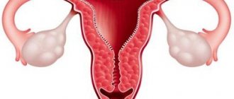
The endometrium, the inner layer of the uterus, develops and thickens every month, creating favorable conditions for the attachment of a fertilized egg. If pregnancy does not occur, menstruation begins, with which excess cells leave the body.
- Endometrium of proliferative type: phases of development, treatment features
Excessive proliferation of hyperplastic cells in the endometrium is called hyperplasia. There are several types of such dishormonal pathology. The most common options are:
- Glandular. The disease is typical for adolescence, it also occurs in women during the menopausal period. The disease is divided into active (acute) and passive (chronic) forms.
- Glandular-cystic. It is similar to the first option, but with a greater degree of severity.
- Atypical (adenomatosis). This form is characterized by intensive growth and, at the same time, structural changes in cells. Depending on the location, diffuse and focal adenomatosis are distinguished.
- Endometrial polyps stand apart - growths of individual areas with underlying stroma. They may be of fibrous or glandular origin.
This classification has been used in medicine for many years. Today WHO has introduced a different definition of the types of hyperplasia. Pathology can be simple or complex; moderate hyperplasia is a transitional stage from one type to the next. If a woman has a benign process, it can be successfully treated with hormonal agents. At what point and why a simple harmless form transforms into a malignant neoplasm is not completely known.
Symptoms of endometrial hyperplasia in postmenopause
The duration of the postmenopausal period is several years, and at this time the function of the ovaries finally fades away. For this reason, there is a decrease in the production of sex hormones (estrogen and progesterone), which affect:
- reproductive organs
- nervous system
- Gastrointestinal tract
- brain
- musculoskeletal system
- hair
- skin covering
When the functioning of the ovaries changes, cysts often develop. They do not manifest themselves as external symptoms, so they are not known until the leg ruptures or bends. But in some cases, on the contrary, very severe pain appears, which is the reason for contacting a doctor. Such cysts and disordered division of endometrial cells often cause malignant tumors.
A warning sign is the appearance of bloody discharge of any type (scanty, too strong, etc.). Symptoms of endometrial hyperplasia in postmenopause include cramping pain in the lower abdomen, which characterize the development of large polyps. In postmenopause, single polyps appear, and the mucous layer also dies.
Symptoms of glandular endometrial hyperplasia
This type of hyperplasia occurs as a result of the characteristic development and enlargement of the endometrial glands of the uterus. This condition is considered precancerous. If adequate treatment is not carried out, an atypical form of hyperplasia occurs with the formation of cells that are similar in structure to cancer.
With glandular hyperplasia, menstrual function is disrupted and bleeding occurs. So-called menorrhagia appears, which is more abundant and lasts longer than normal menstruation. Metrorrhagia also appears, which is acyclic bleeding. Discharge from glandular hyperplasia, as a rule, occurs after a short delay or in the interval between menstruation. During adolescence, active release of blood with clots is possible.
The following manifestations also occur:
- malaise
- weakness
- dizziness
- anemia
- loss of consciousness (fainting)
Signs of endometrial hyperplasia by ultrasound
Ultrasound is done to assess changes occurring in the reproductive organ, to identify the thickness of the mucous membrane with the location of areas of hyperplasia and polyps. The technique is carried out with a special sensor inserted into the vagina. Ultrasound diagnostics for hyperplasia is considered a non-invasive, relatively inexpensive, painless and accurate method for diagnosing endometrial pathologies. Ultrasound gives indicators that must fit into the norms regarding a certain menstrual phase.
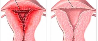
The mucous layer of the endometrium has clear outlines and significant acoustic density compared to the muscle layer, that is, the myometrium. The thickness of the mucous membrane is influenced by the monthly cycle. In the first phase, the wall is only 3-4 mm, in the second phase the figure is 12-15 millimeters. Signs of endometrial hyperplasia according to ultrasound: uniform thickening with pronounced even contours and uniform echogenicity.
Polyps on ultrasound are neoplasms with clear contours, a thin echogenic rim and high acoustic density. If malignant changes are detected, they are characterized by uneven contours and heterogeneous echogenicity.
Diagnostics
Physical research methods
- Survey – characteristics of menstrual dysfunction; during the menopause; from the anamnesis - heredity (presence of neoplasms in the family), infertility.
- General examination – presence of anemia, signs of obesity.
- Deep palpation of the abdomen – pain.
- Examination of the external genitalia.
- Inspection in mirrors.
- Bimanual gynecological examination - mobility, pain, size, consistency of the uterus.
Laboratory research methods
Required:
- determination of blood group and Rh factor;
- general blood test - presence of signs of anemia;
- general urine analysis;
- determination of blood sugar level - presence of diabetes;
- biochemical blood parameters;
- bacterioscopic analysis of secretions;
- hormonal colpocytology;
- cytological examination of aspirates and washings from the uterine cavity (can be used to monitor conservative therapy).
If indicated:
- performing a glucose tolerance test;
- bacteriological examination;
- hormonal study of the state of the pituitary-gonadotropic system;
- study of thyroid function.
Instrumental research methods
Required:
- Ultrasound ultrasound (transabdominal, preferably transvaginal, Doppler) to determine:
- endometrial thickness;
- endometrial structures;
- relief of the uterine cavity;
- the presence of concomitant pathology of the myometrium;
- anatomical features of the uterine appendages;
- endometrial-uterine ratio (the ratio of endometrial thickness to the anteroposterior size of the uterus).
- Separate diagnostic curettage.
If indicated:
- hysteroscopy (visualization of pathological changes in the endometrium, their localization; the ability to perform intrauterine operations using electro-, cryo- or laser surgery);
- endometrial biopsy;
- X-ray examination (hysterosalpingography and bicontrast gynecography - currently rarely used).
Specialist consultations
Required:
- endocrinologist.
If indicated:
- oncogynecologist.
Ultrasound signs of endometrial hyperplastic processes
Differential diagnosis:
- uterine leiomyoma;
- spontaneous abortion;
- trophoblastic disease;
- ectopic pregnancy;
- hormone-producing ovarian tumors;
- endometrial cancer.
Signs of glandular endometrial hyperplasia
With the glandular type of hyperplasia, the endometrium thickens, while the gland cells actively divide and are arranged in groups, unevenly. The pathological condition may occur in the absence of pronounced symptoms. That is why patients do not always describe the symptoms of endometrial hyperplasia, and complaints are received about metabolic and endocrine disorders, such as:
- sleep disorders
- headache
- decreased performance
- weight gain for no reason
- irritability
- extreme thirst
Glandular hyperplasia of the endometrium occurs with uterine bleeding, since the monthly cycle is uneven. The discharge may be too long or too short, too weak or, conversely, abnormally heavy. The development of hyperplasia is also indicated by spotting type discharge that is not associated with menstruation. Failure to achieve pregnancy and pain localized in the lower abdomen often indicate that the uterine mucosa is susceptible to pathological changes at this stage.
If you find yourself with the symptoms described above or some of them, immediately contact a gynecologist for an in-person appointment! The earlier this condition is diagnosed, the lower the likelihood of complications and the easier the treatment.
Development factors
The disease develops under the influence of certain factors and circumstances. The main one is hormonal imbalance, namely, excess production of estrogen - female hormones.
Additional conditions and causes that cause this disease are:
- disruption of the central nervous system;
- PCOS;
- neoplasms in the ovaries;
- pathologies of the endocrine system;
- obesity;
- problems with the immune system;
- hypertension, etc.
Also, this pathology can be caused by mechanical damage to the uterus - abortion, intrauterine device. Uncontrolled use of hormonal medications often leads to dishormonal conditions. Numerous studies in the field of gynecology prove that hyperplasia is also genetic in nature.
The pathology often accompanies infertility. Often the disease is discovered when a woman has been trying to conceive a child for a long time, but to no avail. Hyperplasia is the result of long-term changes in the body; pathological processes can last for years.
Sonographic symptoms of endometrial hyperplasia
To determine the type of hyperplasia and monitor how much treatment helps, an ultrasound is done on the 5-7th day of the monthly cycle. The accuracy of the study of detected hyperplastic transformations is 90%, and of detected endometrial polyps – 60-80%.
The information content of the screening method can be different, depending on the performance characteristics of the device itself, the experience of the diagnostician and the age of the patient. Sonographic signs of endometrial hyperplasia include the following:
- polyps measuring 16.1-17.5 mm
- mid-uterine structure 14.6 to 15.4 mm thick
- adenocarcinoma is suspected at 19.7-20.5 mm
Hyperplasia in the postmenopausal period is diagnosed if the M-echo value is 5 millimeters or higher.
Endometrial hyperplasia is indicated by such echographic symptoms as evenness/unevenness of the M-echo contour, increased sound conductivity, inclusion of echo-negative or echo-positive structures, heterogeneity of the endometrium, altered relief of the uterine mucosa.
Therapy and prevention
Treatment of hyperplasia is prescribed only by an experienced doctor, and sometimes a group of specialists, which includes a gynecologist, reproductive specialist, and endocrinologist, participates in the development of a therapeutic program.
Therapy is selected individually, taking into account factors such as:
- patient's age;
- nature and characteristics of the disease;
- general well-being, etc.
Primary treatment is carried out using conservative methods. It is aimed at restoring hormonal levels, inhibiting cell proliferation and reducing foci of hyperplasia. The basis of therapy includes hormone-containing drugs. The patient is also prescribed COCs (combined oral contraceptives), GnRH agonists, gestagens, enzymes, and vitamins. Additionally, antibacterial and anti-inflammatory drugs may be prescribed.
Often, in addition to the main course of treatment, doctors prescribe herbal medicine. Decoctions, infusions, herbal and natural products have a wonderful auxiliary effect.
IMPORTANT! Traditional recipes should not be used without consulting a doctor, so as not to aggravate the situation.
You can reduce the risk of developing simple hyperplasia or adenomatosis if you take care of prevention. It is enough to be attentive to your health and visit a gynecologist twice a year for a routine examination. It is imperative to promptly treat inflammatory processes of the pelvic organs. Take contraceptives, exercise regularly, and adhere to the principles of proper nutrition.
Symptoms of focal endometrial hyperplasia
In the focal form of hyperplasia, the endometrium grows in areas that are most susceptible to the influence of hormones. Lesions can range in size from 2 mm to several centimeters. External signs of these changes are polyps.
In order for a mature egg to leave the follicle, there must be a certain level of progesterone and luteinizing hormone. If there is more estrogen in the body than normal, then the first phase of the cycle becomes longer, the release of the egg is inhibited, ovulation is delayed, and the uterine mucosa grows. The first symptoms of focal endometrial hyperplasia are usually prolonged and severe bleeding. There are also small bloody discharges when there should not be a period. And such discharge usually lasts 1-3 days.
If there is a lack of estrogen, then the egg does not mature enough to leave the follicle. Partial rejection of the mucous layer occurs, and polyps form in the affected areas. Symptoms of endometrial hyperplasia are similar, stronger or weaker critical days can be from 10 to 14 days.