Cancer diagnosis using ultrasound
Ultrasound is a simple, effective and fast diagnostic method for diagnosing cancer in the early stages. The doctor may diagnose you using other methods of determination. For example, MRI, X-ray or biopsy, etc., an ultrasound examination is painless and completely safe and very informative, which is why it is prescribed to most patients. Screening will 100% show whether you have a tumor in your organ or not and what stage it is at?
Ultrasound technologies are improving and a specialist can do an ultrasound of the intestines and stomach and determine whether there is cancer in those organs or not? This examination is possible due to the fact that ultrasound scanners have an elastography function. Using it, the specialist will record the spectrum of blood flow in the vessels and evaluate the tissue structure in suspicious areas.
Ultrasound of the thyroid gland
Many patients who had problems with the thyroid glands and the doctor suspected a neoplasm were sent by the specialist for an ultrasound scan. The tumor will be detected quickly, but whether it is malignant or benign needs to be determined. In a suspicious node, the ultrasound specialist assesses the condition of the vessels.
The device records the spectral characteristics of the blood flow in the node. Additionally, the doctor will refer you for a biopsy of this area and the established diagnosis will be 100% confirmed by several examination methods. Additionally, lymph nodes are examined. According to the theory, cancer cells can enter them.
Examination of blood vessels in the brain
If the doctor suspects a tumor in the brain, he will most likely prescribe you an MRI rather than an ultrasound. An ultrasound examination with duplex scanning can reveal a lot. What is the condition of the vessels in the brain, are they displaced, is there a developed vascular network that should not be there? If the latter is detected, the patient is given a referral for an MRI. An accurate diagnosis will be established.
Can cancer be diagnosed based on ultrasound?
Ultrasound examination is considered not only simple, but also quite accurate and informative. However, without additional examinations, it is impossible to make an accurate diagnosis of cervical cancer: a specialist can draw up a description and recommend additional examinations.
Ultrasound can reveal certain changes in the structure of the cervix and endometrium, that is, the epithelial lining of the uterus. However, ultrasound cannot answer the question of what causes these changes. Only a doctor can decipher the data received and prescribe additional tests. In addition, whether cervical cancer is visible on ultrasound or not depends on the stage of the disease. At an early stage, the disease can be detected by studying tumor markers (cells with a mutant genome), colposcopy and other studies. If alarming symptoms are detected, a full examination of the body may be required, which will either confirm or refute the preliminary diagnosis.
During an ultrasound, the doctor can determine the following indicators:
- the shape of the uterus, for example, its barrel-shaped state;
- changes in lymph nodes located in the pelvis;
- contours of the uterus;
- presence of changes in veins and arteries;
- type of neoplasm.
Cervical cancer on ultrasound has characteristic signs that will allow the doctor to suspect the presence of the disease. For example, in the early stages, cervical cancer on ultrasound has a rather characteristic appearance: it is visualized as a dense oval neoplasm with clear contours. As the lesion grows, it becomes fuzzy and appears rugged and lumpy.
Important! It is important to remember that the level of detail in ultrasound depends on how modern the equipment is used in the clinic. Modern devices make it possible to detect a tumor whose diameter reaches 3 mm.
Ultrasound of the peritoneum
If the area of cancer localization is in the abdominal cavity, then using an ultrasound scan. They will help to identify, for example, primary tumors - hepatocellular neoplasm, metastases or cholangiocarcinoma.
Gallbladder
When an ultrasound specialist diagnoses the gallbladder, he often finds polyps in it. They need to be monitored once every six months or a year, with repeated ultrasound diagnostics. Polyps can develop into malignant tumors. If the diagnostician has a lot of experience, he will notice on the screen Vaterov’s tumor, which is a nipple.
Pancreas
Is it possible to see a tumor in the pancreas on an ultrasound? It's quite difficult. The picture on the screen is blurry. It is especially difficult to see and difficult to establish a correct diagnosis in obese people. Ultrasound examination is an inexpensive, accessible method and therefore, first of all, patients are referred to it.
When a doctor examines the pancreas, he pays attention to the condition of the lymph nodes at the porta hepatis with the retroperitoneal lymph nodes. If they are enlarged, then it makes sense to prescribe a CT scan to the patient, which will help identify malignant neoplasms.
Spleen
During ultrasound, the spleen is clearly visualized. Fortunately, cancer almost never occurs in this organ, nor do metastases form.
Stomach with intestines
In recent years, ultrasound has often examined the stomach and intestines. These organs are located quite deep in the human body and an ultrasound will accurately show only a grown tumor, or rather, a specialist will recognize it 100%, and smaller tumors can be interpreted as natural processing products.
Will an ultrasound show for sure that a patient has cancer if it is stage 3 or 4? An ultrasound specialist will 100% confirm that a person has neoplasms, because there are already many noticeable pathological changes in the body. In advanced cases, the doctor will see metastases in the stomach and even determine their size.
Can an ultrasound show cancer?
Many people are interested in whether cancer will be visible on an ultrasound? It depends on the stage of the cancer and how deep in the tissue it is located. For example, bone cancer or metastases in them will be shown by x-ray. Let's look at the problem in more detail in the article.
Cancer diagnosis using ultrasound
Ultrasound is a simple, effective and fast diagnostic method for diagnosing cancer in the early stages. The doctor may diagnose you using other methods of determination. For example, MRI, X-ray or biopsy, etc.
, an ultrasound examination is painless and completely safe and very informative, which is why it is prescribed to most patients.
Screening will 100% show whether you have a tumor in your organ or not and what stage it is at?
Ultrasound technologies are improving and a specialist can do an ultrasound of the intestines and stomach and determine whether there is cancer in those organs or not? This examination is possible due to the fact that ultrasound scanners have an elastography function. Using it, the specialist will record the spectrum of blood flow in the vessels and evaluate the tissue structure in suspicious areas.
Ultrasound of the thyroid gland
Many patients who had problems with the thyroid glands and the doctor suspected a neoplasm were sent by the specialist for an ultrasound scan. The tumor will be detected quickly, but whether it is malignant or benign needs to be determined. In a suspicious node, the ultrasound specialist assesses the condition of the vessels.
The device records the spectral characteristics of the blood flow in the node. Additionally, the doctor will refer you for a biopsy of this area and the established diagnosis will be 100% confirmed by several examination methods. Additionally, lymph nodes are examined. According to the theory, cancer cells can enter them.
Examination of blood vessels in the brain
If the doctor suspects a tumor in the brain, he will most likely prescribe you an MRI rather than an ultrasound. An ultrasound examination with duplex scanning can reveal a lot. What is the condition of the vessels in the brain, are they displaced, is there a developed vascular network that should not be there? If the latter is detected, the patient is given a referral for an MRI. An accurate diagnosis will be established.
Ultrasound of the peritoneum
If the area of cancer localization is in the abdominal cavity, then using an ultrasound scan. They will help to identify, for example, primary tumors - hepatocellular neoplasm, metastases or cholangiocarcinoma.
Gallbladder
When an ultrasound specialist diagnoses the gallbladder, he often finds polyps in it. They need to be monitored once every six months or a year, with repeated ultrasound diagnostics. Polyps can develop into malignant tumors. If the diagnostician has a lot of experience, he will notice on the screen Vaterov’s tumor, which is a nipple.
Pancreas
Is it possible to see a tumor in the pancreas on an ultrasound? It's quite difficult. The picture on the screen is blurry. It is especially difficult to see and difficult to establish a correct diagnosis in obese people. Ultrasound examination is an inexpensive, accessible method and therefore, first of all, patients are referred to it.
When a doctor examines the pancreas, he pays attention to the condition of the lymph nodes at the porta hepatis with the retroperitoneal lymph nodes. If they are enlarged, then it makes sense to prescribe a CT scan to the patient, which will help identify malignant neoplasms.
Spleen
During ultrasound, the spleen is clearly visualized. Fortunately, cancer almost never occurs in this organ, nor do metastases form.
Stomach with intestines
In recent years, ultrasound has often examined the stomach and intestines. These organs are located quite deep in the human body and an ultrasound will accurately show only a grown tumor, or rather, a specialist will recognize it 100%, and smaller tumors can be interpreted as natural processing products.
Will an ultrasound show for sure that a patient has cancer if it is stage 3 or 4? An ultrasound specialist will 100% confirm that a person has neoplasms, because there are already many noticeable pathological changes in the body. In advanced cases, the doctor will see metastases in the stomach and even determine their size.
To make the diagnosis as accurate as possible, the following procedures are performed:
- Before the examination, the patient is instructed to drink 300 to 500 ml of plain boiled water on an empty stomach. the main thing is that it does not contain gas. The specialist will ask you to lie down on the couch and you will change positions, and he will examine your stomach from all sides or in different projections.
- The 1st projection is done when the patient assumes a supine position and then lies down on his left side. Later en right. He rises from the couch and the projection needs to be captured while he is standing. The projection will show how large the tumor is and how it has grown into nearby tissues and organs.
Ultrasound of the stomach is considered an auxiliary method. An experienced specialist will notice cancer at an early stage. Especially when it affected the upper layer of the stomach, the muscles. But in the later stages, the lesions are larger and better visualized and interpreted by an ultrasound specialist.
"Advice. If you suspect cancer, after the ultrasound, ask to be referred for an x-ray of the stomach.”
Kidney
Ultrasound diagnostics of the kidneys will show whether the patient has renal cell cancer, transitional cell neoplasms, Wilms cancer with metastases. The diagnosis is accurate. Additionally, a biopsy and other tests are performed.
Bladder
An ultrasound will help you see cancer in the bladder. Benign polyps also grow there, but they can develop into cancer. A cystoscopy is required, then the diagnosis will be considered 100% confirmed.
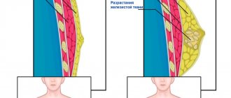
Mammary gland
The doctor will refer you for an ultrasound examination + elastography. These methods for diagnosing breast cancer are very informative. No matter how old you are, ultrasound waves reflected from tissues will show whether there is cancer and at what stage or not? After menopause, try to undergo examinations once a year, or even starting at the age of 40.
Source: https://dermatologpro.ru/info/mozhet-li-uzi-pokazat-rak/
Uterus
Does an ultrasound machine show uterine cancer? Yes. The neoplasm can be either inside the organ or on its neck. When the appendages are examined, it will be established that there are neoplasms in the ovaries or endometrium, since they have excellent echogenicity. Perhaps the patient’s tissues are all healthy.
It is difficult to establish that the endometrium is affected. The doctor may think that it is a benign fibroid, but in fact it is a malignant cancer.
"Advice. Don’t worry prematurely; in addition, you will be referred for a biopsy.”
How often can screenings be done?
If you are over 35 or 40 years old, you can conduct a study of the thyroid gland, peritoneal organs, and urinary tract annually. Women, do not forget to regularly examine your mammary glands. In addition to ultrasound, take blood and urine tests. Even if you do not have bad symptoms, you can be examined preventively.
This or that doctor will give you a referral for an ultrasound of other organs. The examinations are not expensive, and you will have peace of mind that everything is fine with you. Early diagnosis of cancer with timely treatment will save and prolong your life.
Ultrasound is considered a universal method for diagnosing many diseases. Oncology is no exception. An ultrasound can show cancer in 99% of cases. If research becomes difficult, additional methods are used.
Use of ultrasound in oncology
Ultrasound is used to diagnose oncology and benign tumors in the body. Elastography, an advanced study with an ultrasound probe, allows you to view the location of metastases in detail.
Ultrasound can be used to identify neoplasms in the form of cancer, cysts on any organ. The sonologist distinguishes pathological tissue from healthy tissue by density: in malignant or benign tumors, the density is increased and heterogeneous.
Usually the affected part of the organ on ultrasound is called a symptom of PPO, an affected hollow organ. This area of tissue is scanned with high hyperechogenicity, an increased rate of reflection of ultrasound waves by the internal organ. It is hyperechogenicity that indicates the presence of cancer. In the image, the hyperechoic area is lighter than the healthy tissue of the organ.
At what stage is cancer visible?
Cancer varies in stages:
- Initial. Changes in internal tissues are microscopic. The tumor does not spread beyond the site of development.
- Average. A malignant neoplasm extends beyond the boundaries of its original location. But adjacent tissue areas are not affected.
- The last one. Cancer affects adjacent organs and regional lymph nodes. Metastases and secondary foci of the disease appear in organs distant from the site of cancer cell development.
Ultrasound can detect cancer at any stage. It is better to consult a doctor in a timely manner for diagnosis. This way there is a chance of completely curing the disease.
In advanced cases, metastases and cancer cells cover a wide area. This area appears black on the ultrasound machine screen. The results indicate increased echogenicity.
Comprehensive definition of cervical cancer
At the moment, there are several effective ways to detect uterine cancer. The quality of the ultrasound machine influences the correct examination, since it is quite difficult to detect the development of pathology with outdated models. The fact that the specialist performing the ultrasound has experience is also important, since the result of the examination and the diagnosis often depend on him. If there is a suspicion of a cancerous process, then it is worth undergoing additional examinations.
There are several types of cervical cancer, one of them is uterine carcinoma, an insidious disease. The problem is that women sometimes seek help very late, and practically nothing can be done.
Uterine carcinoma is one of the types of malignant tumors, which is distinguished by its strong aggression and rapid course.
Carcinoma can quickly spread metastases throughout the pelvic organs. And when pain appears, precious time is lost. An ultrasound will not determine the origin of the cancer, but will help determine whether a tumor exists. Therefore, it is very important to undergo regular examination.
What do metastases look like?
With cancer, metastases form, new affected areas. Most often they are located in the lungs, liver (see liver cancer) and lymph nodes. Ultrasound shows metastases in the form of black spots of varying sizes. Features of the appearance of metastases on each organ:
- Thyroid. The appearance of cancer cells in the thyroid gland is explained by genetic predisposition or radiation exposure. Diagnostics are carried out annually.
- Vessels in the brain. Using ultrasound, you can see the displacement of blood vessels in the brain, which may indicate the presence of cancerous tumors. Usually a person is prescribed an additional examination - an MRI. The procedure is performed according to indications. The development of cancer cells in the brain is provoked by periodic radiation exposure and heredity.
- Peritoneum. Metastases in the abdominal organs appear due to alcohol abuse, smoking, frequent consumption of fatty foods, and exclusion of plant foods from the diet. Ultrasound diagnostics of this part of the body is recommended once a year.
- Mammary gland. It is recommended to check the condition of the mammary glands annually. But women over 40 years of age are prescribed mammography 2 times a year. Hormonal imbalance, excessive alcohol consumption and smoking lead to the appearance of metastases in the breast.
- Uterus. Cancer can be seen both on the inside and outside of the uterus. An ultrasound of the uterus is performed once a year. It is important to conduct a thorough examination so as not to confuse a cancerous tumor with fibroids, a benign neoplasm. Cancer in the uterine cavity develops mainly due to sexually transmitted infections.
- Prostate. The organ is examined by ultrasound once a year. Cancer cells form in this place due to an inactive lifestyle and sexually transmitted infections.
In other areas of the body, including legs, arms, mouth, salivary glands, ultrasound is performed according to indications.
Watch the video for an example of stomach cancer with metastases to the liver:
Can ultrasound detect cancer?
Ultrasound is considered a universal method for diagnosing many diseases. Oncology is no exception. An ultrasound can show cancer in 99% of cases. If research becomes difficult, additional methods are used.
Use of ultrasound in oncology
Ultrasound is used to diagnose oncology and benign tumors in the body. Elastography, an advanced study with an ultrasound probe, allows you to view the location of metastases in detail.
Ultrasound can be used to identify neoplasms in the form of cancer, cysts on any organ. The sonologist distinguishes pathological tissue from healthy tissue by density: in malignant or benign tumors, the density is increased and heterogeneous.
Usually the affected part of the organ on ultrasound is called a symptom of PPO, an affected hollow organ. This area of tissue is scanned with high hyperechogenicity, an increased rate of reflection of ultrasound waves by the internal organ. It is hyperechogenicity that indicates the presence of cancer. In the image, the hyperechoic area is lighter than the healthy tissue of the organ.
At what stage is cancer visible?
Cancer varies in stages:
- Initial. Changes in internal tissues are microscopic. The tumor does not spread beyond the site of development.
- Average. A malignant neoplasm extends beyond the boundaries of its original location. But adjacent tissue areas are not affected.
- The last one. Cancer affects adjacent organs and regional lymph nodes. Metastases and secondary foci of the disease appear in organs distant from the site of cancer cell development.
Ultrasound can detect cancer at any stage. It is better to consult a doctor in a timely manner for diagnosis. This way there is a chance of completely curing the disease.
In advanced cases, metastases and cancer cells cover a wide area. This area appears black on the ultrasound machine screen. The results indicate increased echogenicity.
With cancer, metastases form, new affected areas. Most often they are located in the lungs, liver (see liver cancer) and lymph nodes. Ultrasound shows metastases in the form of black spots of varying sizes. Features of the appearance of metastases on each organ:
- Thyroid. The appearance of cancer cells in the thyroid gland is explained by genetic predisposition or radiation exposure. Diagnostics are carried out annually.
- Vessels in the brain. Using ultrasound, you can see the displacement of blood vessels in the brain, which may indicate the presence of cancerous tumors. Usually a person is prescribed an additional examination - an MRI. The procedure is performed according to indications. The development of cancer cells in the brain is provoked by periodic radiation exposure and heredity.
- Peritoneum. Metastases in the abdominal organs appear due to alcohol abuse, smoking, frequent consumption of fatty foods, and exclusion of plant foods from the diet. Ultrasound diagnostics of this part of the body is recommended once a year.
- Mammary gland. It is recommended to check the condition of the mammary glands annually. But women over 40 years of age are prescribed mammography 2 times a year. Hormonal imbalance, excessive alcohol consumption and smoking lead to the appearance of metastases in the breast.
- Uterus. Cancer can be seen both on the inside and outside of the uterus. An ultrasound of the uterus is performed once a year. It is important to conduct a thorough examination so as not to confuse a cancerous tumor with fibroids, a benign neoplasm. Cancer in the uterine cavity develops mainly due to sexually transmitted infections.
- Prostate. The organ is examined by ultrasound once a year. Cancer cells form in this place due to an inactive lifestyle and sexually transmitted infections.
In other areas of the body, including legs, arms, mouth, salivary glands, ultrasound is performed according to indications.
Watch the video for an example of stomach cancer with metastases to the liver:
Diagnostic errors
Oncological diseases are almost always accurately determined by ultrasound. The diagnosis can be erroneous if additional studies have not been carried out, which are required if cancer is suspected in certain areas of the body.
On ultrasound, a benign tumor differs from a malignant one in that it does not metastasize.
Daughter cells are not transported through the lymph and bloodstream to distant cells, unlike cancer. Most often, the color of a benign neoplasm in an ultrasound image is also different.
Contraindications
Contraindications to performing an ultrasound in cases of suspected cancer include:
- obesity, due to the thick layer of fat it is impossible to examine and distinguish malignant formations;
- skin diseases at the intended site of the ultrasound probe;
- the person has enuresis or infections of the genitourinary system;
- violations, wounds, burns, skin injuries at the intended examination site.
Ultrasound is a reliable method for diagnosing cancer. The main thing is to undergo timely diagnosis to prevent the development of this deadly disease.
Source: https://za-dolgoletie.ru/info/mozhet-li-uzi-opredelit-rak/
Diagnostic errors
Oncological diseases are almost always accurately determined by ultrasound. The diagnosis can be erroneous if additional studies have not been carried out, which are required if cancer is suspected in certain areas of the body.
On ultrasound, a benign tumor differs from a malignant one in that it does not metastasize.
Daughter cells are not transported through the lymph and bloodstream to distant cells, unlike cancer. Most often, the color of a benign neoplasm in an ultrasound image is also different.
Contraindications
Contraindications to performing an ultrasound in cases of suspected cancer include:
- obesity, due to the thick layer of fat it is impossible to examine and distinguish malignant formations;
- skin diseases at the intended site of the ultrasound probe;
- the person has enuresis or infections of the genitourinary system;
- violations, wounds, burns, skin injuries at the intended examination site.
Ultrasound is a reliable method for diagnosing cancer. The main thing is to undergo timely diagnosis to prevent the development of this deadly disease.
Have you undergone an ultrasound examination? Tell us about the procedure in the comments. Share the article with your friends on social networks. Be healthy.
Ultrasound is widely used in medicine and the scope of its use is expanding every year. This is influenced by the painlessness of the procedure (not like with a biopsy) with an instant result, great information content and safety for the body, unlike, say, an x-ray. Oncology is no exception. The development and improvement of ultrasound equipment makes it possible to detect cancer with 100% accuracy.
Ultrasound helps in making diagnoses for various pathologies
In this article you will learn:
Ultrasound diagnosis of skin tumors in planning the scope of surgical intervention
Ultrasound scanner RS80
A benchmark for new standards!
Unparalleled clarity, resolution, ultra-fast data processing, and a comprehensive suite of advanced ultrasound technologies to solve the most challenging diagnostic problems.
Introduction
In recent years, there has been a rapid increase in the incidence of malignant skin tumors throughout the world; up to 3 million new cases of skin cancer are registered every year.
Malignant skin tumors occupy third place among all human malignant neoplasms, behind stomach and lung cancer. As with other oncological pathologies, the likelihood of developing the disease increases with age: elderly people over the age of 70 are 7 times more likely to develop malignant skin tumors compared to people aged 40 to 49 years and 230 times more likely than those under the age of 40 years [1].
Malignant skin tumors according to their histological structure can be divided into 3 main groups: cancer (basal cell, carcinoma), melanoma, sarcoma. If cancer and melanoma arise from epithelial elements, then sarcoma arises from connective tissue formations of the skin.
Cancer reaches the highest frequency among all other malignant skin tumors - 12-14% (3-5th place); affects men and women almost equally often. Incidence rates range from 50 to 60, and in the elderly 140-145 per 100 thousand inhabitants, the peak incidence occurs at the age of 60-70 years [2].
Sarcomas are rare (less than 1% of all malignant tumors), develop at the age of 35-50 years, and are most often localized on the skin of the trunk and extremities. Sarcomas arise at the site of repeated injuries, scars, against the background of tuberculous lupus, after radiation therapy, as well as in previous fibromas, angiofibromas, lipomas (transformation into sarcoma is noted in 3-12% of cases).
In the structure of malignant neoplasms of the skin, melanoma accounts for about 10%, and the incidence of this tumor worldwide tends to increase by 2.6-11.7% per year, increasing approximately 2 times every 10-15 years, primarily due to persons of young working age [3].
Until recently, cutaneous melanoma was considered a disease with an inevitable fatal outcome, but the introduction of new chemotherapy and immunotherapy drugs into practice in recent years has made it possible to achieve improved treatment results and currently skin melanoma is considered a potentially curable disease, subject to early detection and adequate therapy [4].
The main prognostic factors for cutaneous melanoma are such characteristics of the primary tumor as its thickness according to Breslow and the level of invasion according to Sylven and Clark (Fig. 1), as well as the condition of regional lymph nodes as areas of possible metastasis [5 ].
Rice. 1.
Main prognostic factors for skin melanoma.
Infestation levels according to Sylvain and Clark, thickness according to Breslow.
Currently, the surgical method is the main one in the treatment of primary skin melanoma, and in order to reduce the likelihood of metastasis, the volume of excision should be determined by the thickness of the tumor. Thus, with a Breslow index of 1.0 mm or less, it is necessary to move 1.0 cm away from the edge of the tumor; from 1.0 to 4.0 mm - 2.0 cm; more than 4.0 mm - more than 2.0 cm [6-8].
According to the generally accepted method, the thickness of primary skin melanoma is determined after its removal during histological examination. As a result, either there is a need to perform re-excision in accordance with the value of the Breslow index, or, in order to avoid repeated surgery, a deliberately excessive volume of excision is planned, which is undesirable from a cosmetic point of view, especially when the tumor is located in open areas of the body and face.
Timely assessment of the local spread (thickness and level of invasion) of the tumor and the condition of the regional lymph nodes before treatment is of fundamental importance, since it not only influences the choice of an adequate volume of surgery, but also allows planning adjuvant chemotherapy and immunotherapy at the preoperative stage.
Such a non-invasive method of intravital tissue research as ultrasound (ultrasound) is advantageous in its ability to quantitatively assess skin structures and has begun to actively develop in the last 10-15 years thanks to the advent of high-frequency sensors - 17.5 MHz or more [9], although the use of an excess gel layer allows you to obtain a detailed image of the layers of the skin at operating frequencies from 7.5 to 13.0 MHz. Thus, the introduction of skin ultrasound and standardization of measurements is an urgent task.
The purpose of this work was to clarify the possibility of differentiating skin layers and preoperatively determining the extent of the primary tumor according to the Breslow and Sylvain-Clark criteria when examined with conventional sensors for superficial organs with a frequency of 7.5 to 13.0 MHz.
Materials and methods
The results of ultrasound examination of 52 pigmented skin formations in 21 men and 31 women aged from 24 to 89 years (average age 64.5±3.6 years) were analyzed.
The primary tumor was located on the skin of the head and neck in 7 cases, the trunk - in 25, the arms - in 14, and the legs - in 6. Studies were carried out 8-60 days before the day of surgery (an average of 26 days).
During the study, we used the methodology proposed by G.S. Allahverdyan [10], which consists in using a gel-filled ring as an acoustic window, applied to the skin in the area of the formation under study. The use of a ring instead of a standard gel attachment on the sensor completely eliminates the squeezing factor of an already often extremely thin formation, which leads to improved visualization and the absence of distortions in measurements.
Ultrasound was performed using modern ultrasound machines using linear sensors with a frequency from 7.5 to 13.0 MHz in the Small Parts Superficial scanning mode. To optimize the gray scale image, tissue harmonics and HI ZOOM mode were used. The thickness of the tumor in millimeters was measured from the most superficial to the deepest border of the tumor, the size, shape, echogenicity, structure, contours, localization of the formation relative to the layers of skin and subcutaneous fat, and the presence of additional acoustic effects, such as shadowing or distal amplification of the echo signal, were analyzed. The presence and nature of vascularization were assessed using color Doppler mapping, and the stiffness of the formation was assessed using compression elastography (in 11 patients).
Results and discussion
The epidermis during ultrasound was visualized in the form of a narrow hyperechoic strip with a somewhat uneven outer contour with a thickness of 0.03 to 1.0 mm. The minimum thickness of the epidermis was observed on the inner surface of the shoulder and thigh, the maximum in the area of large joints and the upper back, which is consistent with the literature data on the dependence of the thickness of the human epidermis on localization.
The layering of the epidermis described in the literature when studied with a frequency of 17.5 MHz was not observed at frequencies of 7.5-10.0 MHz. At a frequency of 13.0 MHz, the layering of the epidermis was not clearly determined and only at high magnification in HI ZOOM mode (Fig. 2).
Rice. 2.
The echographic picture of the skin of the anterior chest wall is normal, the sensor frequency is 13 MHz.
1 - epidermis, 2 - dermis, 3 - subcutaneous fat.
The dermis was visualized as a strip of increased or medium echogenicity of a homogeneous structure with a thickness of 0.5 to 4.0 mm (also depending on the study area). The echogenicity of the dermis is due to the presence of collagen and elastic fibers. Vessels in the dermis and epidermis in unaltered areas of the skin were not visualized, which is explained by their small diameter and low blood flow velocity.
When examined in elastography mode, the gel layer inside the ring between the surface of the sensor and the skin was colored in three layers (red-green-blue), the unchanged epidermis was uniformly blue. The dermis was stained according to a mixed type with a predominance of blue and areas of green.
It was not possible to clearly differentiate the layers of the dermis - papillary and reticular, and even more so the superficial and deep zones of the papillary layer when studying with 7.5-13.0 MHz sensors either in gray scale mode or with elastography.
Subcutaneous fat tissue was visualized as a zone of reduced echogenicity with the presence of thin hyperechoic septa, which are a reflection of connective tissue bundles intertwining and forming a large-loop network, in the cells of which there is adipose tissue in the form of lobules. The septa contain collagen fibers, blood and lymphatic vessels and nerves. In the areas of subcutaneous fat adjacent to the dermis, small arterial and venous vessels were visualized when examined in the Color Doppler mode. When examined in elastography mode, subcutaneous fat tissue was stained in a mosaic pattern with equal amounts of blue, green and red. The findings are consistent with the idea that the epidermis and dermis are denser than the subcutaneous fat layer.
Ultrasound obtained 51 images of 52 pigmented skin lesions. In 1 case, no pigmented lesion was visualized and no additional acoustic effects, thickening of skin layers, or changes in vascularity or stiffness were noted.
Morphologically studied pigmented formations turned out to be basal cell carcinoma in 22 cases, skin cancer in 8 cases, melanoma in 18 cases, and pigmented nevus in 4 cases.
Basalioma and skin cancer (Fig. 3, 4) were visualized as hypoechoic formations, often irregular in shape (less often fusiform), had a predominantly moderately heterogeneous structure, the contour was often uneven, the acoustic shadow was not determined, in 1 case there was a dorsal increase in the echo signal (with adenoid cystic basal cell carcinoma of the vulva). The thickness of basal cell carcinoma was on average 4.9 mm (from 2.3 to 7.0 mm), the thickness of cancer was on average 6.1 mm (from 2.1 to 10.0 mm). In all formations, blood flow was recorded in the form of single feeding vessels predominantly with a venous spectrum. When examined in elastography mode, basal cell carcinoma was mosaic-stained; skin cancer was not examined in elastography mode.
Rice. 3.
Sonographic picture of basalioma of the skin of the anterior chest wall, sensor frequency 10 MHz.
1 - epidermis, 2 - dermis, 3 - subcutaneous fat, 4 - basal cell carcinoma 1.0 mm thick.
Rice. 4.
Ultrasound picture of skin cancer of the back, sensor frequency 7.5 MHz.
1 - epidermis, 2 - dermis, 3 - subcutaneous fat, 4 - tumor 7.0 mm thick.
Pigmented nevus and melanoma (Fig. 5-8) more often had a spindle-shaped shape, a homogeneous structure, low echogenicity; no additional acoustic effects—distal amplification of the echo signal or acoustic shadow—were noted; in 1 case with melanoma, lateral shadows were visualized. The thickness of the melanoma was on average 2.45 mm (from 0.1 to 5.8 mm), the thickness of the pigmented nevus was 2.0 mm (from 1.1 to 2.9 mm). The formations had varying degrees of vascularization: from completely avascular to hypervascular. When examined in elastography mode, formations up to 1.5 mm thick were stained in a mosaic pattern, and those over 1.6 mm were stained in three layers.
Rice. 5.
Sonographic picture of melanoma of the skin of the back, sensor frequency 13 MHz.
A)
Gray scale mode. 1 - epidermis, 2 - dermis, 3 - subcutaneous fat, 4 - melanoma with a thickness of 1.6 mm (invasion level according to Sylvain-Clark II, Breslow index - 1.6).
b)
Color Doppler mapping mode.
V)
Compression elastography mode.
Rice. 6.
Sonographic picture of melanoma of the skin of the lumbar region, sensor frequency 10 MHz.
1 - epidermis, 2 - dermis, 3 - subcutaneous fat, 4 - melanoma with a thickness of 5.0 mm (invasion level according to Sylvain-Clark IV, Breslow index - 5.0).
Rice. 7.
Sonographic picture of melanoma of the skin of the back, sensor frequency 13 MHz.
A)
Gray scale mode. 1 - epidermis, 2 - dermis, 3 - subcutaneous fat, 4 - melanoma 8.0 mm thick (V level of invasion according to Sylvain-Clark, Breslow index - 8.0), 5 - lateral shadows.
b)
Color Doppler mapping mode.
Rice. 8.
Sonographic picture of melanoma of the skin of the chest wall, sensor frequency 13 MHz.
A)
Gray scale mode. 1 - epidermis, 2 - dermis, 3 - subcutaneous fat, 4 - melanoma 4.0 mm thick (II level of invasion according to Sylvain-Clark, Breslow index - 4.0).
b)
Energy mapping mode.
V)
Compression elastography mode.
Metastasis of melanoma to the soft tissue of the scapular region (Fig. 9, a-c) was localized in the dermis and subcutaneous fat, had an irregular shape, polycyclic contours, low echogenicity, a rich chaotic vascular network and was mosaic-colored during elastography with a predominance of blue color, corresponding high rigidity.
Rice. 9.
Sonographic picture of melanoma metastasis (arrows) in the soft tissues of the scapular region (a-c) and axillary lymph nodes (d), sensor frequency 13 MHz.
A)
Gray scale mode.
b)
Color Doppler mapping mode.
V)
Compression elastography mode.
G)
Color Doppler mapping mode.
Metastases of melanoma to axillary lymph nodes (Fig. 9, d) were visualized in the form of local thickening of the lymph node cortex up to 5 mm, low echogenicity with the presence of rich chaotic vascularization.
Differential diagnosis of skin tumors based on the analysis of gray scale images and the nature of vascularization seems difficult. The elastographic picture and Doppler measurements require further study.
The thickness of tumor formations measured by ultrasound coincided with the histometric results in the interval of up to 30 days between studies with an accuracy of 0.1 mm; in 2 observations with periods between ultrasound and surgery of 45 and 60 days, the difference in measurement readings was +0.15 and +0.21 mm in the histological specimen, which is most likely due to an increase in the tumor in the interval between measurements.
The high correlation between histometric and ultrasound measurements of tumor thickness when examined with sensors for surface organs of 7.5-13.0 MHz indicates the need to include ultrasound in the mandatory complex of preoperative diagnostics for skin tumors.
Analysis of the level of invasion according to Sylvain-Clark based on the ultrasound picture shows that, as a rule, it is possible to accurately differentiate where the distal border of the tumor is located: in the epidermis (I level of invasion), dermis (II, III, IV) or subcutaneous fat ( Level V). However, since there is no clear difference in the image of the layers of the dermis (superficial and deep zones of the papillary layer, reticular layer), it is impossible to determine whether the distal border of the tumor belongs to these layers.
conclusions
- Ultrasound of the skin with sensors for superficial organs of 7.5-13.0 MHz provides reliable information about the thickness of the tumor, allows the surgeon to plan the volume of excision in accordance with the Breslow index and should be used before surgical treatment for suspected melanoma of the skin in all cases.
- Ultrasound of regional lymph nodes should be included in the standard examination for skin tumors.
Literature
- Oncology: national guide / Ed. Chissova V.I., Davydova M.I. M.: GEOTAR-Media, 2008. 1072 p.
- Oncology: A Practitioner's Handbook / Ed. Poddubnoy I.V. M.: MEDpress-inform, 2009. 768 p.
- Harland GC, Kale SG, Jackson P. et al. Differentiation of common benign pigmented skin lesions from melanoma by high-resolution ultrasound // Br. J. Dermatol. 2000. V. 143. N 2. P. 281-289.
- Balch S.M., Soong S.J., Atkins M. et al. An Evidence-based Staging System for Cutaneous Melanoma // J. Clin. Cancer. 2004. V. 54. P. 131-149.
- Ries L., Kosary S., Hankey B. et al. SEER cancer statistics review, 1973-1996. Bethesda (MD): National Cancer Institute. 1999.
- Breslow A. Thickness, cross-sectional areas and depth of invasion in the prognosis of cutaneous melanoma // Ann. Surg. 1970. V. 172. P. 902-908.
- Clement A., Fayet P., Hoeffel C. et al. Value of high frequency (20 MHz) in the diagnosis of cutaneous tumors // J. Radiol. 1998. V. 79. N4. P. 313-317.
- Rallan D., Harland CC Skin imaging: is it clinically useful? // Clin. Exp: Dermatol. 2004. V. 29. N5. P. 453-459.
- Kudrina M.I. , Makarenko L.A., Markina N.Yu., Nasnikova I.Yu. Ultrasound monitoring of psoriasis // Russian Journal of Skin and Venereal Diseases. 2010. N2. pp. 24-26.
- Allahverdyan G.S. Possibilities of ultrasound diagnostics for skin melanoma: diagnosis of the primary tumor and metastases in regional lymph nodes: Diss. ...cand. honey. Sci. M., 2006.
Ultrasound scanner RS80
A benchmark for new standards!
Unparalleled clarity, resolution, ultra-fast data processing, and a comprehensive suite of advanced ultrasound technologies to solve the most challenging diagnostic problems.
Is ultrasound used in oncology?
Currently, ultrasound is being used more and more in oncology. For example, it can be used to determine whether there is cancer in the stomach or intestines, at what stage, and whether metastases have appeared. This became possible thanks to elastography. Using this method in modern medicine, tissue density is determined. A specialist examining a particular organ uses this method to identify a tumor (the tumor and the healthy part of the organ will have different densities). In addition, the image shows other tissue deformations: enlargement, thickening, curvature, reduction, etc.
Today, treating specialists often choose between traditional biopsy and dynamic observation using ultrasound. A very strong argument in favor of the latter is the fact that it allows one to minimize the number of painful procedures for collecting suspicious organ tissue for examination. Therefore, it is already possible to say with a high degree of probability that ultrasound will soon be the main method for detecting malignant tumors.
The most accessible organ for ultrasound testing is the thyroid gland. Using echolocation (elastography), a suspicious node is examined. If the node is enlarged, darkened, or other changes are visible, the patient is referred for further examination, which will confirm or refute the diagnosis.
Does ultrasound show cancer and what is the information value of the method in oncology?
Ultrasound is widely used in medicine and the scope of its use is expanding every year.
This is influenced by the painlessness of the procedure (not like with a biopsy) with an instant result, great information content and safety for the body, unlike, say, an x-ray.
Oncology is no exception. The development and improvement of ultrasound equipment makes it possible to detect cancer with 100% accuracy.
Ultrasound helps in making diagnoses for various pathologies
Is ultrasound used in oncology?
Currently, ultrasound is being used more and more in oncology. For example, it can be used to determine whether there is cancer in the stomach or intestines, at what stage, and whether metastases have appeared. This became possible thanks to elastography.
Using this method in modern medicine, tissue density is determined. A specialist examining a particular organ uses this method to identify a tumor (the tumor and the healthy part of the organ will have different densities).
In addition, the image shows other tissue deformations: enlargement, thickening, curvature, reduction, etc.
Today, treating specialists often choose between traditional biopsy and dynamic observation using ultrasound.
A very strong argument in favor of the latter is the fact that it allows one to minimize the number of painful procedures for collecting suspicious organ tissue for examination.
Therefore, it is already possible to say with a high degree of probability that ultrasound will soon be the main method for detecting malignant tumors.
The most accessible organ for ultrasound testing is the thyroid gland. Using echolocation (elastography), a suspicious node is examined. If the node is enlarged, darkened, or other changes are visible, the patient is referred for further examination, which will confirm or refute the diagnosis.
Ultrasound of the thyroid gland is performed quite often
Some features of the study of different organs are highlighted:
- when examining the intestines and stomach, a tumor is discovered, unfortunately, already when it becomes large, however, this is the primary study of these organs, allowing the detection of neoplasms;
- Kidney ultrasound primarily diagnoses renal cell malignancy;
- Ultrasound of the kidneys does not give a clear answer when cancer is suspected: there may be both polyps and cancer cells;
- When examining the brain, it is not always possible to detect tumors; only with the help of duplex scanning can abnormal deviations from the norm be noticed;
- in case of pancreatic cancer, the picture shows enlarged lymph nodes, the hilum of the liver with retroperitoneal lymph nodes.
It is important to understand that any change in the organ, deviation from the norm, which is clearly visible on ultrasound, is a reason for further research.
What changes are visible on ultrasound for cancer?
Ultrasound determines the density of organ tissue. When tumors appear, they become denser, which slows down the passage of waves through them. In the picture, these areas are displayed in a darker color.
A tumor is quite easy to detect on ultrasound
It is important to know that before undergoing an ultrasound test, you need to carefully prepare for it. Examinations of the stomach, intestines, and abdominal organs should be performed on an empty stomach.
At what stage can cancer be detected?
Currently, if cancer is detected early, it is more likely to be curable. Therefore, it is very important to identify it at the very beginning of development. For this examination, it is necessary to undergo at least once a year, especially if signs characteristic of malignant neoplasms are observed.
The table provides recommendations on how often it is necessary to examine certain organs in order to identify their diseases, including using ultrasound.
Organ Provoking factors Regularity
| Breast gland in women | Hormonal imbalance, alcohol, smoking, nutrition | Once a year |
| Stomach, pancreas | Little greenery, small amounts of vegetables and fruits, fried and smoked foods, alcohol abuse | Once a year |
| Intestines | Frequent consumption of fatty and meat dishes, few plant foods | Once every two years |
| Lungs | Smoking, frequent inhalation of harmful substances, polluted areas, especially near factories and other air-polluting enterprises | Once a year |
| Prostate | Inactive lifestyle, diet, sexually transmitted infections. | Once a year |
Will ultrasound help detect metastases?
With cancer, metastases very often appear - new foci of malignant neoplasms. Most often they are found in the lymph nodes, liver, and lungs. With various cancerous lesions of organs, metastases appear in certain places. For example, with stomach cancer, metastases usually appear in the lungs, liver, and peritoneum. For lung cancer - in the adrenal glands, another lung, liver, etc.
Ultrasound can also detect metastases
Therefore, first of all, in case of cancer, it is necessary to check the relevant organs. The stages of diagnosing metastases are conventionally divided into two stages:
- initial examination (immediately after cancer diagnosis);
- secondary, observation by a doctor after treatment.
In modern medicine, metastases are detected not only using radiography, tomography, but also ultrasound.
How often can you do an ultrasound for cancer?
It is recommended that men after 40, and women after 45, be examined annually by ultrasound. If there is a risk of cancer, for example, breast cancer, the recommendations are different: – from 20 to 30 years – once every 3 years; from 30 to 40 years - every year.
When examining the breast on an ultrasound, a fibroadenoma (benign tumor) is also detected, which in very rare cases turns into cancer. But there are risks.
Therefore, if you undergo an ultrasound once a year, you have an almost 100% guarantee of defeating this disease.
From this video you will learn more about diagnosing cancer using ultrasound:
When ultrasound is contraindicated
Today there are several types of ultrasound examinations:
- color mapping;
- echography;
- harmonic;
- elastography;
- Sonohystereography.
But, like any other research method, it has its pros and cons. In the following cases, ultrasound is not recommended or completely contraindicated:
- skin diseases in the place where echolaction needs to be done;
- damage to the skin, such as burns;
- obesity (due to thick layers of fat, it is not always possible to see changes in the organ and, thus, not notice malignant formations at an early stage);
- when examining the bladder for urinary incontinence or infectious diseases of the genitourinary system.
Did the article help? Rate it ( 1 votes, average: 5.00 5) Loading…
Source: https://infouzi.ru/ultrazvukovoe-issledovanie/o-procedure/rak.html
What changes are visible on ultrasound for cancer?
Ultrasound determines the density of organ tissue. When tumors appear, they become denser, which slows down the passage of waves through them. In the picture, these areas are displayed in a darker color.
A tumor is quite easy to detect on ultrasound
It is important to know that before undergoing an ultrasound test, you need to carefully prepare for it. Examinations of the stomach, intestines, and abdominal organs should be performed on an empty stomach.
At what stage can cancer be detected?
Currently, if cancer is detected early, it is more likely to be curable. Therefore, it is very important to identify it at the very beginning of development. For this examination, it is necessary to undergo at least once a year, especially if signs characteristic of malignant neoplasms are observed.
The table provides recommendations on how often it is necessary to examine certain organs in order to identify their diseases, including using ultrasound.
| Organ | Provoking factors | Regularity |
| Breast gland in women | Hormonal imbalance, alcohol, smoking, nutrition | Once a year |
| Stomach, pancreas | Little greenery, small amounts of vegetables and fruits, fried and smoked foods, alcohol abuse | Once a year |
| Intestines | Frequent consumption of fatty and meat dishes, few plant foods | Once every two years |
| Lungs | Smoking, frequent inhalation of harmful substances, polluted areas, especially near factories and other air-polluting enterprises | Once a year |
| Prostate | Inactive lifestyle, diet, sexually transmitted infections. | Once a year |
Diagnosis of stomach cancer using abdominal ultrasound: is the pathology visible and how accurate is the result?
Stomach cancer is one of the biggest problems in gastroenterology. In the general structure of oncological pathologies, it ranks 4th in frequency (about 1 million diagnosed cases per year), and in overall mortality it is second only to lung cancer. However, it is often diagnosed only at stages 3-4, when there are already local or distant metastases.
Therefore, the issue of using an imaging technique that shows the disease and makes it possible to make an early diagnosis is very relevant in clinical practice. Today we will consider the role and capabilities of the method in this process, whether ultrasound of the abdominal cavity can show oncology, how to prepare in such a way as to maximize the information content of the examination.
Possibilities of the technique
Ultrasound examination is prescribed for almost all patients with suspected benign or malignant disease in the stomach .
High-frequency waves pass well through human tissue and are partially reflected from them (depending on the density indicator).
They are then captured by a sensor located on the front wall of the abdomen, processed by a computer and displayed as an image on the screen.
Ultrasound is a routine test if there are symptoms of dyspepsia, decreased appetite or pain in the upper abdomen. In addition to the stomach, the condition of the pancreas, liver, gallbladder, spleen, lymph nodes and large vessels of the abdominal cavity is examined.
With high-quality preparation of the patient, ultrasound allows one to clearly visualize the walls of the antrum and body of the stomach . The bottom of the organ in most patients is poorly visible due to the presence of gases in it (even a small amount). If the study is carried out by an experienced specialist, he can also approximately estimate the volume of gastric juice that is in the cavity.
Causes
Gastric cancer is a malignant tumor with uncontrolled growth that develops from the cells of the mucous membrane of the organ. Separately, lymphoma is distinguished - a tumor that is formed from lymphatic tissue (there is a lot of it between the layers of the wall). However, during the initial diagnosis before biopsy, they cannot be separated, since symptoms and signs often overlap.
Among the causes of oncological pathology it is necessary to highlight:
- infection with Helicobacter pylori infection;
- chronic inflammatory and erosive processes (gastritis and peptic ulcer);
- long-term presence of Crohn's disease;
- genetic predisposition;
- decreased reactivity of the immune system;
- smoking and alcohol abuse;
- irregular meals;
- exposure to ionizing radiation and environmental pollution.
Classification
A classification of stomach cancer is often used depending on the degree of progression and spread of the cancer process:
- Stage 0 – the process affects only the mucous membrane;
- Stage 1 – growth of pathologically altered cells into the submucosal layer is observed, distant foci cannot be detected;
- Stage 2 - the process spreads to the muscular and serous layers of the wall, several foci were also found in the nearest lymph nodes;
- Stage 3 – the tumor actively metastasizes through the regional lymphatic system (more than 3 confirmed foci), but without involving other organs;
- Stage 4 - when performing an ultrasound, CT or MRI, it was possible to find foci of the process in other organs (liver, spine, brain, spleen, kidneys or others).
Stages of development of stomach cancer
Clinical picture
The symptoms of the disease also differ greatly depending on the stage of the process. But the problem with this oncological pathology is that the clinical picture, even in stage 3, may not differ from gastritis or peptic ulcer. In patients with first detected stomach cancer, the following complaints are most common:
- periodic pain of varying intensity in the upper abdomen (on an “empty” stomach, or after eating);
- feeling of abdominal discomfort or fullness after a small snack
- nausea;
- a sharp change in diet, the appearance of aversion to certain types of dishes;
- decreased appetite and decreased body weight (even with an unchanged diet);
- black feces;
- belching with an unpleasant odor.
Important
If you have the symptoms listed above, you should immediately seek qualified medical help.
How to prepare for an abdominal diagnosis?
Usually, ultrasound of the stomach is performed routinely, both in outpatient and inpatient settings. After ordering a test, the doctor must tell the patient about all the nuances of preparation and diagnostic methods.
The study is carried out on an “empty” stomach, so on the day of the ultrasound the patient should not eat anything.
It is allowed to drink regular table still water an hour before the start of the diagnosis. All prescribed medications are taken as usual in the morning.
If a patient has problems with increased gas formation (especially in older patients), then a special diet is prescribed a few days in advance , from which all foods and dishes that may contribute to it are removed. Additionally, it is possible to prescribe medications that reduce flatulence (simethicone, absorbents, antispasmodics).
How is the procedure performed?
An ultrasound of the stomach is part of an examination of the abdominal organs and is rarely done separately.
The patient comes to the diagnostic room at the appointed time, takes off his outer clothing, lies down on the couch and exposes the skin of his abdomen. A special gel is applied to it, which increases the permeability for ultrasonic waves, after which the process of examining the patient begins.
For examination, the sensor is placed in a horizontal position in the left upper quadrant of the abdomen, after which its angle of inclination is alternately changed to visualize various parts of the organ. In this case, the left lobe of the liver is used as an acoustic “window”, since a significant part of the stomach is located between it and other anatomical structures (for example, the pancreas).
Reference
Ultrasound examination is absolutely painless and safe. Ultrasound is allowed to be done during pregnancy, in old age and in the presence of any concomitant pathologies.
After the diagnosis is completed, the remaining gel is removed from the skin of the anterior abdominal wall using a disposable towel.
In recent years, endoscopic ultrasound has become more frequent - a diagnostic technique in which a special probe with an ultrasound sensor is inserted through the mouth and esophagus into the stomach cavity (as with FGDS). This allows you to get a more detailed picture of the structure of the walls of the organ (especially in the cardiac region and bottom). The information content of the technique is higher than conventional ultrasound of the stomach through the anterior abdominal wall.
What pathologies will the study show?
Using ultrasound, the following pathological conditions can be determined:
- anatomical abnormalities of the stomach;
- gastritis with hypersecretory state;
- benign and malignant neoplasms of the stomach;
- peptic ulcer;
- metastases of tumors from other localizations;
- intragastric bleeding;
- enlargement of the lower esophageal veins (with increased pressure in the portal vein system of the liver);
- narrowing of the antrum of the stomach;
- the presence of third-party bodies;
- enlargement and inflammation of nearby lymph nodes.
Is cancer visible during the procedure?
Although ultrasound examination of gastric cancer is performed routinely, the informative value of this examination (especially not in specialized oncology clinics) remains low. Whether cancer can be seen on an ultrasound depends on the following factors:
- Sensitivity of information content to the quality of training. In addition, it is not always possible to completely get rid of the presence of gases in the lumen of the stomach (even when using medications).
- Lower quality of visualization of hollow organs. This is the main disadvantage of ultrasound research methods. Therefore, clinical guidelines recommend the use of contrast-enhanced computed tomography if gastric cancer is suspected.
- Depends on the qualifications of the specialist. In most general clinics, gastric examination is carried out by doctors who do not always pay attention to nonspecific signs of the oncological process.
- Use of outdated equipment. In small regional centers, ultrasound is usually performed on devices that are more than 5-7 years old, which reduces its information content.
Whether an ultrasound will show stomach cancer depends on the qualifications of the doctor. A stomach problem can be detected in large oncology clinics, where such patients are diagnosed every day.
note
It is impossible to make a diagnosis of stomach cancer based on ultrasound. It is imperative to supplement the diagnostic program with gastroscopy, biopsy with cytological examination and computed tomography with contrast.
Decoding the results
Ultrasound symptoms, which may indicate oncological pathology, differ at different stages of the process:
- Stage 0 – absence of any ultrasound signs of cancer;
- Stage 1 – an increase in the thickness and layering of the suspicious area of the wall, a moderate decrease in echogenicity;
- Stage 2 – there is a significant change in echogenicity, the surface of the mucous membrane becomes uneven, a defect in the form of a crater or depression may be detected, a sharp change in the diameter of the wall, narrowing of the lumen of the stomach, tuberosity of the external contour, disappearance of the boundary between the different layers (mucosal, muscular and serous);
- Stage 3 - in addition to symptoms of gastric damage, significantly enlarged hypoechoic lymph nodes are detected;
- Stage 4 – additionally, metastases can be detected in neighboring anatomical structures (most often in the liver).
All detected changes are carefully documented in the results, which are given to the patient or attending physician after the end of the study.
Additional Methods
A number of other diagnostic methods are used to diagnose stomach cancer. Their comparative characteristics are given in the table:
| Ultrasound | FGDS with biopsy and cytological examination | CT with contrast | MRI | |
| Characteristic | Non-invasive ultrasound examination | Endoscopic examination with tissue sampling | X-ray examination with intravenous contrast | Using the phenomenon of magnetic resonance |
| Symptoms of stomach cancer | Changes in echogenicity, wall thickness, contour evenness | Violation of the integrity of the mucous membrane, change in its color, proliferation of tissue of various shapes, deformation | “Defect” of contrast accumulation, wall deformation, change in its thickness and shape | Thickening of the wall, changes in the shape of the stomach, changed lymph nodes |
| Information content of primary tumor diagnosis | ++ | ++++ | ++++ | +++ |
| Information content of metastasis detection | ++ | – | +++ | ++++ |
Conclusion
Ultrasound is inferior to other diagnostic methods (FGDS, CT or MRI) in terms of information content for detecting gastric cancer.
Whether an ultrasound can show stomach cancer is determined by the correct preparation of the patient and the qualifications of the specialist. Higher information content of the technique when searching for metastases in regional lymph nodes, or in other organs (liver, spleen, kidneys).
Have you had experience detecting stomach cancer using ultrasound? Under what circumstances did this happen? Share your story with other visitors.
Source: https://gastrosvoboda.com/diagnostika/uzi/vidno-li-rak-zheludka.html
Will ultrasound help detect metastases?
With cancer, metastases very often appear - new foci of malignant neoplasms. Most often they are found in the lymph nodes, liver, and lungs. With various cancerous lesions of organs, metastases appear in certain places. For example, with stomach cancer, metastases usually appear in the lungs, liver, and peritoneum. For lung cancer - in the adrenal glands, another lung, liver, etc.
Ultrasound can also detect metastases
Therefore, first of all, in case of cancer, it is necessary to check the relevant organs. The stages of diagnosing metastases are conventionally divided into two stages:
- initial examination (immediately after cancer diagnosis);
- secondary, observation by a doctor after treatment.
In modern medicine, metastases are detected not only using radiography, tomography, but also ultrasound.
A little about the disease: main symptoms
Before you figure out whether cervical cancer, or, as it is also called, carcinoma, is visible on an ultrasound, it is necessary to say a few words about the specifics of this cancer. Cervical cancer is a malignant neoplasm that develops from the uterine epithelium at the site of its transition to the vaginal epithelium from the cervical epithelium. This type of cancer is one of the most common malignant tumors in women of the childbearing period, ranking second after breast cancer. Unfortunately, the tumor is often detected at a late stage, which is associated both with the rather vague symptoms of the pathology, the full picture of which develops only in the later stages, and with women’s low awareness of the specifics of the disease and methods of its prevention.
The main symptoms of cervical cancer include:
- spotting that is not associated with the menstrual cycle. Uterine bleeding often appears after sexual intercourse;
- weakness, weight loss, pallor, anemia, irritability;
- subfebrile (37-37.5 degrees) temperature, which usually appears in the evening;
- pain in the lower abdomen. Pain may accompany the discharge or occur independently of it. The latter is noted if the cancer is in the last stages of development and the tumor grows into the pelvic organs and ovaries, affecting the nerve endings;
- swelling of the legs and genitals. This symptom indicates that metastases have affected the lymph nodes or blocked the vessels that drain blood and lymph from the lower extremities;
- difficulty urinating, which is associated with tumor growth into the walls of the bladder.
Important! The symptoms described should be a reason to immediately consult a doctor. In advanced stages, cervical cancer cannot be treated, and patients are prescribed palliative care. That’s why it’s so important to know what tests can detect cancer.
How often can you do an ultrasound for cancer?
It is recommended that men after 40, and women after 45, be examined annually by ultrasound. If there is a risk of cancer, for example, breast cancer, the recommendations are different: - from 20 to 30 years - once every 3 years; from 30 to 40 years - every year. When examining the breast on an ultrasound, a fibroadenoma (benign tumor) is also detected, which in very rare cases turns into cancer. But there are risks. Therefore, if you undergo an ultrasound once a year, you have an almost 100% guarantee of defeating this disease.
From this video you will learn more about diagnosing cancer using ultrasound:
When ultrasound is contraindicated
Today there are several types of ultrasound examinations:
- color mapping;
- echography;
- harmonic;
- elastography;
- Sonohystereography.
But, like any other research method, it has its pros and cons. In the following cases, ultrasound is not recommended or completely contraindicated:
- skin diseases in the place where echolaction needs to be done;
- damage to the skin, such as burns;
- obesity (due to thick layers of fat, it is not always possible to see changes in the organ and, thus, not notice malignant formations at an early stage);
- when examining the bladder for urinary incontinence or infectious diseases of the genitourinary system.