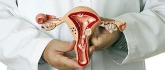
Thanks to the achievements of modern science and medicine, patients who have contraindications to the natural birth of a baby can turn to surgical childbirth. However, despite the fact that cesarean section has been used for many decades, in some cases postoperative complications may occur that impair the health or reproductive function of a woman.
One of the most common problems that can be encountered after surgical delivery is the failure of the uterine scar after cesarean section. Let's consider what the essence of this pathology is, what reasons contribute to its development, and whether it is possible to cure a failing scar.
What distinguishes a normal scar from an incompetent one?
A normal scar always has a sufficient amount of muscle tissue and many blood vessels. It is elastic, contracts well, and can withstand heavy loads. This helps to survive subsequent pregnancies and births without uterine ruptures. As for the incompetent scar, it has few blood vessels and muscle tissue (its place is taken by connective tissue), it is inelastic, contracts very poorly and breaks easily. As the size of the uterus increases during pregnancy, the suture begins to thin and eventually ruptures.
Pronounced (complete) incompetent scar
Scar failure can be considered pronounced (complete) if:
- its thickness does not exceed 1 mm;
- it consists almost entirely of connective tissue;
- it has many poorly fused and thickened areas (the so-called ligatures), mini depressions, cavities and dents;
- Unabsorbed surgical threads are clearly visible at the suture site.
If the incompetence is severe, doctors may even advise the woman to have an abortion or try not to become pregnant until the problem is resolved.
Why does scar failure occur?
The following factors play a major role in the occurrence of insolvency:
- Type of incision made during a cesarean section. Most often, failure occurs in longitudinal sections. They are performed in emergency cases when the life of the mother and fetus is in mortal danger. Conventional transverse lower incisions become insolvent much less frequently.
- Various problems in the abdominal area where stitches are placed after cesarean section. These can be fistulas, inflammatory processes, suppuration and ruptures.
- Poor material from which the seam is made.
- Inflammation of the mucous uterine layer of the endometrium (endometritis). The pathogen that causes the disease enters the body during a CS.
- Repeated operations on the uterus. We are talking not only about cesarean abortions, but also about late abortions, as well as removal of tumor formations of various natures.
- A new pregnancy just a few years after a caesarean section.
In women with a post-cesarean scar, failure of the suture on the uterus can even provoke early abortions. Especially if the embryo was removed by curettage.
Scheduled CS
In some cases, obstetricians and gynecologists recommend that women give birth through a planned cesarean section. Before making a decision, the doctor carefully studies the woman's medical history, analyzes many factors and issues a verdict. Factors in favor of a cesarean section include:
- Narrow pelvis of a woman. The fetus can cause damage and rupture of internal organs if it goes into the world naturally, including tearing the uterus along the scar.
- The woman has already given birth more than twice. This indicates thinning of the walls of the uterus, not only in the area of the scar, but in general.
- There is more than one scar on the uterus. The more scars, the weaker the tissue becomes.
- Incorrect placement of the fetus. If the fetus naturally moves forward with the pelvis, this will create additional stress on the walls of the uterus.
- Reasons why you had a CS for the first time. It is quite possible that this is due to the characteristics of the body.
In these situations, the doctor has no doubts about artificial birth as planned.
Video: Planned and emergency caesarean section. Indications and consequences of cesarean section.
Caesarean section: planned and emergency. Indications and consequences of cesarean section
Symptoms of scar failure
Unfortunately, until a woman becomes pregnant, scar failure rarely reveals itself. The larger the embryo becomes, the more the uterine walls stretch. Thanks to this, the cicatricial incompetence becomes “noticeable” and begins to manifest itself with a number of symptoms:
- Pain sensations appear in the lower abdomen, which radiate to the vaginal area. They look like cramps, they come as if in waves;
- distinct pain when the doctor palpates the area of the scar;
- inability to quickly and completely empty the bladder;
- severe nausea, frequent vomiting;
- low blood pressure;
- general weakness, frequent causeless dizziness;
- A stabbing pain appears in the lower abdomen when a woman stands up or sits down, and also when having sex;
- blood after sexual intercourse;
- the uterus remains “immature” during late pregnancy;
- chronic uterine hypertonicity during pregnancy. The pathology causes pain that resembles labor pains;
- pathological changes in cardiotocographic (CTG) parameters. The fetus often has a low heart rate (bradycardia). A slowdown in its maturation (deceleration) is also typical.
- the fetus is too active - it “squirms” in the womb and moves a lot. At the same time, the woman clearly feels pain.
Emergency caesarean section
In the later stages of pregnancy, a woman should be especially attentive to herself and listen to every movement, every reaction of the body. The scar can break at any moment, even if it seemed strong enough before. Excessive physical activity or a false contraction puts the uterus under such tension that the scar may not be able to withstand it. If the following symptoms appear, you should immediately consult a doctor, as there is a threat of uterine rupture:
- Sharp pain in the lower abdomen
- Increased body temperature
- Slight bleeding
- Tension in the uterus
- Abnormal fetal heart rate
In this case, urgent hospitalization is required. Based on the results of the examination and examination, the doctor can make a verdict about an emergency caesarean section. If a woman does not seek medical help from a doctor in time, a uterine rupture may occur. Internal bleeding will cut off the child's oxygen, and it will not be possible to save him. Due to severe blood loss, the mother may experience hemorrhagic shock, which also leads to irreversible consequences or death.
Why is scar failure dangerous?
The main danger is, of course, uterine rupture during pregnancy or childbirth. This complication often leads to severe bleeding. Without the help of doctors, both mother and fetus quickly die.
Also, an incompetent scar on the uterus can lead to:
- During pregnancy, placenta accreta occurs - a dangerous pathology that can lead to childlessness and even removal of the uterus.
- The fetus will begin to experience hypoxia - it will chronically lack oxygen and nutrients. The complication can result in miscarriage, premature birth, as well as physical and mental abnormalities in the development of the child.
Restoration of the uterus after childbirth
During pregnancy, the uterus stretches greatly due to the elasticity of its walls. Therefore, after childbirth, it should return to its original state - shrink. Full recovery may take up to 2 months.
The suture on the walls of the uterus after a cesarean section does not contribute to rapid contraction, but, on the contrary, slows down the process. The muscle layer is damaged. Nerve endings and blood vessels are dissected. All this interferes with the restoration of the organ.
Timely contraction of the uterus to normal size may be hampered by the presence of adhesions, since due to them the organ may be displaced. With heavy blood loss, the uterus is susceptible to hypotonicity, which affects its contractility. Infections in the uterine cavity pose a danger to its normal recovery.
If the uterus contracts slowly, drug therapy is prescribed, using drugs that have a stimulating effect on its smooth muscles.
Common drugs:
- Ergotal;
- Hyfotocin;
- Desaminooxytocin;
- Pituitrin;
- Oxytacin, etc.
After childbirth, the inner surface of the uterus is a continuous wound, since the mucous membrane is injured. An indicator of a normal recovery process will be uterine bleeding and clots, which are called “lochia”. Such discharge is a wound secretion that is separated from the inner layer of the uterus. The first few days, lochia may be bright red due to a large accumulation of red blood cells, then they become pale.
Important! Normal lochia does not have a pronounced odor. If there is a putrid odor, you must immediately inform your doctor. This indicates their stagnation or inflammatory process.
After a cesarean section, prolonged release of lochia is possible, since postpartum involution is slow.
The external suture and internal sutures on the uterus are painful. However, it is important for a woman to move a lot within a few hours after surgery. Painkillers will be prescribed for several days. Due to prolonged lying or sitting, the uterus, which already contracts poorly, may become bent anteriorly. This will lead to narrowing of the birth canal at the bend and will prevent the removal of lochia. The rejected material is an ideal environment for pathogens, such as saprophytes. The accumulated blood decomposes, toxins and breakdown products enter the general bloodstream, which leads to infection.
Regular bowel movements will also help prevent the uterus from bending anteriorly. If stool abnormalities, such as constipation, are detected, it is necessary to take mild laxatives and review the postpartum diet.
The formation of a scar on the uterus in the postpartum period is important. The connective tissue that forms at the site of the incision does not have sufficient elasticity; this can prevent subsequent spontaneous childbirth. It is necessary that the scar be as thin as possible, and contractions of the uterus, although slightly, still deform it, causing pain. In some cases, a woman in labor may be prescribed physical therapy to better form a postoperative scar.
Diagnostic methods
Ultrasound is the main tool to help identify cicatricial failure. When such a study is carried out, the doctor looks at:
- what is the general condition of the surgical scar;
- are there any voids, dents, bulges (ligatures) and mini-tears in the seam area;
- what is the thickness of the scar tissue;
- whether pathological changes have occurred in the mucous membrane in the area of the suture.
In addition to ultrasound, doctors can also refer you for hysteroscopy. This research method is only suitable for non-pregnant women, as it is unsafe for the fetus. For example, it is necessary when a woman is prescribed plastic surgery for a scar on the uterus after a cesarean section.
In recent years, one of the most pressing problems of modern obstetric and gynecological practice remains the issue of preserving reproductive function in women of childbearing age with uterine fibroids. If previously uterine fibroids were classified as diseases occurring mainly in women at the end of the third - beginning of the fourth decade of life, now uterine fibroids are increasingly diagnosed in young women [2, 4, 7-9, 14-16]. At the same time, recently there has been a clear tendency in society to postpone childbearing to a later date. It is now considered quite common to plan to have your first child at 30-35 years of age. An analysis of the own material from the Department of Operative Gynecology of the Moscow Regional Research Institute of Obstetrics and Gynecology (MONIIAG) confirms the general global trend toward an increase in the number of women of the reproductive period with uterine fibroids who want to preserve their reproductive function. Taking into account all of the above, it is obvious that in the choice of methods of preconception preparation and treatment of fibroids, the woman’s desire to fully preserve her reproductive function is fundamental. Treatment of women with uterine fibroids planning pregnancy should be as organ-preserving as possible. As the number of women with a combination of uterine fibroids and pregnancy, or women with uterine fibroids planning pregnancy, is increasing, obstetricians-gynecologists are increasingly faced with the issue of clarifying the tactics of managing patients with uterine fibroids outside and during pregnancy. Delivery of pregnant women after surgical interventions on the uterus requires a differentiated approach from the obstetrician. A condition for a favorable completion of pregnancy and delivery of a pregnant woman through the natural birth canal is a strong scar on the uterus. To this day, issues remain controversial regarding modern methods for assessing the condition of the uterine scar after organ-saving operations, which serves as the basis for conducting this study.
On the basis of the gynecological department of MONIIAG in 2011-2013. We examined and operated on 150 patients who underwent myomectomy outside pregnancy (Group 1), 15 patients who underwent myomectomy during pregnancy (Group 2), as well as 19 patients who underwent metroplasty due to the failure of the uterine scar after caesarean section at the stage of planning the next pregnancy (group 3).
The age of the patients ranged from 22 to 39 years. 58 (39%) women in the 1st group had a history of pregnancy, 6 (40%) women in the 2nd group and, accordingly, 19 (100%) in the 3rd group, with only 43 (29%) patients in the 1st group. 1st group, 4 (27%) - in the 2nd group and 15 (79%) - 3rd group had children. 21 (9%) patients had a history of spontaneous miscarriages and non-developing pregnancies. Of these, 15 (65%) patients had spontaneous abortions due to uterine fibroids. Moreover, 4 (36%) women had recurrent miscarriage. 6 (35%) patients had a history of ectopic pregnancy.
Of the 165 women of the 1st and 2nd groups, before contacting MONIIAG, 111 (67%) did not know about the presence of uterine fibroids (they were not observed for a long time by a gynecologist): in the 1st group - 98 (65%) patients; in group 2 - 13 (87%). The duration of existence of uterine fibroids ranged from 1 year to 10 years. According to the anamnesis, the growth rate of fibroids was assessed as rapid in 78 (52%) patients of the 1st group and in 12 (80%) patients of the 2nd group. The size of the dominant node in multiple uterine fibroids in the 1st group in 28 (19%) women, in the 2nd group - in 2 (13%) women reached 20 cm in diameter, the number of nodes ranged from 2 to 18 in the 1st group and from 2 to 4 in group 2; in the presence of single myomatous nodes in the 1st group in 78 (52%) patients, in the 2nd group - in 11 (73%) these sizes ranged from 5-15 cm in diameter. According to the location of the nodes, the most common were interstitial uterine fibroids - in 137 (91%), subserous - in 7 (5%), submucosal - in 6 (4%) women of the 1st group. 13 (87%) patients of group 2 had interstitial growth and 2 (13%) had subserous growth. In 67% of the examined patients, there was a combination of different node locations.
All patients underwent clinical and laboratory studies. Despite the variety of research methods, the leading diagnostic method used after general clinical examination in all patients with uterine fibroids was ultrasonography.
Ultrasound examination (ultrasound) of the pelvic organs was performed on a Medison accuvix V20 device using sensors in two-dimensional imaging mode using transabdominal scanning with a frequency of 2-6 MHz and transvaginal examination with a sensor pulse frequency of 4-9 MHz. The location, number and size of myomatous nodes, their relationship to the uterine cavity were assessed. The results of an ultrasound scan performed before surgery were compared with the patients’ medical history data on the growth rate of myomatous nodes.
Principles of surgical treatment of patients with uterine fibroids outside and during pregnancy
The indications for surgical treatment of patients of group 1 outside pregnancy were the following [1, 5]:
1. The presence of heavy, prolonged menstruation or acyclic bleeding leading to anemia.
2. Large tumor sizes (exceeding the size of the uterus corresponding to 12 weeks of pregnancy), even in the absence of complaints, since large tumors disrupt the anatomical relationships in the pelvis and abdominal cavity, often leading to dysfunction of adjacent organs.
3. Tumors of any size in the presence of symptoms of compression of neighboring organs (frequent urination, defecation disturbance) in the presence of atypical isthmus and cervical nodes of various locations (with growth towards the bladder, rectum, parametrium). In the latter cases (intraligamentary location of fibroid nodes), pain appears due to compression of the nerve plexuses.
4. Cervical localization of uterine fibroid nodes.
5. Rapid tumor growth - this is considered to be an increase in the size of the uterus during the year by an amount corresponding to 4 weeks of pregnancy or more. In most cases, with fibroids, so-called false growth of the tumor is observed, associated with the development of an active inflammatory process in it or impaired circulation in the myomatous node and its swelling.
6. The presence of subserous myomatous nodes larger than 4-5 cm. Such nodes must be removed routinely, since there is a danger of torsion of the node’s stem.
7. Necrosis of myomatous node.
8. Presence of submucosal uterine fibroids. Such fibroids usually cause heavy bleeding, leading to severe anemia.
9. The presence of a nascent submucosal myomatous node.
10. Infertility associated with the presence of uterine fibroids.
Indications for myomectomy in group 2—during pregnancy—in all cases were situations posing a high risk of impairment of the health of the mother and fetus.
1. Rapid tumor growth.
2. Large and gigantic sizes of nodes, leading to dysfunction of the abdominal and pelvic organs.
3. Necrosis of the node.
4. Torsion of the leg of the knot.
Thus, ⅓ of pregnant women had symptoms of dysfunction of the urinary system due to cervical isthmus and intraligamentary nodes (urinary retention requiring bladder catheterization); 8 (53%) pregnant women had clinical and ultrasound signs of circulatory disorders in the node (pain, soft consistency, swelling, destruction); 6 (40%) patients had large and gigantic nodes filling the abdominal cavity during pregnancy up to 16 weeks of gestation.
Preoperative preparation
In all cases, we performed organ-saving operations as planned. Preoperative preparation of patients with uterine fibroids for surgical treatment is of great importance for the outcome of surgery. Before a planned operation, a standard clinical examination, extended colposcopy, and cytological examination of smears from the cervical canal and vaginal part of the cervix for the presence of atypical cells are carried out. In addition, examinations are carried out for the presence of sexually transmitted infections (STIs), and mandatory sanitation of foci of infection is carried out, which is especially important before performing organ-preserving operations. In pregnant women of group 2, the content of hormones of the fetoplacental complex in the blood plasma is determined before myomectomy.
Considering the high risk of miscarriage after surgery, intensive prevention of this complication should be carried out in the preoperative period (5-7 days before surgery). It includes the administration of adrenergic agonists (hexoprenaline 5 mcg in 10-20 ml of isotonic sodium chloride solution), 30 ml of a 25% solution of magnesium sulfate in 200 ml of isotonic sodium chloride solution. The drugs are administered intravenously by slow drip. To prevent the side effects of β-adrenergic agonists on the cardiovascular system (tachycardia), the drugs are administered together with calcium antagonists (verapamil). Infusion therapy continues for 7-10 days after surgery with a gradual transition to tablet forms. The dose is selected individually depending on the severity of symptoms of threatened miscarriage. At the end of intravenous administration of drugs, if there is a pronounced threat of miscarriage, 5 ml of baralgin is administered intravenously as a bolus, since combined analgin preparations have an antiprostaglandin effect and should be included in a complex of therapy aimed at prolonging pregnancy. In addition, for oral administration, antispasmodic drugs are included in the complex of therapy aimed at prolonging pregnancy. Considering that uterine fibroids negatively affect fetoplacental blood flow, especially in cases where the placenta is localized in the area of the myomatous node, we carry out therapy aimed at improving blood flow. For this, patients are prescribed chimes 0.025 g 3 times a day, as well as drugs for the prevention of intrauterine fetal hypoxia (piracetam, cocarboxylase, ascorbic acid, 40% glucose solution).
Myomectomy technique
According to the results of various studies, myomectomy is an appropriate surgical procedure to restore the reproductive system in women with uterine fibroids.
In any clinical situation, an important problem in the formation of a successful scar is the activity of tissue repair in the area of the wound on the uterus. The course of the healing process is determined by a large number of factors, which include the state of the macroorganism, the surgical technique, the suture material used, the duration of the operation and blood loss, and the course of the postoperative period.
For the formation of full-fledged scars on the uterus and favorable gestation of a subsequent pregnancy, important points of the surgical technique are the choice of an incision on the uterus, removal of all nodes, enucleation of nodes with opening of the capsule, careful hemostasis by compressing the vessels with tissue without extensive use of electrocoagulation, layer-by-layer sutures without leaving “dead” space, the use of non-reactive synthetic long-absorbable suture material. An important stage of the operation is anti-adhesion measures. We include thorough drainage of the pelvis and abdominal cavity, and reliable hemostasis.
To create the most gentle conditions for the pregnant uterus and fetus, as well as optimal access to atypically located fibroid nodes, it is advisable to use lower median laparotomy. In this case, the body of the uterus with the fetus located in it is brought out into the wound and held by an assistant, which helps reduce blood loss. The surgical technique is similar to that of myomectomy outside pregnancy. However, it should be noted that we only remove problematic nodes.
Postoperative rehabilitation
Postoperative rehabilitation is aimed at restoring hemostasiological parameters, treating anemia, preventing purulent-septic complications and, ultimately, forming a full-fledged scar on the uterus and carrying a pregnancy. To prevent infectious postoperative complications, we use antibiotic prophylaxis with protected penicillins in the form of a single (during laparotomy) or three-time administration of the drug every 8 hours. A five-day course of antibiotic therapy was administered to patients with extensive adhesions, when the uterine cavity was opened during surgery and the scope of the operation was expanded before the intervention on adjacent organs. Postoperative management of pregnant women who have undergone myomectomy has its own specific features, due to the need to create favorable conditions for tissue repair, prevent purulent-septic complications, adequate intestinal functioning, eliminate the threat of miscarriage and improve uteroplacental blood flow.
Diagnosis of the consistency of the uterine scar
Diagnosis of an incompetent scar on the uterus is always complex and ambiguous, especially at the stage of pregnancy planning or in the early stages of an already established and desired pregnancy. As a rule, neither patients nor clinicians are willing to accept a diagnosis based on the results of a single ultrasound examination. Verification of the diagnosis outside of pregnancy is carried out in all cases during a consultative examination, using hydrosonography and hysteroscopy.
Echography is an important method for assessing the course of reparative processes and the consistency of the scar on the operated uterus.
In the postoperative period, all patients underwent ultrasound monitoring of the condition of the uterine scar on the 5-7th day of the postoperative period, after 2 and 6 months in the 1st and 3rd groups and by trimester of pregnancy in the 2nd group.
Criteria for failure of the uterine scar at the stage of pregnancy planning (Fig. 1)
.
Figure 1. Incompetent uterine scar after cesarean section.
+ — myometrial thickness less than 2 mm. Absolute signs:
— visualization of a complete myometrial defect in the projection of the scar in the form of a niche on the side of the uterine cavity, reaching the wall of the bladder in case of failure of the uterine scar after cesarean section or to the serous membrane of the uterus after myomectomy;
— visualization of an incomplete defect in the myometrium in the projection of a scar in the form of a niche on the side of the uterine cavity with thinning of the lower uterine segment to 2 mm or less;
- deformation of the myometrium with retraction on the side of the serous membrane of the uterus and a niche on the side of the uterine cavity, with thinning of the unchanged myometrium to 2 mm or less.
If these signs are identified, surgical treatment—metroplasty—should be recommended. Signs of partial failure of the uterine scar include visualization of niches and deformations in the projection of the scar with thinning of the myometrium to 4-5 mm or less. Very often, such defects have an irregular slit-like shape and are located both centrally and eccentrically. Sometimes it is possible to visualize niches and defects along the parametrium. The absence of convincing vascularization in the myometrium during energy mapping also suggests partial failure of the uterine scar. If partial scar failure is suspected, office hysteroscopy and hydrosonography are recommended [10, 13].
It should be noted that the criteria for assessing the consistency of the scar are absolutely identical after any operation on the uterus (cesarean section, myomectomy outside and during pregnancy, metroplasty, after plastic surgery on the uterus for developmental anomalies).
As criteria for the consistency of a uterine scar in the early postoperative period (6-7 days), we consider the following signs (Fig. 2)
:
Figure 2. A well-developed scar on the uterus on the 7th day after metroplasty.
- absence of deformations, niches, areas of retraction from the outer integument and the uterine cavity;
— the thickness of the myometrium in the area of the postoperative scar is equal to the thickness of the intact myometrium;
— absence of hematomas in the structure of the scar, connective tissue inclusions, and fluid structures;
— visualization of well-structured ligatures in the myometrium;
- adequate blood flow: poor on days 2-3 and increased on days 4-7 (reparative processes in the myometrium);
— the thickness of the vesicouterine fold of the peritoneum is less than 5 mm, the absence of hematomas or liquid contents under this fold [10, 11].
Ultrasound signs of a healthy scar after myomectomy in the long-term period (6 months after surgery) include:
- evenness and clarity of the contours of the uterus;
- ideal scar - determination of localization is not possible;
— the thickness of the myometrium in the projection of the scar is equal to that of the unchanged myometrium;
- absence of niches and deformations of the uterine walls;
- absence of ligatures in the myometrium or single ligatures;
— good vascularization of the scar with Doppler measurements (moderate number of vascular loci, comparable to intact myometrium) (Fig. 3)
[10, 12, 13].
Figure 3. Well-defined uterine scar 6 months after myomectomy of multiple nodes.
In 7 (5%) patients of the 1st group in the postoperative period, the uterine scar was visualized in the form of single hyperechoic (connective tissue) inclusions against the background of unchanged myometrium, and also in 9 (6%) patients - in the form of deformation of the outer contour or uterine cavity and in the scar area. At the same time, among patients with active vascularization in the area of the bed of removed nodes on days 5-7 of the postoperative period, only 4 (3%) were able to visualize a scar on the uterus after 6 months.
Assessment of scar consistency 6 months after metroplasty (Fig. 4)
:
Figure 4. A well-defined uterine scar 6 months after metroplasty.
— the position of the scar corresponds to the incision on the uterus during surgery;
- absence of deformations, niches, areas of retraction from the outer integument and the uterine cavity;
— the thickness of the myometrium in the region of the lower uterine segment is equal to the thickness of the anterior wall of the uterus;
— absence of organized hematomas in the structure of the scar, connective tissue inclusions;
— adequate blood flow (the amount of increase in loci is equal in the intact myometrium);
— condition of the vesicouterine fold, pouch of Douglas, parametrium — without edema, hematomas and free fluid [10].
In some cases, hydrosonography and hysteroscopy were used to confirm the diagnosis.
Office hysteroscopy was performed 6 months after surgery on the 4th or 5th day of the menstrual cycle, when the functional layer of the endometrium is completely rejected, and the underlying tissue is clearly visualized through the thin basal layer. If the scar fails, retractions or thickening in the scar area are noted. The whitish color of the scar tissue and the absence of blood vessels indicate a pronounced predominance of the connective tissue component, and retractions indicate thinning of the myometrium as a result of inadequate regeneration. In the absence of these signs, the scar is considered anatomically complete. But with anatomical consistency, it can be morphologically defective with a predominance of connective tissue elements. The following hysteroscopic types of uterine scar condition are distinguished [6]:
Type I: the scar on the uterus is practically not visualized;
Type II: among the muscle elements, individual elements of connective tissue are visible, poorly vascularized;
Type III: wide connective tissue avascular scar.
Office hysteroscopy was performed on 6 patients of group 1, whose uterine cavity was opened during surgery, and 7 patients of group 3. In all cases, the uterine scar was not visualized.
Assessment of the condition of the uterine scar during pregnancy
A necessary condition is the availability of information about the localization of nodes before myomectomy (a clear information description). A thorough examination of all the walls of the uterus is carried out at all stages of perinatal observation. In the second and third trimesters, it is not possible to determine the localization of the scar in all cases, so it seems more rational to assess the thickness and structure of the myometrium in the myomectomy area. The main research method is transabdominal scanning, but additional vaginal ultrasound is also required for low localization of nodes. It is advisable to begin examining the pregnant uterus with a transabdominal sensor, which allows you to obtain an overview of the anatomy of all the walls of the uterus as a whole, and detail it with segmental scanning. It is more difficult to examine the posterior wall of the uterus, especially at the end of the 2nd-3rd trimester. Knowing the topic of the removed nodes, the doctor scans the expected area of scars (“zone of interest”) at high magnification. If technical capabilities are favorable (absence of massive subcutaneous fat, good sound conductivity of tissues), it is advisable to use a linear high-frequency sensor. Its high resolution provides differentiation of the layers of the anterior abdominal wall and the anterior wall of the uterus. When scanning from the level of the navel to the symphysis pubis in transverse sections, then from the right to the left iliac bones in multiplanar sections, we evaluate the thickness of the myometrium in the “zone of interest,” homogeneity, and the presence of inclusions. We consider a myometrial thickness of at least 0.3 cm to be an unambiguous criterion for the anatomical consistency of a scar. This ultrasound picture, in combination with clinical signs, suggests the possibility of delivery through the natural birth canal. We consider the thickness of any part of the myometrium to be less than 0.2 cm as an unambiguous criterion for anatomical failure (Fig. 5)
.
Figure 5. Incompetent scar on the uterus during pregnancy.
When the localization of the scar is low, the use of vaginal scanning is mandatory. The use of this technique allows for diagnostic accuracy of up to 97% [10, 12]. Thus, the echographic criteria for the consistency of a uterine scar during pregnancy are:
— thickness and homogeneity of the myometrium;
— absence of deformations, niches, areas of thinning, connective tissue inclusions [10].
Method of delivery for pregnant women with a scar on the uterus after organ-preserving operations
Further observation of the operated patients of all 3 groups showed that among 150 patients of the 1st group who had reproductive problems, pregnancy occurred in 93 (62%): 81 women carried it to the due date, while 3 patients had urgent spontaneous births, 78 patients were delivered by cesarean section; in 8 women, pregnancy is currently progressing. In 4 patients, unfavorable pregnancy outcomes were noted: 1 woman had a spontaneous miscarriage in early pregnancy, in 3 cases a non-developing short-term pregnancy was diagnosed. The remaining patients are not currently planning pregnancy.
Among 15 patients of the 2nd group, one had an urgent spontaneous birth and 11 were delivered by cesarean section; in 3 women of the 2nd group, pregnancy is currently progressing.
Among 19 patients of the 3rd group, pregnancy occurred in 11 - 8 were delivered by cesarean section, in 1 case a non-developing short-term pregnancy was diagnosed. In 2 women, pregnancy is currently progressing.
One woman of the 2nd group had a repeat myomectomy during a cesarean section (a myomatous node, localized in the projection of the placenta, was removed; therefore, it was decided to abstain from myomectomy at 16 weeks of gestation due to the high risk of miscarriage). One patient of group 1 underwent repeat myomectomy in a delayed period after cesarean section, since during surgical delivery, due to the large size of the tumor and the atypical location of the myomatous node, there was a high risk of expanding the scope of the operation before hysterectomy.
A strictly individual approach to the method of delivery in women after organ-preserving operations is necessary. The method of delivery depended on the age of the pregnant woman, the presence of concomitant extragenital pathology, the course of the present pregnancy, the condition of the fetus, the condition of the scars on the uterus, the number of removed myomatous nodes, the localization of the scars on the uterus, and the method of surgical treatment.
97 (52.7%) pregnant women were delivered by cesarean section.
During cesarean section, the condition of the uterine scar after organ-preserving operations in all groups, the presence of adhesions, myomatous nodes and their size in the 2nd group of patients were assessed. The scars on the uterus looked like star-shaped retractions or longitudinal depressions of varying lengths depending on the size of the removed nodes, from 1 to 2.5 cm wide, whitish in color.
No deformations of the uterine cavity were detected in any group. In 12 cases, an adhesive process was noted in the pelvis and abdominal cavity in the form of a soldered distal edge of the greater omentum or intestinal loops and appendages to the scar area on the uterus. Macroscopically, in no case was there an incompetent scar after myomectomy. During cesarean section, 16 patients of group 1 and 5 patients of group 2 underwent a knife biopsy of the scar after myomectomy; in 8 women of group 3, the scar was excised along its entire length, followed by histological examination of the excised tissue. All placentas were also subjected to histological examination. In all patients, the placentas were of normal weight, and the villous tree corresponded to the gestational age; in 2 cases, signs of chronic mild uteroplacental ischemia and a slight decrease in placental weight were noted.
In all excised scar fragments, the smooth muscle component was well expressed and represented by hypertrophied muscle fibers. Stromal fibrosis is noted to be predominantly diffuse in the form of thin layers of connective tissue between smooth muscle fibers. In some cases, large-focal fibrosis of subserous areas in combination with adhesive periprocess was noted. The latter was noted in a small number of cases and was represented by individual well-vascularized fibrous adhesions. In the thickness there were small focal perivascular lymphohistiocytic infiltration and single small granulomas of foreign bodies.
Macroscopically, during cesarean section, thinning of the myometrium in the scar area was not detected in any case, which was confirmed by ultrasound before surgery and subsequently by morphological examination of scar biopsies obtained during surgical delivery of such patients.
Thus, the data obtained indicate that a rational surgical technique for removing nodes during myomectomy and scar formation during metroplasty, an uncomplicated course of the postoperative period with actively occurring processes of neovascularization and reparation in the area of the scar and the bed of the removed node, as well as improving the diagnosis of the consistency of the uterine scar through the use of a complex of diagnostic methods (clinical, echography using Doppler ultrasound and three-dimensional ultrasound, endoscopic) make it possible to improve reproductive outcomes after organ-saving operations even in the most difficult clinical situations.
Treatment
The method of treating the pathology depends on the diagnosis:
- Partial failure. With this diagnosis, doctors usually order the woman to undergo additional tests. For example, it may be referred to check the patency of the fallopian tubes (hydrosonography), echohysteroscopy and magnetic resonance imaging.
- Complete failure. If the diagnosis showed that a woman has a pronounced (complete) scar failure, she should urgently undergo surgery. Surgery is usually open (laparotomy). This is due to the fact that the scar is located under the bladder, so the closed (laparoscopic) option is inconvenient for the surgeon and is accompanied by excessive blood loss. The purpose of this operation is to completely excise (remove) the scar tissue and then apply stitches to create a new scar. After such an operation, doctors do not recommend becoming pregnant for several years.
Types of suture after cesarean section
Subsequent pregnancy after cesarean directly depends on the type of incision made - each type requires appropriate time for normal healing. But the doctor cannot influence the choice of the safest suture for the subsequent birth of a child - this is determined by the course of the woman’s pregnancy, complications that arise during pregnancy and during surgery. If complications arise during a cesarean section, the doctor can quickly change the decision on the choice of incision. As a result, the time for scar formation can increase significantly, causing subsequent pregnancies to be delayed.
The following types of cesarean section suture are distinguished:
- Transverse is the most popular type today, since it is performed in the lower abdomen, above the pubis, and does not exceed 10-12 cm in length. Moreover, this arrangement leads to faster scar formation on the skin. It is also noted that after childbirth, it is much easier for women to care for a child due to the anatomical location of the muscles on the abdomen.
- Longitudinal - differs from the first type in location, since a suture is made on the upper part of the uterus. In terms of location on the stomach, it is located vertically under the navel. Such an incision entails damage to a large number of blood vessels, which is why the scar takes a long time to form, and the woman is forced to experience severe pain and discomfort.
- Vertical - this suture is made only in an emergency situation when labor has already begun. It is also often used in the presence of uterine pathology. It is difficult for a scar to form with such an incision, since the woman constantly “disturbs” it in a sitting position.
Caring for the suture after childbirth by cesarean section occurs within 1.5-2 months - this is the time of formation of the external scar. The internal suture on the uterus heals within six months - it becomes somewhat denser, but remains vulnerable.
Preventive measures
In order not to become childless, a woman who has had a cesarean section:
- It is undesirable to become pregnant in the first few years after a CS;
- She should also check the condition of her uterine scar with an ultrasound at least once a year.
In the postpartum period, the risk of complications increases due to the increase in CS operations. A failed suture is the most well-known pathology, which often results in surgical intervention. To avoid repeated surgery, a woman should study the nature of possible complications, not violate the prohibitions that the doctor warned about, and carefully monitor her condition.
Surgical interventions on a woman’s body and possible consequences
Nature’s wonderful idea is to create a female body with the aim of fulfilling a holy and noble mission, to bear and give birth to full-fledged offspring! The norm of a full-fledged family is the creation of conditions for fertilization, gestation and the birth of healthy offspring. However, not every representative of the fair half of humanity can boast of complete women's health during pregnancy. On the path to healthy motherhood, various obstacles may arise, which, with a competent, correct, timely, qualified approach, can be overcome and resolved. We are talking about forced surgical interventions in a woman’s body, necessary to correct her health.
For example, an operation such as a conservative myomectomy can restore a patient's ability to conceive. The fibroid is removed, but the organ is preserved. However, after surgery, as a rule, a scar always forms. A scar can also occur during various necessary plastic reconstructive surgeries (when the uterine horn is removed, a tubal or cervical pregnancy is operated on simultaneously with the uterine angle). The wall of the uterus is perforated during surgery to forcefully remove the fertilized egg; if labor is overstimulated, rupture may occur. For some women during pregnancy, doctors prescribe a planned caesarean section if they are unable to deliver on their own. As a result, the integrity of the uterus is compromised. After suturing the incisions, punctures or ruptures made, a scar is formed on the uterus. All of this can have consequences during pregnancy.