Red blood cells in a smear, the reasons for their appearance
A smear test, or smear test, is a doctor-ordered test that collects a small amount of material from the mucosal surface. Smear analysis takes place for men - in urology, and for women - in gynecology. A study carried out to study the microflora allows you to check for the presence of unpleasant pathogenic bacteria or cancer cells; allows for a generally correct assessment of hormonal levels, as well as the general condition of tissues. A smear is taken during a preventive examination. In the event that you are undergoing any treatment, a smear for infection is taken only after completing the full course of treatment, thereby increasing the accuracy of the analysis.
- Red blood cells in a smear
- Microcytosis
Red blood cells in a smear
A smear is the same test as all other tests. You should take into account the fact that a smear taken from the cervix or vagina is a completely painless procedure that allows you to find out about the state of women's health. Laboratory research is carried out with the help of an experienced medical specialist and a microscope. A smear is applied to glass and stained with special dyes, which allow bacteria to be very clearly seen and distinguished under a microscope.
To prepare for a smear test, you need the following:
- Abstinence from sexual intercourse for 1 or 2 days
- Do not douche or douche for 2 days
- Do not use vaginal suppositories or creams
- It is not recommended to urinate for approximately 2 or 3 hours before taking a smear test.
- It is advisable to take a smear immediately after menstruation on the 4th or 5th day of the cycle
A small number of red blood cells may normally be present on a smear test. The normal rate of red blood cells during analysis is no more than two cells in the field of view. In women, the presence of red blood cells can increase significantly during menstruation or in case of injury to the mucous membrane of the vagina or uterus during inflammation, hormonal disorders and various inflammatory processes.
The reasons for the presence of red blood cells in a smear can be very different, ranging from the place where the analysis was taken and ending with dependence on the situation.
Below we will try to consider for which diseases and what the presence of red blood cells in the analysis can tell about.
Exceeding the norm in an analysis taken from the urethra can indicate a tumor, the presence of crystals or pebbles in the urinary tract, or traumatic urethritis.
An increase in red blood cells in tests taken from the cervical canals or cervical canal may indicate the development of inflammatory processes and the development of cervical erosion. Women who have an intrauterine device or hormonal disorders that are accompanied by bleeding are at risk. Also, this can happen just before or after menstruation.
A smear examination taken by a doctor will allow him to learn about the content of red and white blood cells in the analysis. Red cells or red blood cells, as is known, can be contained only in small single quantities. Therefore, tests are best done in the middle of the cycle.
Microcytosis
To take a closer look at changes in the size of red blood cells, let's familiarize ourselves with some scientific terms, one of which is microcytosis. Microcytosis is the so-called predominance of red blood cells in a blood smear with small sizes ranging from 5 to 6.5 microns. The causes of microcytosis are:
- Iron-deficiency anemia
- Hereditary spherocytosis
- Thalassemia
Video about the disease anemia:
Another scientific term, macrocytosis, refers to the predominance of red blood cells in a blood smear with sizes greater than 9 microns. The causes of macrocytosis are:
- Liver diseases
- Physiological feature in newborns
- Decreased thyroid function
- Malignant neoplasms
- Myeloproliferative diseases
- Macrocytic anemia (adults)
- Anemia in pregnant women
Another scientific term associated with the presence of red blood cells in smear tests, megalocytosis refers to the predominance of oval-shaped red blood cells with sizes equal to 11 or 12 microns. The causes of megalocytosis are:
- Worm infestation
- Anemia in pregnant women
- Folate deficiency anemia
- Dyserythropoiesis
Schizocytosis is the presence of only small, insignificant fragments of red blood cells or altered cells at the degenerative level, having an irregular shape with a size of 2 or 3 microns. The causes of schizocytosis are:
- Uremia
- Vasculitis
- Microangiopathic hemolytic anemia
- March hemoglobinuria
- Myelodysplastic syndrome
- DIC syndrome
- Hemoglobinopathy
- Glomerulonephritis
Anisocytosis is the presence of red blood cells of different sizes in the analysis. The causes of anisocytosis are:
- Paroxysmal hemoglobinuria (nocturnal)
- Thalassemia
- Myeloproliferative diseases
- Iron-deficiency anemia
It should be remembered that with microanacitosis, small erythrocytes predominate, and with macroanacitosis, large erythrocytes predominate.
Noticed a mistake? Select it and press Ctrl+Enter to let us know.
Liked? Like and save on your page!
mirbodrosti.com
No red blood cells were found in the smear
Principle. Examination of stained blood smears using an immersion microscope system.
Reagents: 1) immersion oil; 2) diethyl ether.
Progress of the study. A slide with a stained and dried blood smear is placed on the microscope stage and using low magnification (7× eyepiece, 8× objective) the edge of the smear is found.
Without changing the position of the glass, apply a drop of immersion oil to the edge of the smear in the place located under the lens.
Place the immersion lens in a vertical position relative to the smear, and the lens is immersed in a drop of oil.
Using a macroscrew, carefully obtain an image in the field of view of the microscope. Then, using a microscrew, a clear visibility of the drug is established. The criterion for a correctly selected focal length for each eye will be a clear image of cells with clear boundaries and intracellular structure.
After this, they begin to study the morphology of red blood cells, paying attention to their shape, size, color intensity, the presence of pathological forms, intracellular inclusions, etc.
Since cells have a certain volume, to better view them it is necessary to constantly change the focal length using a microscrew.
To obtain a more complete picture of cell morphology, it is necessary to view several fields of view by moving the smear by hand or using a cross-shaped device.
It is necessary to study the morphology of erythrocytes in thin areas of the smear, where they are located singly, without forming “coin columns,” usually at the edge of the smear near the marginal “panicle.”
At the end of microscopy, use a macroscrew to lift the microscope tube, remove a smear from the stage, and wipe off the immersion oil from the lens and slide with gauze soaked in ether.
In healthy people, red blood cells have the shape of a biconcave disk, their sizes are approximately the same. In stained preparations, red blood cells are round in shape, pink in color, with uniform coloring and a slight clearing in the center. Oxyphilia is caused by hemoglobin, so the intensity of the color can be used to judge the degree of saturation of red blood cells with hemoglobin.
Normal red blood cells
Unified microscopic technique for measuring the diameter of red blood cells using an eyepiece micrometer in a stained blood smear
Special equipment: 1) microscope; 2) eyepiece-micrometer - an eyepiece mounted on the microscope tube, with a round glass plate and a scale applied to it, divided into 50 divisions; 3) object-micrometer - a glass slide with a scale 2 mm long, divided into 200 divisions, each of which is equal to 10 microns.
Progress of determination. Before starting work, determine the price of one division of the eyepiece micrometer scale, which depends on the length of the microscope tube and the magnification of the lens. The determination is carried out using an object micrometer. It is installed on the microscope stage so that the scales of the micrometer eyepiece and the micrometer object coincide.
Then count the number of divisions on the eyepiece-micrometer scale that coincide with one or another number of divisions on the scale of the object-micrometer, and determine the price of one division. For example, 40 divisions of the eyepiece-micrometer scale coincided with 6 divisions of the object-micrometer, which corresponds to 60 microns, i.e. one division of the eyepiece-micrometer will be equal to: 60: 40 = 1.5 microns.
This determination is carried out once for a specific microscope, on which erythrocytometry is subsequently carried out.
In a thin blood smear, using an immersion microscope system with a maximum illuminated field of view, measure the diameter of at least 100 red blood cells, noting how many divisions of the eyepiece-micrometer scale the red blood cell occupies. The measurement result for each red blood cell is noted. Knowing the cost of division and the number of red blood cells with the same number of divisions, express the result as a percentage.
Example: red blood cells with a diameter of 4.5 microns - 5%, 6.0 microns - 10%, 7.5 microns - 70%, 9.0 microns - 11%, 10.5 microns - 4%.
The result can be presented in the form of an erythrocytometric curve (Price-Jones curve), with the size of erythrocytes plotted along the abscissa axis, and the number of erythrocytes of a given size along the ordinate axis. In routine practice, the Price-Jones curve is rarely used due to the high complexity of its implementation. Modern hematology analyzers draw the Price-Jones curve automatically.
If necessary, the result is expressed as the average size of red blood cells.
Clinical significance of the study of erythrocyte morphology
Size. The diameter of normal red blood cells (normocytes) is 7.0–8.0 μm, mostly 7.2–7.5 μm. There are deviations in the range of 4.7–9.5 µm. Microcytes have a size of 6.7 microns or less, macrocytes - more than 7.7 microns, and megalocytes - more than 9.5 microns (Abramov M. G., 1985).
According to E. A. Cost (1975), the normal size of erythrocytes ranges from 7.1 to 7.9 microns in diameter, microcytes have a diameter of less than 6.5 microns. Red blood cells larger than 8 microns are called macrocytes, those larger than 12 microns are called megalocytes or gigantocytes.
Microcytosis is a condition when 30–50% of the total number of red blood cells are microcytes. A shift of the erythrocytometric curve to the left often occurs in iron deficiency anemia, microspherocytosis, thalassemia, and lead poisoning.
Microspherocytes
Macrocytosis is a condition when 50% or more of the total number of red blood cells are macrocytes. A shift of the erythrocytometric curve to the right is observed in B12 and folate deficiency anemia, alcoholism, and diffuse liver damage.
Macrocytes
Anisocytosis . The presence of red blood cells of different sizes in a blood smear is called anisocytosis. There are three degrees of anisocytosis:
- 1st degree: 50% of red blood cells in the field of view are represented by cells of different sizes (micro- or macrocytes);
- 2nd degree: 75% of red blood cells in the field of view are represented by cells of different sizes;
- 3rd degree: more than 75% of red blood cells in the field of view are represented by cells of different sizes. When assessing the size of red blood cells in a blood test, it is advisable to indicate the predominance of micro- or macroanisocytosis.
Form. Red blood cells can change shape, becoming oval, pear-shaped, stellate, jagged, etc.
Pathological forms of red blood cells are described: microcytes (small red blood cells); microspherocytes (spherical red blood cells with increased thickness and reduced diameter, without clearing in the center), which are a pathognomonic sign of hereditary hemolytic microspherocytic anemia; ovalocytes or elliptocytes - found in up to 10% of healthy people, in patients with hereditary elliptocytosis they account for 25–75% of the total number of erythrocytes; target-shaped red blood cells (with a colored area in the center of the cell against the background of an unstained zone) are often found in thalassemia and iron deficiency anemia; schizocytes (small fragments 2–3 µm in size); acanthocytes (Burr Cells) - red blood cells with a jagged edge, with “burrs”; drepanocytes (sickle-shaped red blood cells), found in sickle cell anemia; planocytes, or leptocytes (flat red blood cells).
Ovalocytes
Target-shaped red blood cells
Drepanocytes (sickle-shaped red blood cells)
The presence of red blood cells of different shapes is called poikilocytosis . There are four degrees of poikilocytosis:
- 0 degree: less than 10% of red blood cells of different shapes in the field of view;
- 1st degree: in the field of view 10-25% of red blood cells of various shapes;
- 2nd degree: in the field of view up to 50% of red blood cells of different shapes;
- 3rd degree: more than 50% of red blood cells of different shapes in the field of view.
Aniso- and poikilocytosis are nonspecific signs of anemia of various origins. As the severity of anemia increases, the number of red blood cells of different shapes and sizes increases.
Coloring. Red blood cells are stained pinkish-red with acidic dyes. The degree of oxyphilia of a cell is determined by the presence of hemoglobin and its quantity. Red blood cells of healthy people have a uniform color and a slight clearing in the center (normochromia).
Pale coloration of red blood cells with a wide, uncolored central part is called hypochromia. Hypochromia of erythrocytes is caused by a low hemoglobin content in erythrocytes and is more often characteristic of iron deficiency, but also occurs with lead poisoning and thalassemia.
Hypochromia is usually combined with microcytosis. There are three degrees of hypochromia:
- 1st degree: clearing in the center is slightly greater than normal;
- 2nd degree: the painted part is presented in the form of a narrow ribbon;
- 3rd degree: the colored part is presented in the form of a very narrow ring.
Increased coloration of red blood cells is called hyperchromia, caused by an increase in the volume of red blood cells and is usually combined with macro- and megalocytosis. Microspherocytes can also be hyperchromic.
Macrocytes are large red blood cells with preserved clearing in the center, megalocytes are giant red blood cells without clearing. These changes in red blood cells indicate pathological hematopoiesis associated with a deficiency of vitamin B12 and folic acid.
Deficiency of these hematopoietic factors often occurs with diphyllobothriasis, organic diseases of the stomach, alcoholism, and pregnancy.
Immature forms . Anisochromia is a different intensity of staining of individual red blood cells or sections of one red blood cell, often found in iron deficiency anemia.
Polychromatophilia (polychromasia) is insufficient accumulation of hemoglobin in red blood cells with remnants of basophilic substance.
Polychromatophilia is caused by the mixing of two highly dispersed colloidal phases, one of which (with an acidic reaction) is a basophilic substance, and the other (with a slightly alkaline reaction) is hemoglobin.
Thanks to this, the immature erythrocyte perceives both acidic and alkaline dyes and, depending on whether the basophilic component of the cytoplasm or hemoglobin predominates in them, is colored from blue to grayish-pink. Reticulocytes are also usually polychromatophilic.
Normally, single polychromatophilic erythrocytes are found. Their number may increase with increased erythropoiesis (posthemorrhagic, hemolytic anemia). Anemia occurring with polychromatophilia has a favorable course. There are three degrees of polychromasia:
- P1: single polychromatophils every 2–3 fields of view;
- P2: 1-4 polychromatophils in each field of view;
- RZ: more than 10 polychromatophils in each field of view.
Polychromatophils can be determined not only in a regular preparation, but also in a thick drop of blood (Kost E. A., 1975):
- normally, 1–2 red blood cells with a basophilic mesh are not detected in every field of view and are designated P+ (polychromasia);
- P2: 3–5 polychromatophils;
- RZ: 5–10 polychromatophils;
- P4: more than 10 polychromatophiles.
The first degree of polychromasia is more common.
In thalassemia and other forms of anemia, so-called target-shaped red blood cells are found - with a colored area in the center of the cell against the background of an unstained area.
During a morphological examination of red blood cells, it is necessary to determine the presence of pathological forms of red blood cells or inclusions in red blood cells in the smear. Nucleated red blood cells (normoblasts, erythroblasts) are found in a variety of conditions.
The highest degree of normoblast content occurs in hemolytic anemia at the time of hemolytic crisis, in chronic myelofibrosis, and metastases of malignant tumors in the bone marrow.
A moderate amount is observed in acute erythromyelosis; in myelodysplastic syndrome (MDS), the number of normoblasts ranges from 1 to 4 per 100 red blood cells (Yavorkovsky L.I. et al., 1992); in vitamin B12-deficiency anemia, transient normoblastosis is diagnosed after blood loss.
Inclusions . In vitamin B12-deficiency anemia and after splenectomy, red blood cells with remnants of nuclei in the form of Cabot rings, Jolly bodies, and Weidenreich dust particles are found. Jolly bodies are remnants of nuclear chromatin of a round shape, 1 micron in size or more, in the amount of 1 to 3 in an erythrocyte, red-violet in color.
Cabot rings are remnants of the nuclear membrane in the form of thin thread-like rings, figures of eight or ellipses, painted red. They are sometimes found in polycythemia vera, leukemia, and heavy metal poisoning.
Weidenreich dust particles are remnants of nuclear substance of pink, sometimes blue color, found in severe anemia, mainly megaloblastic, similar to basophilic punctuation of erythrocytes.
Taurus Jolly
Cabot's rings
Heinz-Ehrlich bodies are usually one round inclusion (rarely 2-3) 1-2 µm in size, located along the periphery of the erythrocyte. Rarely, bodies are found outside the cell. With conventional Romanovsky staining, they are not visible. They are determined according to the Deutsche method (Todorov I., 1963) with methyl violet.
The corpuscles are colored purple-red. It is believed that these are denatured lipoproteins of the erythrocyte membrane.
The appearance of Heinz-Ehrlich bodies is evidence of severe toxic damage by substances that oxidize hemoglobin (nitrobenzene, aniline, nitroglycerin, Bertholet salt, sulfonamide drugs) and leading to hemolysis.
Heinz-Ehrlich bodies
Source: onlab.info
Source: https://nikitazhilyakov.com/jeritrocity-v-mazke-ne-obnaruzheny/
Red blood cells are normal in women
Determining the number of individual blood components is the most important task of the analysis. It is especially important to know the number of red blood cells that perform the function of transporting oxygen to all tissues. The norm of red blood cells in women is slightly higher than in the male half, and based on their number, conclusions are drawn about the presence of inflammation, infections, and also judge whether the chosen treatment helps. Therefore, determining the number of blood cells is one of the main blood tests.
The level of red blood cells in the blood is normal in women
Normal values for the number of blood components are determined based on the age and gender of the patient. For patients, indicators within the range of (3.4-5.1) x 10^12 g/l are considered normal. Any minor deviations are considered a consequence of pathological processes occurring in the body.
If a pregnant woman's blood test for red blood cells turns out to be low (up to 3-4.7), then this is considered the norm for pregnant women. However, if there is a drop in hemoglobin levels along with this, this indicates anemia, which can jeopardize pregnancy.
In addition, a drop in the number of blood cells occurs during hydremia (injection of excess fluid). A decrease in the indicator also occurs due to:
- dysfunction of the respiratory system;
- erythremia;
- for heart disease;
- for infections accompanied by blockage of the respiratory tract.
The average volume of red blood cells may exceed the permissible norm in women, but this phenomenon does not occur often. This usually happens:
- when experiencing serious stress;
- in case of blood composition disorders (erythremia);
- with insufficient oxygen supply caused by prolonged diseases of the heart and respiratory organs.
Red blood cells in urine are normal in women
In an absolutely healthy person, red blood cells are practically not detected in the urine or are found, but in very small quantities. The norm for women is slightly higher than for men and is up to 3 units.
If blood cells are detected in the urine, the woman is sent for a repeat test, which is taken using a catheter. If after this a high level of red blood cells is also noted, the doctor will prescribe a full examination of the urinary system. After all, this phenomenon indicates a number of pathologies:
- cystitis;
- pyelonephritis;
- urolithiasis disease;
- injuries of the urinary system.
Red blood cells in a smear are normal in women
Sometimes blood cells may be found in the smear. Normally, there should be no more than two of them in the field of view. The number of red blood cells increases due to:
- inflammation;
- menses;
- injuries to the vaginal mucosa;
- with hormonal imbalance.
Articles
Determining the number of individual blood components is the most important task of the analysis. It is especially important to know the number of red blood cells that perform the function of transporting oxygen to all tissues. The norm of red blood cells in women is slightly higher than in the male half, and based on their number, conclusions are drawn about the presence of inflammation, infections, and also judge whether the chosen treatment helps. Therefore, determining the number of blood cells is one of the main blood tests.
The level of red blood cells in the blood is normal in women
Normal values for the number of blood components are determined based on the age and gender of the patient. For patients, indicators within the range of (3.4-5.1) x 10^12 g/l are considered normal. Any minor deviations are considered a consequence of pathological processes occurring in the body.
If a pregnant woman's blood test for red blood cells turns out to be low (up to 3-4.7), then this is considered the norm for pregnant women. However, if there is a drop in hemoglobin levels along with this, this indicates anemia, which can jeopardize pregnancy.
In addition, a drop in the number of blood cells occurs during hydremia (injection of excess fluid). A decrease in the indicator also occurs due to:
- dysfunction of the respiratory system;
- erythremia;
- for heart disease;
- for infections accompanied by blockage of the respiratory tract.
The average volume of red blood cells may exceed the permissible norm in women, but this phenomenon does not occur often. This usually happens:
- when experiencing something serious;
- in case of blood composition disorders (erythremia);
- with insufficient oxygen supply caused by prolonged diseases of the heart and respiratory organs.
Red blood cells in urine are normal in women
In an absolutely healthy person, red blood cells are practically not detected in the urine or are found, but in very small quantities. The norm for women is slightly higher than for men and is up to 3 units.
If blood cells are detected in the urine, the woman is sent for a repeat test, which is taken using a catheter. If after this a high level of red blood cells is also noted, the doctor will prescribe a full examination of the urinary system. After all, this phenomenon indicates a number of pathologies:
A smear test, or smear test, is a doctor-ordered test that collects a small amount of material from the mucosal surface. Smear analysis takes place for men - in urology, and for women - in gynecology. A study carried out to study the microflora allows you to check for the presence of unpleasant pathogenic bacteria or cancer cells; in general, it allows you to correctly assess hormonal levels, as well as the general condition of the tissues. A smear is taken during a preventive examination. In the event that you are undergoing any treatment, a smear for infection is taken only after completing the full course of treatment, thereby increasing the accuracy of the analysis.
What does a smear show on flora in women: main analysis indicators
A study of vaginal flora is necessary to identify and identify bacteria and determine the degree of their danger.
This method is the least expensive and popular for diagnosing inflammatory processes and diseases that can be contracted during sexual intercourse. The advantage of this method is that taking a smear for analysis is completely painless. When taking a smear, minor subjective discomfort is possible. A smear is taken during an examination by a gynecologist, then a sample of vaginal contents is placed on a laboratory glass.
Later, upon arrival at the laboratory, the sample is stained to distinguish and identify microbes.
What does a gynecological smear show on vaginal flora in women, what infections can we talk about? Let's discuss further.
Main indicators in gynecological analysis
The vaginal environment in women is not normally sterile.
Many microorganisms constitute the vaginal flora. Among these microorganisms there is constant competition for space and food.
The most common representatives of the vaginal flora are lactobacteria and bifidobacteria .
In many ways, they themselves determine the microclimate of their habitat, releasing alcohol, peroxide, and acids, including lactic acid, in the process of their life, as a result of which the pH of the vaginal environment has a value in a smear on the flora, which characterizes an acidic environment.
These bacteria secrete enzymes that prevent other microbes from multiplying.
So what does a smear reveal for flora in women?
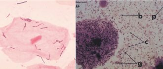
Leukocytes and red blood cells
The number of white and red blood cells is determined in the selected smear. The norm for a healthy woman is 10–15 leukocytes, and about 2 erythrocytes.
If the analysis is taken immediately after menstruation , then the number of leukocytes can be up to 25. If the patient is pregnant, then the leukocyte level is allowed to be no more than 30.
A lot of white blood cells indicate an infectious disease, then you need to retake the test.
Here you need to take into account all conditions that additionally affect the level of leukocytes - pregnancy, menstruation, colds. If all these cases are excluded, then an inflammatory process is suspected.
Phagocytosis
What does phagocytosis mean in a flora smear? Phagocytosis is the process of capture and destruction of foreign cells (for example, pathogenic bacteria) by leukocytes.
The smear may reveal incomplete phagocytosis - the presence of undigested microbial cells inside leukocytes. This happens when there is an infection or if the body is exposed to:
- stress;
- unfavorable ecology;
- poor nutrition.
Cytolysis
Cytolysis in the analysis means the presence in the vagina of a non-inflammatory process caused by the excessive proliferation of a special type of lactobacilli that produce hydrogen.
Their uncontrolled growth leads to alkalization of the vaginal environment and cytolysis (destruction) of epithelial cells.
Epithelium
What does a large amount of epithelium in a smear on the flora in women indicate, what to do if there is a lot of it?
The presence of squamous epithelium in a smear for flora in women is the norm, provided that it has a certain type and quantity.
It is considered normal to have 5 to 10 squamous epithelial cells in the selected sample.
When taking a smear test for flora at a gynecologist, deviations from the norm:
- Decreased or absent epithelial cells, indicating death of the epithelium due to increased testosterone levels and/or decreased estrogen levels.
- A significant excess of epithelial cells in the smear may appear for the following reasons:
- inflammation in the mucous layer of the vaginal walls;
- diseases that can be contracted during coitus;
- vaginitis;
- candidiasis;
- inflammation of the cervix and urethra;
- renal and genitourinary infectious and other diseases.
Ectopia is not a disease, but requires observation.
Fibrin
Fibrin is a blood plasma protein. Its detection in a smear means the presence of an inflammatory process.
Slime
In healthy patients, mucus can be found exclusively in a vaginal smear. If mucus is found, for example, in the urethra, then this indicates inflammation. A vaginal sample may normally have moderate or low amounts of mucus, designated ++ and +, respectively.
In addition, excess mucus may indicate poor hygiene or improper smear sampling.
Doderlein sticks
Doderlein bacilli should normally predominate in the vaginal microflora.
These are lactobacilli that secrete lactic acid , which is necessary for the formation of normal vaginal microflora.
If lactobacilli are present in the vagina in sufficient quantities, infections do not develop there, since lactic acid prevents other bacteria from multiplying.
If the number of these beneficial microorganisms is reduced, then the vaginal pH shifts to the alkaline side. Inflammation may occur.
In a normal smear, lactobacilli make up 95% of the total flora.
Opportunistic flora
In gynecology, opportunistic microbes are those microbes in a flora smear that may not cause any diseases until they begin to multiply excessively. The main types of such microbes:
- cocci;
- ureaplasma;
- diphtheroids;
- Candida mushrooms.
Cocci
Cocci are round shaped bacteria.
According to the staining method, there are gram-positive and gram-negative.
Gram-positive cocci (staphylococci, streptococci and enterococci) are opportunistic, their presence is considered normal within the following limits:
- staphylococci – up to 104;
- streptococci – up to 105;
- enterococci – up to 105.
Gram-negative cocci are causative agents of dangerous diseases. These include gonococci, the causative agents of gonorrhea.
Ureaplasma
These are very small bacteria that, when multiplied above 104 units, cause inflammatory diseases. They prevent conception.
Leptothrix
This is a representative of the anaerobic UP flora, a gram-positive rod-shaped microorganism. Leptothrix itself in a flora smear is dangerous only during pregnancy and can cause a miscarriage.
In non-pregnant patients, the discovery of leptothrix is not a cause for concern.
Diphtheroids
As representatives of the UP flora, they can be present in a smear in small quantities. When their level is exceeded, vaginosis begins . Diphtheroids are similar to the bacteria that cause diphtheria, Corynebacterium.
If corynobacteria were found in the flora smear, their quantity is determined. In case of moderate amounts, a course of drug treatment is prescribed.
If corynebacteria are more than a certain threshold, then additional research needs to be carried out for the presence of other infections, since these bacteria are often associated with others.
Klebsiella
A representative of the flora, Klebsiella under certain conditions (with decreased immunity, pregnancy) can cause urinary tract diseases .
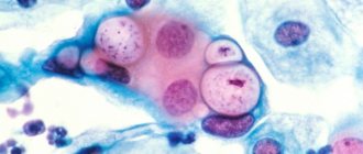
Candida mushrooms (soor)
What does a positive soor in a flora smear give? If fungi of the genus Candida are isolated from a smear, this indicates candidiasis.
These fungi are also representatives of the UP flora; they are introduced into the vagina from the outside. Candidiasis is caused by decreased immunity.
With candidiasis, a smear on the flora can be found:
- filaments of pseudomycelium (mycelium). Pseudomycelium is represented by filamentous growths of blastospores with constrictions.
- blastospores (spores). If blastospores are detected in the analysis of a vaginal smear for flora, it means that the woman’s body is affected by candidiasis. The number of blastospores depends on the prevalence and stage of the disease. Blastospores are embryonic forms of cells formed by budding from maternal forms of cells. If blastospores are found in a smear on the flora, it is necessary to examine the female body for yeast spores of fungi.
Key (atypical) cells
The key cells in a smear for flora are epithelial cells on the surface of which microorganisms have collected (this is especially typical for gardnerella in women, which causes bacterial vaginosis). The presence of atypical cells in a smear is a sign of an infectious disease .
Scarcity and abundance
Based on a smear sample, the type of microflora is determined:
- scanty - contains only lactobacilli;
- medium – lactobacilli + up to 10 leukocytes;
- mixed – lactobacilli + up to 30 leukocytes + cocci;
- abundant - almost no lactobacilli, a lot of leukocytes, mucus and cocci.
What determines the degree of purity
Based on the results of analyzing a smear sample for flora, the degrees of vaginal cleanliness are distinguished:
- The flora is mainly represented by lactobacilli. Few white blood cells, moderate amount of epithelial cells, moderate amount of mucus. Normal, good local immunity.
- The flora is represented by lactobacilli + cocci + yeast. Few white blood cells, moderate amount of epithelial cells, moderate amount of mucus. Variant of the norm.
- The number of leukocytes is increased. Flora – cocci, yeasts, fungi, lactobacilli are few. Lots of epithelial cells and mucus. An inflammatory process that requires treatment.
- White blood cells are everywhere. Flora – cocci, yeast, fungi, lactobacilli no. Lots of epithelial cells and mucus. Severe inflammatory process requires treatment.
With 1 and 2 degrees of purity, the environment in the vagina is acidic and slightly acidic, in the case of 3 and 4 - slightly alkaline and alkaline, respectively.
Source: https://beautyladi.ru/chto-pokazyvaet-mazok-na-floru/
Red blood cells in a smear
A smear is the same test as all other tests. You should take into account the fact that a smear taken from the cervix or vagina is a completely painless procedure that allows you to find out about the state of women's health. Laboratory research is carried out with the help of an experienced medical specialist and a microscope. A smear is applied to glass and stained with special dyes, which allow bacteria to be very clearly seen and distinguished under a microscope.
To prepare for a smear test, you need the following:
- Abstinence from sexual intercourse for 1 or 2 days
- Do not douche or douche for 2 days
- Do not use vaginal suppositories or creams
- It is not recommended to urinate for approximately 2 or 3 hours before taking a smear test.
- It is advisable to take a smear immediately after menstruation on the 4th or 5th day of the cycle
A small number of red blood cells may normally be present on a smear test. The normal rate of red blood cells during analysis is no more than two cells in the field of view. In women, the presence of red blood cells can increase significantly during menstruation or in case of injury to the mucous membrane or uterus during inflammation, hormonal disorders and various inflammatory processes.
The reasons for the presence of red blood cells in a smear can be very different, ranging from the place where the analysis was taken and ending with dependence on the situation.
Below we will try to consider for which diseases and what the presence of red blood cells in the analysis can tell about.
Exceeding the norm in an analysis taken from the urethra can indicate a tumor, the presence of crystals or pebbles in the urinary tract, or traumatic urethritis.
An increase in red blood cells in tests taken from the cervical canals or cervical canal may indicate the development of inflammatory processes and the development of cervical erosion. Women who have an intrauterine device or hormonal disorders that are accompanied by bleeding are at risk. Also, this can happen immediately before or after.
A smear examination taken by a doctor will allow him to learn about the content of red and white blood cells in the analysis. Red cells or red blood cells, as is known, can be contained only in small single quantities. Therefore, tests are best done in the middle of the cycle.
Red blood cells in a smear for flora in women: normal, reasons for the increase
Secondly, red blood cells in a smear can be detected if the material collection technique is incorrect, when the gynecologist puts too much pressure on the instrument and injures the tissue, which causes slight bleeding and, accordingly, red blood cells getting into the smear.
If there are few squamous epithelial cells in the cytology smear - up to 5 in the field of view, then this is a sign of estrogen deficiency in the woman’s body and the development of atrophic processes in the mucous membranes of the vagina, cervix, etc.
Normally, in a cytology smear, squamous epithelial cells should be present in small numbers (
Next, the gynecologist wipes the cervix with a sterile swab moistened with saline to remove mucus. If a mucus plug is visible in the cervical canal, the doctor also removes it with a cervical brush or scraper.
Cytological examination of the cervical canal or Pap test in women
If cancer cells are detected in large numbers in the smear, then this simple analysis allows you to identify the tumor at an early stage and carry out the necessary treatment as soon as possible.
If the cytology smear result is negative, then it is also called normal or good, since this indicates the absence of pathologically altered cells in the cervix and pathogenic microorganisms (for example, herpes viruses, human papilloma, etc.) that can provoke inflammatory processes.
Metaplasia is not cancer or even a precancerous process, but it is not the norm either. Therefore, women with epithelial metaplasia are recommended to undergo examination to identify the causes of the degeneration of one type of epithelium into another.
), therefore, if leukocytes are detected in a cytology smear, it is necessary to undergo tests for sexually transmitted infections and bacteriological culture of vaginal discharge for flora in order to identify the causative agent of the inflammatory process in a particular case and carry out the necessary treatment.
What do the various abnormal cells in a cytology smear indicate?
Next, this container of liquid is sent to the cytology laboratory, where all the liquid is centrifuged to obtain a pellet of cells at the bottom of the tube.
However, if there are many metaplastic epithelial cells or they are located in clusters, then this indicates that the single-layer squamous epithelium on the outer part of the cervix is replaced by multilayered squamous epithelium.
Endocervicitis or ectocervicitis can be caused by various pathogenic microorganisms (for example, Trichomonas, chlamydia, human papillomavirus, etc.
With a lack of red blood cells, the body receives less oxygen, fatigue increases, apathy appears, and performance decreases.
Indications for a smear test for cytology
Remember that violation of these rules can lead to false results of a smear on the flora, such as increased leukocytes when they are normal, as well as a normal number when there are a large number.
Cytology smear can be positive or negative. A negative result is a normal smear with no pathological changes.
Thirdly, if the smear was taken correctly and a sufficient time after menstruation, then the presence of red blood cells in it indicates an inflammatory process in the tissues of the cervix.
In such a situation, the presence of red blood cells in the smear also does not play any role and has no diagnostic value. It is very easy to understand that there was an incorrect technique for collecting material - after the manipulation, the woman had bloody discharge from the vagina for several hours.
Microcytosis
To take a closer look at changes in the size of red blood cells, let's familiarize ourselves with some scientific terms, one of which is microcytosis. Microcytosis is the so-called predominance of red blood cells in a blood smear with small sizes ranging from 5 to 6.5 microns. The causes of microcytosis are:
- Hereditary spherocytosis
- Thalassemia
Video about the disease anemia:
Another scientific term, macrocytosis, refers to the predominance of red blood cells in a blood smear with sizes greater than 9 microns. The causes of macrocytosis are:
- Liver diseases
- Physiological feature in newborns
- Decreased thyroid function
- Malignant neoplasms
- Myeloproliferative diseases
- Macrocytic anemia (adults)
- in pregnant women
Another scientific term associated with the presence of red blood cells in smear tests, megalocytosis refers to the predominance of oval-shaped red blood cells with sizes equal to 11 or 12 microns. The causes of megalocytosis are:
- Worm infestation
- Anemia in pregnant women
- Folate deficiency anemia
- Dyserythropoiesis
Schizocytosis is the presence of only small, insignificant fragments of red blood cells or altered cells at the degenerative level, having an irregular shape with a size of 2 or 3 microns. The causes of schizocytosis are:
- Uremia
- Vasculitis
- Microangiopathic hemolytic anemia
- March hemoglobinuria
- Myelodysplastic syndrome
- DIC syndrome
- Hemoglobinopathy
- Glomerulonephritis
Anisocytosis is the presence of red blood cells of different sizes in the analysis. The causes of anisocytosis are:
- Paroxysmal hemoglobinuria (nocturnal)
- Thalassemia
- Myeloproliferative diseases
- Iron-deficiency anemia
It should be remembered that with microanacitosis, small erythrocytes predominate, and with macroanacitosis, large erythrocytes predominate.
A small amount of red blood cells in a smear indicates a good state of women's health.
Exceeding the normal level of red blood cells in vaginal discharge is considered a symptom of hormonal disorders and inflammation, which can externally manifest itself in unpleasant and sometimes dangerous diseases.
A flora smear is a diagnostic method that involves studying material collected from the surface of the mucosa under a microscope. The analysis is done for everyone: both men and women.
Men undergo a smear to detect urological diseases. Material for research is taken from the urethra. In women, a smear can be taken from the urethra and vagina.
The study of the microflora and cytological composition of the biomaterial obtained by scraping or imprinting the mucous membrane makes it possible to diagnose infectious, including sexually transmitted and hormonal diseases, and to detect cancer, precancerous and background conditions.
A smear is taken for both therapeutic and preventive purposes. When undergoing a course of treatment for genitourinary diseases, a smear is usually taken before and after treatment.
Taking a smear is a completely painless procedure that helps to correctly assess the condition of a woman’s reproductive system.
The biomaterial is applied to a glass slide and a thin smear is made - a microslide suitable for study under a microscope.
The material for research is selected with a glass pipette or a sharp spoon, and then applied to the edge of a glass slide and smeared with the edge of a coverslip. The smear is lightly dried in air or in a burner flame and stained.
There are two ways to stain a vaginal smear. Monochrome is used in cytological analysis, polychrome is used for cytological and hormonal studies.
After all the manipulations performed, the microspecimen is considered ready for examination under a microscope.
Violation of the technology for preparing a smear will lead to unreliable results, but this situation is rare, since a vaginal smear is a standard test that does not require special qualifications from a health worker.
A woman should come prepared for a smear test. For the analysis to be reliable, you need to abstain from sexual intercourse for two days, not douche, and not be treated with vaginal suppositories, ointments and creams.
Why do they take a smear?
A flora smear is a diagnostic method that involves studying material collected from the surface of the mucosa under a microscope. The analysis is done for everyone: both men and women.
Men undergo a smear to detect urological diseases. Material for research is taken from the urethra. In women, a smear can be taken from the urethra and vagina.
The study of the microflora and cytological composition of the biomaterial obtained by scraping or imprinting the mucous membrane makes it possible to diagnose infectious, including sexually transmitted and hormonal diseases, and to detect cancer, precancerous and background conditions.
A smear is taken for both therapeutic and preventive purposes. When undergoing a course of treatment for genitourinary diseases, a smear is usually taken before and after treatment.
Taking a smear is a completely painless procedure that helps to correctly assess the condition of a woman’s reproductive system.
The biomaterial is applied to a glass slide and a thin smear is made - a microslide suitable for study under a microscope.
The material for research is selected with a glass pipette or a sharp spoon, and then applied to the edge of a glass slide and smeared with the edge of a coverslip. The smear is lightly dried in air or in a burner flame and stained.
There are two ways to stain a vaginal smear. Monochrome is used in cytological analysis, polychrome is used for cytological and hormonal studies.
After all the manipulations performed, the microspecimen is considered ready for examination under a microscope.
Violation of the technology for preparing a smear will lead to unreliable results, but this situation is rare, since a vaginal smear is a standard test that does not require special qualifications from a health worker.
READ Properties of red blood cells in the MCHC blood test
Video:
A woman should come prepared for a smear test. For the analysis to be reliable, you need to abstain from sexual intercourse for two days, not douche, and not be treated with vaginal suppositories, ointments and creams.
It is recommended to urinate no earlier than two or three hours before visiting the gynecological chair. It is advisable to take the test on the 5th day of the cycle, as soon as your period ends.
Reasons for appearance
What is the normal number of red blood cells in a vaginal smear? The norm is when there are no red cells in the smear at all.
It is considered normal to have two or three red blood cells in the field of view of a laboratory technician looking through the eyepiece of a microscope.
An increased number of red blood cells in a smear indicates an inflammatory process in the cervix. This sign is not direct, but indirect.
A smear is taken from the cervix with a brush with silicone bristles. With severe inflammation of the cervix, the tissue becomes so vulnerable that the elastic bristles scratch it until it bleeds, and a large number of red blood cells get into the smear.
Red blood cells are non-epithelial elements of a vaginal smear. The norm for red blood cells is not to be in mucus, but in the blood, where they carry oxygen from the lungs to body tissues and transport carbon dioxide in the opposite direction.
Red blood cells are the most numerous cells in the human body: every 4 cells are an erythrocyte.
Every second, more than two million new red blood cells are formed in the bone marrow, which enter the bloodstream and begin to perform their important functions.
Red blood cells of human blood are extremely small cells, shaped like a disk, slightly concave on both sides.
This shape and size allows red blood cells to move freely through the smallest capillaries and still have a large enough surface area to allow gas exchange.
Red blood cells can only enter the vaginal or cervical mucosa along with blood—they do not leave the bloodstream on their own.
In most cases, an increase in the number of red blood cells occurs during the collection of biomaterial, since the analysis is taken with a shaggy brush, which can accidentally scratch the cervix.
Accordingly, some amount of blood will get into the material. In such cases, a large number of red blood cells is not a pathology, but the norm.
Blood can enter the vagina not only due to injury, but also for natural reasons, during menstruation. Red blood cells found in a smear at this time are the absolute norm, even if they are in huge quantities.
Single red blood cells may appear in a vaginal smear on certain days of the cycle:
- during the period of ovulation (usually 13–15 days of the cycle);
- on day 28 (before the start of menstruation).
A large number of red blood cells in the material taken from the cervical canal indicates the development of cervical erosion or inflammatory processes in this part of the vagina.
The appearance of red blood cells can be caused by intrauterine contraceptives and hormonal disorders accompanied by bleeding.