The functioning of a woman's body changes greatly during pregnancy. This happens for many reasons and factors that make the body more vulnerable. The most common problems arise from hormonal disorders, because the body adapts and redistributes forces to develop a new life.
The endometrium, which lines the surface of the uterus and its cervix, is very sensitive to hormonal changes. During menstruation, the endometrium is renewed and removed from the body. But during pregnancy and hormonal changes during it, the endometrium does not have time to remove the excess, which makes it possible for polyps to appear in the cervical canal and cervix.
A polyp that appears before pregnancy does not make it possible to conceive a child. Such formations must be removed or, if possible, cured. Before and during pregnancy, polyps are detected using ultrasound.
Detection of a polyp during pregnancy does not harm the health of the mother or child. The exception is inflammatory processes that can accompany polyps. In this case, treatment for the infection is prescribed.
Very often, after childbirth, when hormonal levels are restored, polyps disappear without a trace. If the polyp does not disappear, it is removed after childbirth.
Why does a polyp bleed during pregnancy?
The most important source of blood is a polyp formed in the cervical canal, which is located between the uterus and its cervix. The appearance of blood can occur during sexual intercourse or a gynecological examination due to the weakness of the walls of such a polyp. Bleeding usually occurs with a large polyp, due to various types of external contacts.
A polyp may also appear if removal was performed before pregnancy, and immediately after that it was possible to conceive a child.
If blood appears due to a cervical polyp, the doctor may prescribe antimicrobial therapy, since blood can become a source of inflammatory processes. Prevention is also recommended.
Prevention for severely bleeding polyp:
• Timely visit to the doctor. • Reduce physical activity. • Stop sexual intercourse. • You cannot visit the bathhouse. • Maintain hygiene. • Use panty liners.
The birth of a child with a polyp that bleeds occurs normally and without any abnormalities. The most important thing is that if blood appears due to a polyp, monitor the appearance of other symptoms and, if necessary, consult a doctor immediately.
How to treat
Polyps that a doctor can detect during a gynecological examination are not dangerous.
They are characterized by independent resorption at certain stages of pregnancy. They can also come out during childbirth. If a cesarean section had to be performed, the patient must be offered a surgical method to remove the formation. To accurately determine whether treatment is necessary, the patient must undergo smears from the cervical canal. The test results will help determine whether a tumor is benign or malignant. If it does not threaten the health of the mother and fetus, no treatment will be required.
There are cases when there is no need to treat a polyp, since the polyp disappears on its own or is removed after childbirth. In any case, medical supervision is necessary and includes prescribing and taking certain medications. As a rule, this is a progesterone substitute - Duphaston. Antibacterial treatment is possible if the cause of growth is inflammation of the genital organs.
To avoid miscarriage or premature birth, doctors do not perform surgery during pregnancy. In this case, patients are prescribed regular ultrasound, which allows them to monitor indicators of transformation, changes in parameters, and are also recommended to maintain sexual rest and avoid excessive stress.
Cervical polyp and childbirth
If a formation is detected before birth, the tactics depend on whether any symptoms appear or not, and whether the polyp interferes with the natural passage of the child through the birth canal. When the birth process begins, it is most often not touched.
Tactics of labor management with a polyp on the cervix
The vast majority of births in women with this pathology are carried out naturally. The tactics of labor management depend on whether the tumor bleeds or interferes with the birth of the fetus.
There are the following types of management tactics:
- When formed from the mucous layer of the cervical canal, there is no interference during childbirth. After the baby is born, the cervix and polyp are examined. Then it is removed and sent for histology (malignant cells are searched for).
- For decidual polyp, a different tactic is used. It usually bleeds during childbirth. Detachment of the edge of the placenta occurs and severe bleeding occurs. Usually a puncture of the amniotic sac is performed. Amniotic fluid is released, labor is stimulated, and the fetal head compresses the edge of the formation. Through mechanical action, bleeding stops. After birth, the formation comes out with the placenta and the bleeding stops.
- During childbirth, in rare cases, a situation arises when tumors bleed heavily. They need to be removed urgently. Due to delay, there may be large blood losses.
If all recommendations and rational tactics for labor management are followed, a caesarean section may not be necessary.
How it all happens
In general, the implementation of these mini-operations is similar. The woman is positioned approximately on this gynecological table.
Her legs, which are wearing anti-embolic stockings (a requirement of anesthesiologists!), are fixed. A sensor is placed on one arm to monitor heart rate and blood pressure levels. Medicine is injected into a vein in the other arm. Intravenous anesthesia for such gynecological interventions is practiced almost everywhere. Although in some remote corners of Russia, curettage of the uterine cavity and endometrial polyp is still carried out under local anesthesia. More details about this in our other article.
The anesthesiologist talks to the woman, asks about all sorts of nonsense, as if talking to her teeth, calming her down. Then the nurse injects another medicine into the IV, which literally makes the woman “fall asleep” within a minute. She does not notice how long the operation to remove an endometrial polyp takes. Usually this is about 20 minutes. Moreover, anesthesia is given only when the patient is already lying in a chair, the doctor and the honey. personnel are ready to begin the operation. Awakening occurs almost immediately after disconnection from the IV. Usually at this time the woman is on a gurney in the department. After which she is transported to the ward. You may feel dizzy for the first 1-2 hours. At this time, it is not recommended to get up, drink, or eat. Then all this is possible. And if there are no complications, the woman goes home the same day.
The histological report will be ready in 7-10 days. If the conclusion does not contain the phrase “atypical cells,” everything is normal. For example, in “histology” the diagnosis “glandular fibrous endometrial polyp” may be indicated.
Consequences
Will there be consequences after curettage, especially if it is performed by a nulliparous woman? None! Only positive ones. The woman stops intermenstrual bleeding. Menstruation becomes less heavy. Hemoglobin increases, which means well-being, mood, disease resistance, skin and hair condition improves. And pregnancy occurs quickly, provided there are no other reasons for infertility.
Treatment after curettage of endometrial polyps is usually not required. The doctor only gives recommendations on abstaining from sexual activity for one month, recommends injecting antibiotics and taking a couple of tablets of an antifungal drug so that the anti-inflammatory drug does not provoke thrush.
Main reasons
A polyp in the cervix during pregnancy is a rare occurrence. If such a pathology is diagnosed, conception becomes complicated and the risk of early miscarriage is high. Although polyposis is one of the most common gynecological diseases, the exact causes of its occurrence are still unknown.
It is believed that the formation of polyps can be triggered by factors such as:
- chronic inflammatory processes in the cervix or in the organ cavity;
- history of sexually transmitted infections;
- pathologies in which the endometrium grows;
- hormonal imbalances;
- previous curettage;
- pathological proliferation of blood vessels;
- autoimmune diseases;
- endocrine disorders;
- hereditary predisposition.
Growths in the cervical canal can be caused by other reasons. Uncontrolled use of medications, excess weight and metabolic disorders contribute to the development of pathology. The tumor may appear as a result of an unsuccessful pregnancy, ending in miscarriage or death.
Long-term wearing of an intrauterine device changes the normal structure of endometrial tissue. A pregnancy that occurs soon may be complicated by the formation of polyps.
Causes and symptoms of appearance
Polyps in the uterus are a proliferation of endometrial cells. They have a stroma, a stalk with a blood vessel and other cellular elements. During pregnancy, such growths often appear in the cervical canal.
The reason for the formation of this pathology in pregnant women should be considered:
- infectious and inflammatory processes occurring in the uterus;
- hormonal disorders, when there is an excess of estrogen with a lack of progesterone;
- mechanical injury to the endometrium after medical termination of pregnancy, surgical abortion, childbirth, etc.
Various immune disorders, heredity, sexually transmitted diseases, diabetes mellitus, hypertension, etc. can contribute to excessive growth of the endometrium in the uterus and the formation of growths.
The pathology has pronounced symptoms. During pregnancy, a polyp bleeds when exposed to external influences - during sexual intercourse, during a gynecological examination. There are cramping pains in the lower abdomen and lumbar region. The amount of natural discharge increases, often with an unpleasant odor, due to the attachment of a bacterial infection to the source of inflammation. In this case, cervical bleeding is accompanied by purulent discharge.
Benign growths, depending on the cells that form them, can be:
- glandular;
- placental;
- fibrous;
- adenomatous, etc.
Fibrous and adenomatous polyps are urgently removed during pregnancy, as they tend to quickly degenerate into a cancerous tumor. Other types of formations have a small risk of malignancy, so they are observed throughout the entire period of gestation.
If a polyp is diagnosed and pregnancy is just planned, then to reduce adverse risks, the doctor performs a planned removal of this formation.
Manifestations during pregnancy
If a polyp appears while carrying a baby, the woman should immediately consult a doctor. He will determine the type of formation and select an effective treatment regimen. Such formations can be recognized by a number of characteristic features.
If it bleeds
When the walls of the formations are damaged, the woman experiences bloody discharge. Their number increases after a routine examination by a gynecologist or sexual intercourse. It is worth noting that pregnant women experience spotting on the dates of their expected menstruation. This symptom is considered alarming, since when the polyp is injured, the chances of developing an infectious process increase.
Pain
The pain is aching in nature and intensifies after sexual intercourse or examination by a gynecologist. The pregnant woman also has other signs:
- burning sensation in the vagina;
- severe itching;
- nagging pain in the lower back.
At the first unpleasant symptoms, you should immediately consult a doctor.
What it is
A cervical polyp is a benign neoplasm that develops as a result of excessive growth of the epithelium lining the cervical canal of the cervix. In gynecology, this is a common phenomenon, and patients with this diagnosis can be of any age. The disease is detected in young girls, in women during pregnancy, in older patients (before, after and during menopause).
A benign tumor rarely degenerates into oncology, which makes the further prognosis favorable. The neoplasm can be different:
- on a thin leg;
- with a wide base;
- single;
- multiple (polyposis);
- small (from a few millimeters);
- large (several centimeters);
- different in shape, color and consistency.
It can develop as an independent disease or occur as a concomitant with other diseases, both gynecological (for example, infectious diseases) and systemic (more often with pathologies of the endocrine system). The disease can be successfully treated, but can recur. It is often asymptomatic and does not appear for several years, and then is detected during examination, ultrasound, or during pregnancy.
Preventive routine examinations and contacting a specialized specialist at the first signs of malaise, menstrual irregularities or other problems usually accompanying gynecological diseases help in timely detection.
Discharge from a polyp of the cervical canal during pregnancy
Most often, polyps form on the cervix or in the cervical canal. Let us note that the body cannot always quickly respond with visible symptoms to gynecological diseases (and to a serious infection!) - and this entirely applies to polyps, since at their initial stage even a visible allergy may not be observed. Only laboratory tests of discharge can be used as a means of reliable diagnosis, since external signs of discharge may not differ at all from normal, and symptoms such as menstrual irregularities or abdominal pain may be absent.
In the presence of a polyp, the mucous membrane of the cervical canal, as a rule, grows greatly, which causes periodic bleeding, which can become the root cause of subsequent anemia. Symptoms of anemia are as follows: dizziness and general weakness of the body, a sharp decrease in appetite. In the absence of pregnancy, bleeding can be disguised as heavy menstruation, and its cyclicity is often disrupted (until it disappears completely). In this case, discharge can vary greatly: from very scanty and ichorous to profuse leucorrhoea - the body’s reaction here is hardly predictable, you need to pay attention to any “deviations from the established personal norm.”
It is easier to physically detect a cervical polyp with a simple gynecological examination on a chair, using colposcopy and mirrors (no difficulties arise here even in the earliest stages of the disease), and a hysteroscopic examination is performed to confirm the diagnosis.
Methods for removing uterine polyps
Removal of polyps is performed using several methods, which are selected based on:
- general clinical history of the woman,
- laboratory and instrumental research data,
- nature of polyposis,
- risks of malignancy of growths.
The following types of intervention are distinguished:
- Polypectomy. Removal occurs by twisting the polyp until it is torn off. Afterwards, the wound is cauterized with electrodes or liquid nitrogen. Next, the polyp is sent for histological examination. A week after the operation, a control ultrasound is performed.
- Curettage. A surgical procedure known as curettage involves scraping out an endometrial polyp in the uterus. The procedure is often complicated by postoperative infection and relapses of polyposis.
- Ablation of the uterine cavity. Ablation is used in women of mature age who have achieved motherhood, or in women at risk of polyp degeneration into a malignant formation. Removal is carried out with laser, radio frequency waves, liquid nitrogen, and electric currents. After ablation, a woman is unable to bear a child on her own.
- Hysteroscopy of uterine polyp. The procedure is of a therapeutic and diagnostic nature and refers to endoscopic methods for removing polyps. The main advantage is the ability to diagnose and simultaneously remove polyps using powerful optical equipment. In addition, recovery after such manipulation is much faster.
- Hysterectomy or high amputation. A radical method of treating polyposis in cases of suspected oncogenic degeneration of tumors and the growth of metastases. The uterus is removed along with the appendages.
The most common method is polypectomy - an organ-preserving surgical technique that allows a woman to realize the desired motherhood in the near future.
Treatment of polyps during pregnancy
Treatment of small tumors is not required: the mother and child do not suffer from them. A polyp during pregnancy is dangerous in the following cases:
- it has been established that the growth is of an oncological nature (it must be removed surgically);
- the neoplasm provokes infectious processes (the woman is prescribed antibacterial drugs);
- the growth causes dilatation of the cervix and threatens premature onset of labor (the woman is inserted a pessary or the cervix is sutured).
Treatment of polyps in the early stages
If neoplasms in the uterine cavity or in the cervical canal bother a woman, the following options for a medical solution to the problem are possible:
- Removal of the tumor surgically for a period of 12-14 weeks. Indications are the presence of an inflammatory process, rapid growth of the polyp, and its bleeding.
- Antibacterial treatment. Indicated in the early stages of fruiting with an active increase in cervical growth.
- Prescribing drugs with progesterone to prevent the tumor from increasing in size.
As preventive measures in the early stages of bearing a child, women are advised to refrain from intimacy with a partner, excessive physical activity, and to monitor the activity of the growth and the condition of the fetus through systematic ultrasound monitoring.
Polyp removal during pregnancy
Performing surgical procedures on pregnant women is undesirable - it can harm the fetus. But in some cases, the benefits of surgery outweigh the risks. Indications for surgical intervention for cervical canal disease are:
- large and multiple growths;
- a persistent tendency towards an increase in the size of neoplasms (the polyp increases in size by more than 2 mm per month);
- severe bleeding, creating a threat of miscarriage;
- threat of infection in the membranes.
The operation is done through hysteroscopy.
In the endometrium of the uterus, surgical removal of growths is incompatible with pregnancy.
Categories
AllergistAnesthesiologist-resuscitatorVenereologistGastroenterologistHematologistGeneticGynecologistHomeopathDermatologistPediatric gynecologistPediatric dermatologistPediatric cardiologistPediatric ENTPediatric neurologistPediatric ophthalmologistPediatric pulmonologistPediatric rheumatologistPediatric urologistPediatric surgeonPediatric endocrinologistNutrologistImmunologistInfection specialist ist Cardiologist Clinical psychologist Cosmetologist Speech therapist ENT Mammologist Medical lawyer Narcologist Neuropathologist Neurosurgeon Nephrologist Nutritionist Oncologist Oncourologist Orthopedic traumatologist Ophthalmologist Parasitologist Pediatrician Plastic surgeon Proctologist Psychiatrist Psychologist Pulmonologist Rheumatologist Radiologist Reproductologist Sexologist-Andrologist Dentist Therapist RychologistUrologistPharmacistPhytotherapistPhlebologistPhysiatristSurgeonEndocrinologist
Should I delete it?
As a rule, when polypous neoplasms are detected, gynecologists advise removing the pathology.
However, patients do not always agree to this procedure, and often ask the doctor to prescribe conservative methods.
A woman should understand that it is possible to completely get rid of a polyp only through surgical treatment, however, if the polyp is small in size, and if there is no risk of degeneration into a malignant formation, drug treatment is possible.
The choice of therapy is influenced by the following factors:
- polyp size – neoplasms up to 1 cm can be treated with medication,
- type of polyp - with a fibrous polyp, conservative tactics are acceptable, but if an adenomatous formation is diagnosed, surgery is mandatory,
- stage of the disease - the sooner the pathology is detected, the easier it will be to get rid of it,
- age of the woman - for women of reproductive age, doctors try to preserve the cervix,
- individual reaction of the patient’s body to medications.
If a woman follows all the specialist’s recommendations and takes the right medications, conservative therapy can give good results.
But the presence of a large polyp that has atypical cells is an absolute indication for surgical intervention.
Purulent discharge.
Purulent discharge from cervical polyps is a very rare occurrence. This symptom can be observed if polyps of significant size block the cervical canal. The fact is that pus is most actively formed in infected closed cavities. When the cervical canal closes, the uterus becomes such a cavity. As pus accumulates in it, a strong increase in temperature occurs ( more than 39 degrees
). Pure pus can gradually leak from the uterine cavity into the vagina, and from there be released out. Such a serious complication requires urgent surgical intervention.
However, mucopurulent or purulent discharge is not a typical symptom of cervical polyps. They can appear only in the event of an exacerbation of sexually transmitted diseases, or the proliferation of pathogenic microflora in favorable conditions.
What determines the length of the cervical canal
The length of the cervical canal changes in each period of a woman’s life. In an adult nulliparous woman, the length of the cervical lumen does not exceed 40 mm, its width on average is 8 mm.
During pregnancy, the cervical cavity decreases in size - both in length and width. This is necessary in order to form a compacted muscle barrier for the growing fetus. Under the influence of gravity, the fetus, increasing in size, tends downward, towards the exit from the uterine cavity. The thickened and decreased lumen of the cervix does not allow the fetus to leave the uterine cavity and keeps it there until the time of physiological labor.
The muscular layer of the cervix is also slightly different from the muscular layer of the uterus. It has an increased content of elastic fibers, which also contributes to the greater extensibility of the canal.
The length of the cervix depends on a variety of external and internal environmental conditions. It can change under various pathological and physiological conditions.
- With congenital pathology of the development of the genitals, for example, atresia of the cervical canal. In this case, the lumen of the cervix is partially or completely closed, and its length may decrease.
- Expansion of the cervical canal is observed during various inflammatory processes in it.
- The length of the cervical lumen can be affected by such a pathological condition as its stenosis.
- A polyp of the cervical canal in most cases leads to its expansion. The length of the channel, as a rule, does not change. The polyp can be located in any part of the cervical canal mucosa.
- The length of the cervical canal often changes due to various injuries - childbirth with a large fetus, surgery, abortion.
- During pregnancy, throughout its entire length, the length of the cervical canal should not be less than 35 mm. A decrease in this length indicates the development of cervical incompetence—isthmic-cervical insufficiency. This condition threatens premature birth.
If the length of the cervical canal during pregnancy decreases to 22 mm, this indicates a possible risk of premature birth. The length of the cervical canal is considered critical if it is less than 15 mm. This is an indication for suturing the uterine isthmus.
A pregnant woman is prescribed several ultrasound examinations throughout her pregnancy. This is done precisely in order to monitor the condition of the cervical canal and timely notice the developing signs of isthmic-cervical insufficiency. Ultrasound allows you to accurately determine the length of the isthmus.
Important! If signs of isthmic-cervical insufficiency appear, the woman is hospitalized in the hospital to maintain the pregnancy. If conservative treatment fails, several stitches are placed on the cervix
These sutures hold the opening external os in the desired position and prevent the development of premature birth. The sutures are removed at the time when physiological labor is possible.
In the first trimester of pregnancy, the physiological length of the cervical canal is 40 mm. In the second trimester, the canal is shortened to 35 mm. At 36 weeks of pregnancy, the length of the canal is 30 mm. In the last week before childbirth, the cervix ripens. The channel reaches a length of 10 mm. There is an expansion of the canal and opening of the uterine pharynx. In the first stage of labor, the cervix smoothes and the external and then internal os gradually open. At this time, every half hour the obstetrician assesses the condition of the cervix and the woman’s readiness for childbirth.
If labor is weak, prostaglandins can be injected into the cervical canal - substances that enhance the contractility of the uterus.
When giving birth to a large fetus or with a narrow pelvis, a rupture of both the perineum and the cervix may occur. In this case, the lumen of the cervical canal increases and bleeding develops. To restore the integrity of the cervix, the obstetrician-gynecologist applies stitches. Within a month after the rupture, sexual rest is prescribed for complete healing. But even under such conditions, the shape and length of the cervical canal may change.
WHAT TO DO IF YOU HAVE A FIBROID, CYST, INFERTILITY OR OTHER DISEASE?
- You are experiencing sudden abdominal pain.
- And I’m already quite tired of long, chaotic and painful periods.
- You do not have enough endometrium to become pregnant.
- Discharge that is brown, green or yellow.
- And for some reason the recommended medications are not effective in your case.
- In addition, constant weakness and ailments have already become a firm part of your life.
Opening of the cervical canal
The formation of polyps in the cervix during pregnancy can lead to serious problems. The growths provoke the opening of the canal ahead of schedule. This leads to premature birth, miscarriage, and in the early stages to miscarriage.
The cervical region serves as protection for the developing fetus from the environment. The cervix is normally closed, the canal is thin and tightly blocked by a mucus plug. As the fetus grows and its mass increases, the pressure on the cervix increases, the polyp is pushed lower, causing dilatation.
Abnormalities in the mucous structures and opening of the canal by more than 6 mm for primiparous women are corrected by using a pessary or suturing the uterus. If assistance is not provided after the cervix opens, a miscarriage will occur or labor will begin (depending on the period).
Recovery period after
After the operation, it will take some time for the body to recover. The main signs of a normal process in the postoperative period will be:
- absence of any discharge;
- cycle normalization;
- reduction in the amount of blood released during menstruation;
- the duration of menstrual bleeding is reduced (reviews from women confirm this).
Recovery usually occurs quite quickly. If the signs of the development of the disease were unpleasant symptoms (they are listed above), then they usually disappear immediately after surgery.
Rehabilitation most often involves a number of restrictions:
- more careful hygiene to eliminate the risk of germs;
- It is not recommended to use tampons during menstruation (the first 2 - 3 months);
- limiting physical activity, lifting heavy objects is especially not recommended;
- It is prohibited to visit baths, saunas, take hot baths, or swim in open water;
- After surgery, sexual contact is excluded for 2 weeks.
Antibiotics in the postoperative period are taken only when indicated on the recommendation of a doctor. Contraceptive methods should be discussed with your gynecologist to avoid pregnancy in the first six months. In case of any deviations from the norm (appearance of pain, discharge of any type, unscheduled menstruation), you should see a doctor immediately. The patient is being seen by a gynecologist because... there is a risk of relapse, especially in the presence of certain concomitant diseases (most often hormonal).
Pregnancy after removal of a polyp in the uterus
If the cause of infertility is the presence of a polyp in the uterine cavity, then after polypectomy pregnancy, as a rule, occurs quite quickly. In this case, based on the results of histology, the specialist prescribes hormonal therapy aimed at reducing the likelihood of relapse. Such drug treatment can last from 3 to 6 months, after which the patient can begin planning a pregnancy.
In some cases, the patient manages to become pregnant even in the presence of a fairly large endometrial polyp. Such a pregnancy requires especially careful monitoring, since such neoplasms in some cases can provoke miscarriages. In this regard, it is better to undergo a thorough examination at the planning stage in order to eliminate risks to the health and life of the child and the expectant mother.
Otherwise, pregnancy after removal of a polyp in the uterus, contrary to numerous rumors, is not only possible, but also more likely.
Make an appointment with a gynecologist
Gynecologists cannot give a definite answer to the question of whether it is possible to get pregnant with a polyp in the uterus. Much depends on the individual characteristics of the woman, her age, the presence or absence of concomitant diseases of the genitourinary system.
Medical statistics contain facts of conception and successful childbirth with growths. However, stopping the development of the fetus due to a polyp is also a common occurrence. In cases where the pathology grows, the woman will not be able to give birth on her own.
When there is growth in the endometrium, a woman needs to carefully plan her pregnancy. She should:
- go for an ultrasound;
- undergo a gynecological examination;
- pass the necessary tests;
- treat the growth and, if necessary, remove it.
The presence of a foreign tumor in the uterine cavity can negatively affect the early stages of pregnancy:
- there is a high risk of placental abruption;
- high risk of miscarriage;
- the placental layer experiences a lack of blood supply - the child receives less nutrients than he needs;
- the fetus receives insufficient oxygen (this is dangerous due to the onset of hypoxia);
- the consequences of a uterine growth can be detected both in the first trimester and after childbirth in the form of developmental delay.
A woman should know that a polyp not only affects pregnancy, but can also lead to complications in the postpartum period. Due to the growth, the following happens:
- ovarian dysfunction;
- endometriotic inflammation of an acute or chronic nature;
- sepsis;
- infertility;
- uterine oncology;
- ectopic pregnancies;
- uterine rupture;
- uterine bleeding.
Will it be possible to get pregnant with an endometrial polyp? A question that often worries nulliparous women does not have a clear answer. According to statistics, there are many cases of successful conception in the presence of a polyp in the uterus. If the endometrium grows unnaturally, pregnancy is possible; the polyp does not interfere with conception. If the pregnancy is planned in advance, you will first need to undergo an ultrasound, visit a gynecologist, and undergo tests.
If a polyp is detected, you need to undergo a course of treatment, and, if necessary, remove the polyp. If conception occurred in the presence of minor foreign bodies in the uterus, then regular visits to the doctor will not hurt you. The problem must be kept under control, since the growth can lead to placental abruption, which can lead to a decrease in the flow of blood into it.
A polyp bleeds during pregnancy: how dangerous is it?
Approximately 25% of diagnosed women have benign changes in the cervix.
They can be provoked by stressful situations, hormonal imbalances, as well as large production of estrogen. In this case, the gynecologist identifies erosions, leukoplakia, condylomas, and polyps in the uterus. The latter can cause unpleasant and painful sensations, and also lead to serious complications.
Why is a polyp dangerous? What treatment methods exist in medicine?
Polyp in the uterus
Polyp in the uterus - what is it?
A polyp is a mushroom-shaped benign neoplasm that develops from the mucous uterine layer, “having a stalk” or “a wide base” .
Polyps of the uterine body (endometrial) and cervix (cervical) are distinguished.
The size of polyps varies from a few millimeters to 4-5 centimeters in diameter.
The causes of polyps in the uterus are not fully known; their appearance is associated with :
- inflammatory processes of the endometrium (endometritis, salpingo-ophritis, oophritis)
- hormonal disorders (ovarian dysfunction with increased secretion of estrogen, fibroids, ovarian cysts)
- hormonal surges (pregnancy, menopause)
- systemic diseases (thyroid, immune, obesity, diabetes)
- mechanical injuries (frequent abortions, prolonged use of the IUD, unsuccessful surgical operations, damage to the uterus during childbirth)
- psycho-emotional conditions leading to a malfunction of the immune system (depression, stress, chronic fatigue)
- chronic pathologies of the reproductive system (mastopathy, fibroma)
- sexually transmitted infections (herpes, candidiasis, chlamydia, mycoplasmosis and others)
- preservation of a piece of the placenta in the uterus after childbirth, frozen pregnancy or abortion
What is a uterine polyp?
A polyp is a pathological formation in the uterine cavity, which is a growth of the uterine mucosa, or endometrium. A uterine polyp is not the same as a fibroid.
Unlike uterine fibroids, which are the result of an overgrowth of the muscles of the uterus, polyps appear when the endometrium (the inner lining of the uterus) grows.
Typically, polyps are round or oval in shape and can reach different sizes: from a few millimeters to several centimeters.
Reasons for the formation of polyps in the uterus
The exact cause of uterine polyps is unknown. Scientists suggest that the development of uterine polyps is promoted by hormonal imbalance, namely, an increase in the level of estrogen in the blood.
Uterine polyps can be detected at any age, but the frequency of their appearance increases over the years (the older the woman, the higher the risk). Excess body weight, high blood pressure, hormone replacement therapy during menopause, and taking the drug Tamoxifen (used in the treatment of breast cancer) also increase the risk of uterine polyps.
Symptoms of polyps in the uterus
Uterine polyps are sometimes asymptomatic and are discovered accidentally during an ultrasound of the uterus for another reason. However, with endometrial polyps, the following symptoms often appear:
- Irregular menstrual cycle
- Excessively heavy periods
- Prolonged menstruation (lasting more than 7 days)
- Bloody discharge in the middle of the cycle or throughout the cycle
- Bloody discharge after sexual intercourse
- Women in menopause may experience bleeding from the vagina again.
Can polyps cause infertility?
Polyps are often found in women suffering from infertility, even if they have no other complaints other than infertility. This fact suggests that the presence of uterine polyps may cause difficulties in conceiving a child and infertility.
Can polyps develop into uterine cancer?
Unlike uterine fibroids, which almost never develop into cancer, uterine polyps sometimes become cancerous. The risk that a uterine polyp will develop into cancer increases in women who have reached menopause (menopause). In women under 40 years of age, the risk of cancer cells developing in a polyp is very low.
Diagnosis of uterine polyps
The following methods are used to diagnose uterine polyps:
As a rule, endometrial polyps are first detected during an ultrasound scan of the uterus. The doctor performing the ultrasound may notice one or more masses in the uterine cavity, the size of which can vary greatly between women.
However, such a diagnosis is never made solely on the basis of an ultrasound of the uterus. The fact is that even the best ultrasound machine cannot distinguish a uterine polyp from other formations (for example, from the same uterine fibroids, endometrial hyperplasia or uterine cancer). That is why an ultrasound specialist can only suspect the presence of polyps and recommend a more thorough examination.
During hysteroscopy, a small camera is inserted into the uterus, allowing the doctor to examine the uterine cavity in detail and detect any abnormalities, including endometrial polyps.
During hysteroscopy, an experienced doctor can more or less accurately distinguish a uterine polyp from other conditions. However, an external examination alone is not enough to clarify the diagnosis.
The thing is that the doctor cannot determine by eye what the polyp consists of and whether it contains malignant (cancerous) cells.
In order to clarify the nature of the polyp and make sure that it does not pose a danger, right during hysteroscopy, the doctor can remove the polyp and send it for histological examination (under a microscope). It is histology that allows us to finally clarify the diagnosis and formulate further tactics.
Can polyps resolve on their own?
Small polyps (less than 10 mm) sometimes disappear without treatment. The larger the size of the polyp, the less likely it is that it will go away on its own.
Source: https://gpk1.ru/bolezni/mozhet-li-polip-krovit.html
Classification
According to the developed classification, depending on the morphology - the relationship of elements of different tissues in the structure of the formation, the following types of true abnormal outgrowths are distinguished:
- Glandular polyp of the cervical canal. This neoplasm does not grow more than 10 mm and doctors often find it in patients of childbearing age. The glandular elements produce mucus, which is manifested by increased secretions and increased monthly bleeding.
- Fibrous cervical polyp. Due to the large number of dense fibers of connective tissue, such outgrowths are rarely injured, bleed or become inflamed. This form of the disease is diagnosed in premenopause.
- Glandular fibrous polyp of the cervical canal. The structure of this seal contains both connective fibers and glands. It is characterized by more pronounced symptoms - pain, intermenstrual bleeding, painful sexual intercourse and bleeding. During menopause, a glandular polyp is formed in women 2 times less often than during childbearing age.
- Adenomatous polyp. This is a rare type of cervical tumor, which experts classify as atypical formations. Adenomatous polyps of the cervical canal of the uterus are characterized by active proliferation of epithelial cells, which makes them dangerous from the point of view of malignant degeneration (malignization). Such formations occur at any age, reach a large size and are considered a precancerous pathology.
- Endometriotic polyp of the cervical canal. This growth occurs only in young women, usually in the presence of adenomyosis - pathological growth of the uterine mucosa (endometrium) and the spread of endometrial cells to the area of the cervical canal. Such a polyp contains a functional layer of endometrial cells and may bleed during menstruation. At the base of the tumor-like outgrowth there is a plexus of blood vessels that are easily damaged.
In addition to true polyps of the cervical canal, a false decidual polyp is isolated. Such a pseudopolyp of the cervical canal appears during gestation (pregnancy), has no stalk and consists of placental cells. In most patients, such formations resolve on their own after childbirth. But since the pseudopolyp contains many thin vessels, it bleeds during physical exertion and the slightest damage, and easily becomes infected.
Symptoms of the disease
Polyposis is often asymptomatic. While the tumor is small in size, it may not cause discomfort. If pregnancy has already occurred, the woman should know how this pathology manifests itself.
Most often, a formation in the cervical canal area prevents normal conception. This is partly due to the inability of sperm to penetrate the genital tract, and due to hormonal changes, ovulation may not occur at all.
Frequent menstrual irregularities occur, and menstrual periods come at different intervals. Even attempts at in vitro fertilization remain unsuccessful.
If pregnancy does occur, the formation of a polyp may be indicated by the following clinical signs:
- cramping or aching pain in the lower abdomen;
- brown discharge from the cervical canal;
- leucorrhoea with a characteristic unpleasant odor;
- minor bleeding after performing physical activity;
- the appearance of bloody mucus after visiting a gynecologist.
A polyp in the cervical area in pregnant women bleeds infrequently. But if it is large, then the risk of injury increases, and then reddish or brownish discharge may appear.
A burning sensation appears only when, in addition to polyposis, there is an infection in the woman’s body. This is especially dangerous during pregnancy, since pathogenic pathogens can adversely affect the development of the fetus and its nutrition.
Polyp bleeds during pregnancy
The presence of polyps usually interferes with conceiving a child and successfully carrying a pregnancy to term. When the tumor is large, it is more likely to be damaged, causing bleeding.
The location of the polyp in the cervical canal requires constant monitoring by a doctor. During sexual intercourse, the formation is injured, causing pain and bleeding.
Polyp removed - factors influencing pregnancy delay
In the vast majority, removal of uncomplicated polyps does not lead to serious consequences and problems with subsequent childbearing. Many women succeed in successfully conceiving within a month after the operation. However, there are factors that can delay the long-awaited pregnancy.
Hormonal disorders
Unstable levels of sex hormones are a common cause of delayed conception. Typically, clinicians are faced with a significant increase in the level of estrogen - female sex hormones. On the one hand, in such a situation the chances of conception increase. On the other hand, instability in the hormonal balance can provoke rejection of the fertilized egg.
If attempts are unsuccessful due to hormonal disorders, there is a risk of relapse of polyposis.
Infectious diseases
The postoperative condition of women is often complicated by infection. If infection has become a trigger for the formation of polyps, then the penetration of pathogenic media into the postoperative wound is quite large.
Infection can occur due to insufficient sanitation of equipment and insufficient antiseptic treatment of the genitals after manipulation.
Infections affect the possibility of pregnancy, which is expressed in the following manifestations:
- Recurrent polyposis;
- Intrauterine infection of the embryo and growing fetus;
- Defects and anomalies in the development of organs and systems of the unborn child;
- Risks of miscarriage in early and late stages.
To avoid infectious complications, pregnancy should be planned after completion of antibacterial therapy.
Along with the usual examinations, you should donate blood for polymerase chain reaction and bacteriological analysis to determine the absence of active infection.
Adhesive process
Adhesions are possible after curettage of the uterus. Curettage causes severe trauma to the uterus, so recovery requires several months. The formation of adhesions is caused by the formation of cords, which can become an obstacle to successful pregnancy planning. Adhesions require repeated removal.
To prevent the risk of adhesions, you must:
- follow all doctor's recommendations;
- attend sessions of electrophoresis, magnetic therapy, laser therapy, therapeutic exercises, and ultrasound therapy sessions.
Anemic bleeding
After polyps are removed, internal bleeding is possible.
Due to blood loss:
- iron deficiency anemia develops,
- hemoglobin levels and red blood cell volume decrease.
In this situation, conception and normal pregnancy are difficult.
If pregnancy occurs, the fetus develops chronic hypoxic syndrome.
To prevent anemia, the following recommendations must be followed:
- Drug rehabilitation therapy with iron preparations;
- Elimination of bleeding;
- Nutrition correction.
Women need to undergo regular examinations by a doctor, ultrasound examinations, blood and urine tests to fully realize healthy motherhood.
General malaise, weakness
Fatigue and stress can weaken a woman’s body. Menstrual irregularities often occur, and therefore problems with normal conception.
To solve the problem, a course of vitamin and mineral complexes and an active lifestyle are recommended. An important aspect is a balanced diet and the elimination of bad habits.
In the absence of serious complications, experts advise planning pregnancy in the next 2-3 months after removal of polyps.
Firstly, gentle techniques eliminate severe trauma to the uterine cavity. Secondly, there is always a risk of recurrence of polyposis, after which a new removal procedure will be required.
How does a cervical polyp affect pregnancy?
Tumor growth poses a threat to the fetus and the health of the expectant mother. If pregnancy occurs against the background of growth, the woman should be examined by a gynecologist more often compared to healthy pregnant women. This situation is dangerous due to the development of severe complications.
These include:
- placental abruption;
- fetal hypoxia;
- slowing down the development of the embryo;
- infection of the membranes;
- squeezing the unborn baby;
- intrauterine fetal death;
- increasing the possibility of miscarriage.
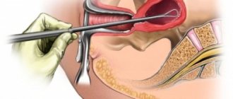
How is curettage of the uterus or cervix done for a polyp?
Curettage of the uterine endometrium for polyps is carried out in the following ways:
- Separate diagnostic curettage (in the abbreviation DVR). This refers to the traditional expansion of the cervical canal and curettage of the polyp. Despite constant progress in medicine, curettage is still used in centers where the latest equipment and advanced technologies are not yet available. The manipulation is carried out almost blindly.
- Separate diagnostic curettage and hysteroscopy. Curettage is performed using a hysteroscope, which provides excellent visualization, precise removal of polyps and prevention of relapses. Another type of manipulation is hysteroresectoscopy of a uterine polyp.
Preparation for the procedure
Image courtesy of meepoohyaphoto on FreeDigitalPhotos.net
Traditionally, a pelvic examination and pap smears are performed to assess the sterility of the vagina and cervix.
The following studies are mandatory before surgery:
- blood, urine, stool tests;
- Ultrasound of the pelvic organs, bladder, uterus and appendages;
- chest x-ray, fluorogram;
- ECG or ultrasound of the heart.
The data obtained help to assess the woman’s general clinical history and choose the optimal method of removal. If necessary, specialists of the relevant profile are involved: endocrinologists, cardiologists, gastroenterologists, infectious disease specialists.
A week before the expected curettage, exclude all medications (vaginal suppositories, tablets, except for vital ones), and stop sexual intercourse.
Algorithm of actions
The technique of performing the operation with and without hysteroscopy does not differ from each other. The main difference is the organization of good visualization of the entire course of the manipulation. The operation is performed in a small operating room on a gynecological chair. The woman is seated with her legs wide apart and anesthesia is administered.
The algorithm is as follows:
- Introduction of expanders;
- Assessing the condition of the uterine mucosa, detecting a polyposis focus;
- Removal of the tumor along with the endometrium;
- Curettage of the entire cavity with a curette in case of multiple lesions.
The removed polyp is sent for histological examination. The histology result after curettage of the endometrial polyp indicates whether there are potential risks of oncological degeneration of the cells.
After cleaning, the woman is taken to the ward until she awakens and stabilizes her condition.
Recommendations after endometrial polyp curettage
Image courtesy of Naypong on FreeDigitalPhotos.net
It is mandatory for a woman to be under the supervision of a doctor for 3-5 hours after waking up from anesthesia. The early postoperative period is accompanied by regular examination of the patient and assessment of her general condition.
The following symptoms are normal:
- nagging pain in the lower abdomen;
- slight bleeding;
- increase in temperature (not higher than 37.5 degrees);
- confusion, nausea;
- lack of appetite.
The discharge after curettage of the polyp should resemble the last day of menstruation. It is normal to change 2 pads per day. After 3-4 days, such symptoms disappear, and the woman returns to her normal life.
Drug therapy
After removal, drug correction is of great importance.
To prevent relapses and secondary complications, the following drugs are prescribed:
- antibiotics (cephalosporins, penicillins);
- antispasmodics and painkillers (Ketorol, Ketanov, Xefokam, No-shpa);
- local antimicrobial drugs (Betadine suppositories, solutions of Furacilin, sodium chloride, chlorhexidine);
- vitamin complexes.
Hormonal correction is selected individually. If a woman plans to give birth to a child, then Duphaston must be prescribed after removal of the endometrial polyp, as well as a course of Utrozhestan and Norkolut. If you do not want to become pregnant in the coming years, a hormonal IUD is installed.
Usually, women are prescribed drugs Janine, Logest, Diane, Tri Mercy and others to restore hormonal levels, regardless of the desire to realize motherhood.
Pregnancy and polyp
The functioning of the female body in general, as well as the reproductive system in particular, is very dependent on the level of sex hormones. Under certain conditions, due to hormonal imbalance, a growth called a polyp can form in a woman’s genitals.
This is a pathological formation, which, of course, should not exist normally. But sometimes its treatment has to be postponed if a woman is pregnant. Let's discuss how a polyp and pregnancy are combined and what every woman needs to know about it.
Is it possible to get pregnant with a polyp?
Essentially, a polyp is a growth of the mucous membrane lining the uterus. Normally, it is constantly updated during monthly menstruation. But with hormonal imbalances and as a result of surgical interventions in the uterine cavity (previous births, abortions), the tissues can grow, forming an endometrial polyp in the uterus or cervix. If it extends beyond the cervix, a polyp of the cervical canal is diagnosed.
Gynecological practice shows that women with this diagnosis often experience difficulty conceiving a child. After all, such an endometrial process impairs the patency of the genital tract, thereby preventing the advancement of the fertilized egg to the uterine cavity. It is also believed that due to hormonal disorders, against the background of which these processes are formed, ovulation is often absent, without which conception cannot occur. If the egg has been fertilized, it may not take root in the uterus, the endometrium of which is affected by a polyp, due to dysfunction of the mucous membranes.
That is why all women planning a pregnancy need to undergo a full examination, which necessarily includes an assessment of the condition of the mammary tract and genital tract.
If a polyp is discovered at the planning stage, it will be removed
Source